Review
. 2025 Apr 3;26(1):26.
doi: 10.1186/s12865-025-00685-9.

Modulating immune responses in alopecia: therapeutic insights and potential targets of antisense oligonucleotides

Affiliations
Review

Modulating immune responses in alopecia: therapeutic insights and potential targets of antisense oligonucleotides

Shahnaz Begum et al. BMC Immunol. .

Abstract

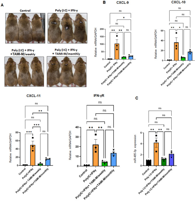
Background: Alopecia areata (AA) are hair loss disorders with distinct pathogenetic mechanisms involving immune dysregulation and microRNA modulation. AA, a T cell-mediated autoimmune disease, is characterized by sudden hair loss, with interferon-gamma (IFN-γ) playing a pivotal role in pathogenesis. The upregulation of IFN response genes, including IFN-inducible chemokines CXCL9, CXCL10, and CXCL11, in lesional skin reflects the activation of the IFN response pathway and contributes to immune cell recruitment and inflammation.

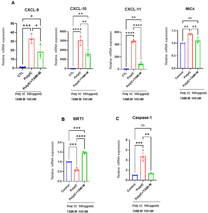
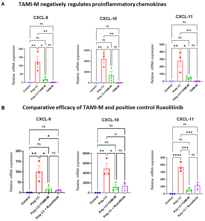
Results: Recent research highlights the role of SIRT1, a class III histone deacetylase, in modulating immune responses in AA. SIRT1 inhibition promotes the production of Th1 cytokines and chemokines, impairing inflammation, while SIRT1 activation suppresses autoreactive responses through NF-κB deacetylation and STAT3 phosphorylation. Additionally, antisense oligonucleotides (ASOs) targeting miR-485-3p show therapeutic potential in promoting hair regrowth and mitigating inflammation in murine models of androgenic alopecia (AGA) and AA.

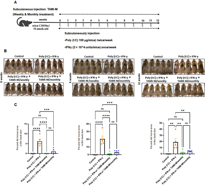
Conclusion: Understanding chemokine dysregulation provides key insights into AA pathogenesis and highlights TAMI-M as a potential therapy for reducing inflammation and promoting hair regeneration. These findings advance the exploration of immune, microRNA, and SIRT1 pathways as targets for novel hair loss treatments.

Keywords: Alopecia areata; Antisense oligonucleotides; C3H/HeJ mice; Outer root sheath; SIRT1.


Conflict of interest statement

Declarations. Ethics approval and consent to participate: The animal experimentation conducted in this study adhered to the National Institutes of Health Guide for the Care and Use of Laboratory Animals and was approved by the Institutional Animal Care and Use Committee (IACUC) of Biorchestra (permit numbers: BOIACUC-20230531-0002, BOIACUC-20220908-0001). Consent for publication: Not applicable. Competing interests: The authors declare no competing interests.

Figures

Fig. 1
Fig. 1
Efficacy of TAMI-M weekly and monthly treatments in C3H/HeJ AA model Mice: A Schematic representation of the experimental design. Fifteen-week-old C3H/HeJ mice were injected subcutaneously with poly I: C and IFN-γ to induce AA-like symptoms. TAMI-M was administered subcutaneously at a dose of 0.8 mg/kg either weekly or monthly, while control animals received PBS (vehicle). Each treatment group consisted of 5 mice (n = 5). B Representative images of hair regrowth and alopecia severity were taken at defined intervals post-treatment on 8, 9 and 11 weeks. Images show the extent of lesional area before and after treatment in the TAMI-M weekly, monthly, and vehicle groups. C Quantitative analysis of AA lesion areas in the dorsal skin of treated and control mice was performed. Statistical significance was determined using one-way ANOVA followed by post hoc comparisons (Tukey’s test). The effects of TAMI-M treatment against AA lesion formation were significant for both dosing regimens in the presence of poly I: C and IFN-γ treatment. Statistical significance: *p < 0.05, **p < 0.01, ***p < 0.001 versus vehicle (before poly I: C induction); *p < 0.05, **p < 0.01, ***p < 0.001 versus vehicle (after poly I: C induction)
Fig. 2
Fig. 2
Effect of TAMI-M treatment on proinflammatory chemokines, and cytokines in C3H/HeJ AA model mice: A C3H/HeJ mice were induced with AA-like symptoms using poly I: C and IFN-γ, mimicking the inflammatory milieu observed in human alopecia areata. TAMI-M was administered subcutaneously at a dose of 0.8 mg/kg weekly or monthly. B Relative expression levels of proinflammatory chemokines and cytokines were measured in dorsal skin samples via qPCR following TAMI-M treatment. Data reflect significant reductions in inflammatory markers in treated groups compared to vehicle controls. C miR-485-3p expression levels were evaluated in the skin of treated and control mice. Weekly and monthly TAMI-M dosing regimens both resulted in substantial downregulation of miR-485-3p expression compared to vehicle controls. Statistical significance was determined using one-way ANOVA followed by post hoc comparisons (Tukey’s test). Statistical significance is indicated as follows: *p < 0.05, **p < 0.01, ***p < 0.001 versus vehicle (before poly I: C induction); *p < 0.05, **p < 0.01, ***p < 0.001 versus vehicle (after poly I: C induction)
Fig. 3
Fig. 3
Effect of TAMI-M on Poly I: C-induced production of proinflammatory chemokines, SIRT1, and Caspase-1 in HORS: A HORS cells were treated with Poly I: C for 3 h to simulate an inflammatory environment resembling alopecia areata pathology. TAMI-M was applied to evaluate its potential effects. Relative mRNA expression levels of proinflammatory chemokines were quantified using qPCR. Treatment with TAMI-M significantly reduced chemokine production compared to Poly I: C-induced controls. B TAMI-M treatment restored SIRT1 expression levels, which were downregulated following Poly I: C induction. C Caspase-1 activity, a marker of inflammasome activation, was measured by qPCR. TAMI-M treatment significantly reduced Caspase-1 activity in HORS cells compared to Poly I: C-treated controls. Statistical significance was determined using one-way ANOVA followed by post hoc comparisons (Tukey’s test). Significant differences are denoted as *p < 0.05, **p < 0.01, and ***p < 0.001 between groups
Fig. 4
Fig. 4
Effects of TAMI-M on Polyinosinic: polycytidylic acid-induced production of proinflammatory chemokines in HORS Cells. A mRNA expression levels of CXCL9, CXCL10, and CXCL11 in human ORS cells treated with Poly I: C (10 µg/mL) and TAMI-M (100 nM) for 24 h. B mRNA expression levels of CXCL9, CXCL10, and CXCL11 in human ORS cells treated with Poly I: C, TAMI-M, and the positive control Ruxolitinib (100 nM) for 24 h. Statistical significance was determined using one-way ANOVA followed by post hoc comparisons (Tukey’s test). Significant differences are denoted as *p < 0.05, **p < 0.01, and ***p < 0.001 between groups. This figure illustrates the differential impact of TAMI-M and Ruxolitinib on the expression of chemokines associated with AA pathogenesis
Fig. 5
Fig. 5
The counteractive effect of SIRT1 on NF-κB signaling by TAMI-M in vivo. A Immunoblotting analysis of SIRT1, AR, and CD36 expression in control, DHT-induced AGA mice, and TAMI-M treated mice (2 × 0.6 mg/kg, subcutaneously). B Expression of inflammatory markers: IL-6, P-STAT3, and P-NF-κB in the same groups. C Expression of TNF-α, HSP70, and NLRP3 in the control, DHT-induced AGA, and TAMI-M treated groups. Relative protein expression levels were quantified and normalized to β-actin. Individual data points are shown to illustrate variation within groups. Statistical significance was determined using one-way ANOVA followed by post hoc comparisons (Tukey’s test). Significant differences are indicated as *p < 0.05, **p < 0.01, and ***p < 0.001

Similar articles

References

    1. Suchonwanit P, Kositkuljorn C, Pomsoong C. Alopecia Areata: an autoimmune disease of multiple players. ImmunoTargets Therapy. 2021;10:299–312. - PMC - PubMed
    1. Strazzulla LC, Wang EHC, Avila L, Lo Sicco K, Brinster N, Christiano AM, et al. Alopecia Areata: Disease characteristics, clinical evaluation, and new perspectives on pathogenesis. J Am Acad Dermatol. 2018;78:1–12. - PubMed
    1. Xing L, Dai Z, Jabbari A, Cerise JE, Higgins CA, Gong W, et al. Alopecia Areata is driven by cytotoxic T lymphocytes and is reversed by JAK inhibition. Nat Med. 2014;20:1043–9. - PMC - PubMed
    1. Rathnayake D, Sinclair R. Male androgenetic alopecia. Expert Opin Pharmacother. 2010;11:1295–304. - PubMed
    1. Yip L, Sinclair RD. Antiandrogen therapy for androgenetic alopecia. Expert Rev Dermatol. 2006;1:261–9.

LinkOut - more resources