Review
. 2025 Apr 3;23(1):392.
doi: 10.1186/s12967-025-06392-8.

Gene therapy shines light on congenital stationary night blindness for future cures

Affiliations
Review

Gene therapy shines light on congenital stationary night blindness for future cures

Yi Zhang et al. J Transl Med. .

Abstract

Congenital Stationary Night Blindness (CSNB) is a non-progressive hereditary eye disease that primarily affects the retinal signal processing, resulting in significantly reduced vision under low-light conditions. CSNB encompasses various subtypes, each with distinct genetic patterns and pathogenic genes. Over the past few decades, gene therapy for retinal genetic disorders has made substantial progress; however, effective clinical therapies for CSNB are yet to be discovered. With the continuous advancement of gene-therapy tools, there is potential for these methods to become effective treatments for CSNB. Nonetheless, challenges remain in the treatment of CSNB, including issues related to delivery vectors, therapeutic efficacy, and possible side effects. This article reviews the clinical diagnosis, pathogenesis, and associated mutated genes of CSNB, discusses existing animal models, and explores the application of gene therapy technologies in retinal genetic disorders, as well as the current state of research on gene therapy for CSNB.

Keywords: Animal models; Congenital stationary night blindness; Full-field electroretinography; Gene editing; Gene therapy; Inherited retinal disease; Retina.


Conflict of interest statement

Declarations. Ethics approval and consent to participate: Not applicable. Consent for publication: Not applicable. Competing interests: Part of our work was supported by the grant from Hangzhou Bipolar Biotechnology Co., Ltd. Qingyang Ye from Hangzhou Bipolar Biotechnology Co., Ltd. has a role in the preparation of the manuscript.

Figures

Fig. 1
Fig. 1
Diagram of the visual pathway in retina. When light passes through the cornea and vitreous body of the eye and is focused onto the retina, the photoreceptor cells convert the light signal into an electrical signal. This signal is then transmitted step by step via BCs and other structures, eventually being relayed to the brain through ganglion cells, where it generates visual perception.
Fig. 2
Fig. 2
Full-field electroretinography (ffERG) testing. (A). ERG testing diagram: Two electrodes are placed on the patient’s forehead and lower eyelid. When the patient is exposed to stimuli from a flash stimulator, the eye generates corresponding electrical currents. These currents are amplified by a signal amplifier and displayed as waveforms in the program. (B). ERG of a normal individual: According to the standards set by the ISCEV, the CSNB waveform of a normal individual is shown as depicted
Fig. 3
Fig. 3
Clinical and molecular characteristics of CSNB. (A). Upper: Based on clinical characteristics, CSNB can be categorized into three main types: normal fundus type, abnormal fundus type, and GNB3 type; Lower: Although CSNB is a rare disease, it exhibits multiple inheritance patterns, including X-linked CSNB, autosomal recessive (arCSNB) and autosomal dominant (adCSNB). (B). CSNB related genes and their distribution: Mutations associated with CSNB are primarily located in rod cells and BCs, which explains why the visual impairment in CSNB patients mainly occurs in the signal transmission between photoreceptor cells and BCs
Fig. 4
Fig. 4
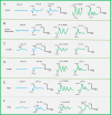
Electrophysiological findings of different CSNB patients. (A). ERG of OD patients: OD patients exhibit abnormal fundus. The ERG shows a complete disappearance of the b-wave under dark-adapted (DA) 0.01 conditions. Subsequent tests reveal a reduction in both a- and b-waves, while the light-adapted (LA) response is generally normal [14]. (B). ERG of FA patients: FA patients have white spots in the fundus. The ERG is characterized by a severely reduced b-wave under DA 0.01 conditions, and both a- and b-waves are reduced under DA 10.0 conditions [17]. (C). ERG of icCSNB patients: icCSNB patients have a normal fundus. Under DA 0.01 conditions, the b-wave is significantly reduced but still detectable. Under DA 10.0 conditions, the b-wave shows further reduction while the a-wave remains normal. Under LA 3.0 30 Hz conditions, both a- and b-waves are reduced and delayed. Similarly, under LA 3.0, both a- and b-waves show reductions [34]. (D). ERG of cCSNB patients: cCSNB patients have a normal fundus. The b-wave is completely absent under DA 0.01 conditions, while under DA 10.0, the b-wave is significantly reduced but the a-wave remains normal. Under LA 3.0 30 Hz conditions, the waveform broadens, and under LA 3.0, the a-wave is normal but the trough broadens and the b-wave becomes steeper [34]. (E). ERG of Riggs patients: Riggs patients have a normal fundus. Under DA 0.01 conditions, the b-wave is severely reduced or absent, and both a- and b-waves are reduced under DA 10.0 conditions. Their light-adapted response is mostly normal [28]. (F). ERG of GNB3 related CSNB: GNB3 patients are rare and show varying phenotypes. In the cases mentioned, the ERG mainly shows a reduction in the b-wave under DA 0.01 or DA 10.0 conditions. Under LA 3.0 30 Hz, the ERG may show weakening or delays, while under LA 3.0, the a-wave is normal but may be delayed, and the b-wave is weakened and delayed [33]
Fig. 5
Fig. 5
Rod phototransduction cascade. Upon absorbing photons, rhodopsin becomes activated and interacts with the G protein transducin (GNAT1), leading to the activation of the α subunit of transducin. The activated α subunit then binds to the γ subunit of phosphodiesterase-6 (PDE6), subsequently activating the αβ subunits of PDE6. The activated phosphodiesterase reduces the intracellular levels of cGMP, causing the closure of cGMP-regulated cation channels [35]
Fig. 6
Fig. 6
Signal transmission between photoreceptors and BCs. (A). A cone pedicle, located at the synaptic terminal of cone cells, features synaptic ribbons situated on the invaginated dendrites of HCs and ON bipolar cells (ON BC). This synaptic arrangement is referred to as the “triad” structure. Multiple synaptic ribbons are present at the cone cell terminal. The dendrites of OFF bipolar cells (OFF BC) form contacts at the base of the cone pedicle. (B). A rod spherule, located at the synaptic terminal of rod cells, contains a single synaptic ribbon at the terminal, positioned on the invaginated dendrites of HC and ON BC. The dendrites of OFF BC form contacts at the base of the rod spherule. (C). Transmission of light signals between photoreceptors and BCs. In well-lit conditions, the release of glutamate into the synaptic cleft decreases. This leads to the depolarization of ON BCs, while the reduction in glutamate signals received by the AMPA or kainate receptors of OFF BCs causes their hyperpolarization. In contrast, under dark conditions, glutamate release increases. The mGluR6 receptors on ON BC receive the glutamate, causing hyperpolarization of ON BC, while OFF BC depolarize. The specific mechanism of Lrit3 remains unclear
Fig. 7
Fig. 7
Possible gene therapy strategies for CSNB. (A). CRISPR-mediated gene editing: Taking Cas9 as an example, gene editing occurs by creating a double-strand break at the target site, followed by editing through the DNA repair mechanisms, including HDR (Homology-Directed Repair), NHEJ (Non-Homologous End Joining), or HITI (Homology-Independent Targeted Integration). (B). Single-base editing: Classic single-base editing does not require cutting the DNA strand; instead, it achieves single-base conversion through deamination reactions. Currently, various types of base editors have been developed, as illustrated in the image, including CBE (Cytosine Base Editor), ABE (Adenine Base Editor), and CGBE (Cytosine-Guanine Base Editor). (C). Prime editing: A novel editing approach that introduces specific sequences from the RT template of pegRNA through reverse transcription after cutting one strand of DNA, theoretically allowing the introduction of any mutation at any position. (D). RNA regulation: The process of correcting erroneous RNA expression by regulating RNA splicing or processing through ASO (Antisense Oligonucleotides) or siRNA (small interfering RNA)
Fig. 8
Fig. 8
Different ocular drug delivery methods
Fig. 9
Fig. 9
Comparison of different delivery carriers in ophthalmic diseases. The primary vectors for gene therapy in the eye are either viral or non-viral. Viral vectors include lentivirus, adenovirus, AAV, and herpes simplex virus, with AAV being widely used in clinical applications. Among non-viral vectors, nanoparticles such as lipid nanoparticles (LNPs) and virus-like particles (VLPs) are prominent. However, since these technologies have emerged more recently, most remain in the preclinical stage
Fig. 10
Fig. 10
Current results of gene therapy for CSNB. The currently published successful cases of gene therapy for CSNB primarily involve animal models, including Nyxnob mice, Lrit3−/− mice, and Beagle dog models

References

    1. Masland RH. The fundamental plan of the retina. Nat Neurosci. 2001;4:877–86. - PubMed
    1. Baden T, Euler T. Retinal physiology: Non-Bipolar-Cell excitatory drive in the inner retina. Curr Biol. 2016;26:R706–8. - PubMed
    1. Zeitz C. Molecular genetics and protein function involved in nocturnal vision. Expert Rev Ophthalmo. 2007;2:467–85.
    1. Russell S, Bennett J, Wellman JA, Chung DC, Yu ZF, Tillman A, Wittes J, Pappas J, Elci O, McCague S, et al. Efficacy and safety of voretigene neparvovec (AAV2-hRPE65v2) in patients with RPE65-mediated inherited retinal dystrophy: a randomised, controlled, open-label, phase 3 trial. Lancet. 2017;390:849–60. - PMC - PubMed
    1. Kim AH, Liu P-K, Chang Y-H, Kang EY, Wang H-H, Chen N, Tseng Y-J, Seo GH, Lee H, Liu L, et al. Congenital stationary night blindness: clinical and genetic features. Int J Mol Sci. 2022;23:14965. - PMC - PubMed

Supplementary concepts

LinkOut - more resources