

A non-fluorescent immunohistochemistry method for measuring autophagy flux using MAP1LC3/LC3 and SQSTM1 as core markers
- PMID: 40181489
- PMCID: PMC12127880
- DOI: 10.1002/2211-5463.70014
A non-fluorescent immunohistochemistry method for measuring autophagy flux using MAP1LC3/LC3 and SQSTM1 as core markers
Abstract
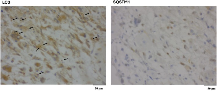
Macroautophagy/autophagy is a crucial cellular process for degrading and recycling damaged proteins and organelles, playing a significant role in diseases such as cancer and neurodegeneration. Evaluating autophagy flux, which tracks autophagosome formation, maturation, and degradation, is essential for understanding disease mechanisms. Current fluorescence-based methods are resource-intensive, requiring advanced equipment and expertise, limiting their use in clinical laboratories. Here, we introduce a non-fluorescent immunohistochemistry (IHC) method using MAP1LC3/LC3 and SQSTM1 as core markers for autophagy flux assessment. LC3 levels reflect autophagosome formation, whereas SQSTM1 degradation and a decrease in the number of its puncta indicate active flux (i.e., lysosomal turnover). We optimized chromogenic detection using diaminobenzidine (DAB) staining and developed a scoring system based on puncta number and the percentage of stained cells. This accessible, cost-effective method enables reliable autophagy quantification using a standard light microscope, bridging the gap between experimental research and clinical diagnostics. Our protocol allows accurate autophagy evaluation in fixed tissues, offering practical applications in biomedical research and clinical pathology assessment.
Keywords: autophagometer; autophagy flux measurement; cellular homeostasis analysis; chromogenic detection; cost‐effective autophagy assay; non‐fluorescent immunohistochemistry.
© 2025 The Author(s). FEBS Open Bio published by John Wiley & Sons Ltd on behalf of Federation of European Biochemical Societies.
Conflict of interest statement
M.C. has received honoraria/consultation contracts from EQA Certificados and OCA Global. The other authors declare no conflict of interest.
Figures
Update of
-
Unlocking a New Path: An Autophagometer that Measures Flux Using a Non-Fluorescent Immunohistochemistry Method.bioRxiv [Preprint]. 2024 Jun 30:2024.06.26.600741. doi: 10.1101/2024.06.26.600741. bioRxiv. 2024. Update in: FEBS Open Bio. 2025 Jun;15(6):898-905. doi: 10.1002/2211-5463.70014. PMID: 38979364 Free PMC article. Updated. Preprint.
Similar articles
-
Immunohistochemical Detection of the Autophagy Markers LC3 and p62/SQSTM1 in Formalin-Fixed and Paraffin-Embedded Tissue.Methods Mol Biol. 2017;1560:189-194. doi: 10.1007/978-1-4939-6788-9_13. Methods Mol Biol. 2017. PMID: 28155154
-
Redundancy of human ATG4 protease isoforms in autophagy and LC3/GABARAP processing revealed in cells.Autophagy. 2019 Jun;15(6):976-997. doi: 10.1080/15548627.2019.1569925. Epub 2019 Feb 1. Autophagy. 2019. PMID: 30661429 Free PMC article.
-
Immunohistochemical Detection of the Autophagy Markers LC3 and p62/SQSTM1 in Formalin-Fixed and Paraffin-Embedded Tissue.Methods Mol Biol. 2023;2566:133-139. doi: 10.1007/978-1-0716-2675-7_10. Methods Mol Biol. 2023. PMID: 36152247
-
Size, organization, and dynamics of soluble SQSTM1 and LC3-SQSTM1 complexes in living cells.Autophagy. 2016 Sep;12(9):1660-74. doi: 10.1080/15548627.2016.1199299. Epub 2016 Jul 21. Autophagy. 2016. PMID: 27442348 Free PMC article.
-
The Molecular Mechanism and Therapeutic Application of Autophagy for Urological Disease.Int J Mol Sci. 2023 Oct 4;24(19):14887. doi: 10.3390/ijms241914887. Int J Mol Sci. 2023. PMID: 37834333 Free PMC article. Review.
References
-
- Vargas JNS, Hamasaki M, Kawabata T, Youle RJ and Yoshimori T (2023) The mechanisms and roles of selective autophagy in mammals. Nat Rev Mol Cell Biol 24, 167–185. - PubMed
-
- Cordani M, Strippoli R, Trionfetti F, Barzegar Behrooz A, Rumio C, Velasco G, Ghavami S and Marcucci F (2024) Immune checkpoints between epithelial‐mesenchymal transition and autophagy: a conflicting triangle. Cancer Lett 585, 661. - PubMed
-
- Alizadeh J, da Silva Rosa SC, Weng X, Jacobs J, Lorzadeh S, Ravandi A, Vitorino R, Pecic S, Zivkovic A, Stark H et al. (2023) Ceramides and ceramide synthases in cancer: focus on apoptosis and autophagy. Eur J Cell Biol 102, 337. - PubMed
-
- Alizadeh J, Kochan MM, Stewart VD, Drewnik DA, Hannila SS and Ghavami S (2021) Inhibition of autophagy flux promotes secretion of chondroitin sulfate proteoglycans in primary rat astrocytes. Mol Neurobiol 58, 6077–6091. - PubMed
MeSH terms
Substances
Grants and funding
LinkOut - more resources
Full Text Sources
Miscellaneous
