. 2025 Apr;15(4):e70308.
doi: 10.1002/brb3.70308.

Multiplex Immunofluorescent Batch Labeling of Marmoset Brain Sections

Affiliations

Multiplex Immunofluorescent Batch Labeling of Marmoset Brain Sections

Daryan Chitsaz et al. Brain Behav. 2025 Apr.

Abstract

Purpose: The common marmoset is a small nonhuman primate that has emerged as a valuable animal model in neuroscience research. Accurate analysis of brain tissue is crucial to understand marmoset neurophysiology and to model neurodegenerative diseases. Many studies to date have complemented magnetic resonance imaging (MRI) with histochemical staining rather than immunofluorescent labeling, which can generate more informative and higher resolution images. There is a need for high-throughput immunolabeling and imaging methodologies to generate resources for the burgeoning marmoset field, particularly brain histology atlases to display the organization of different cell types and other structures.

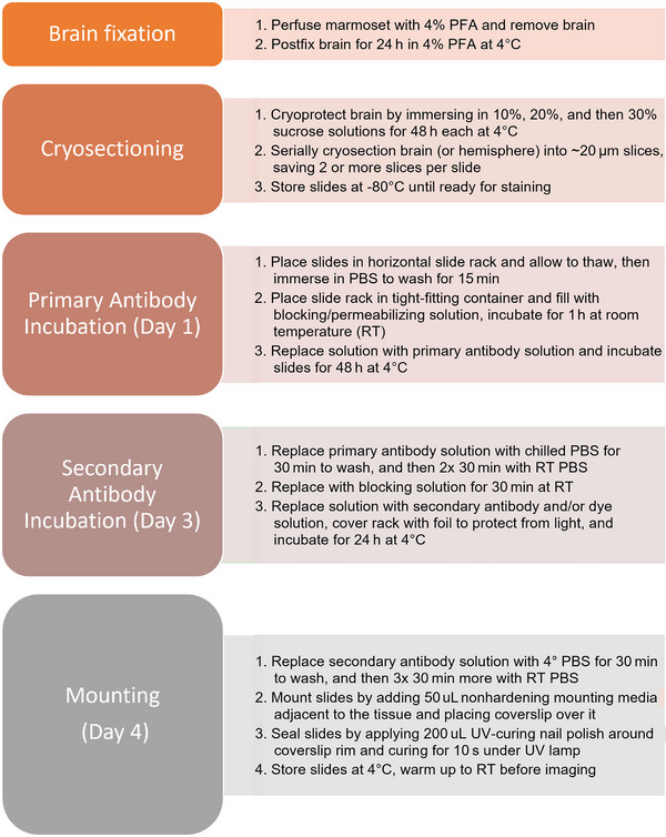
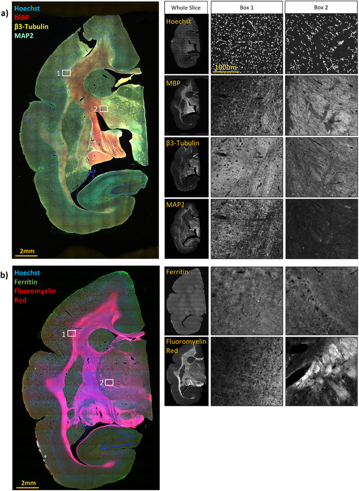
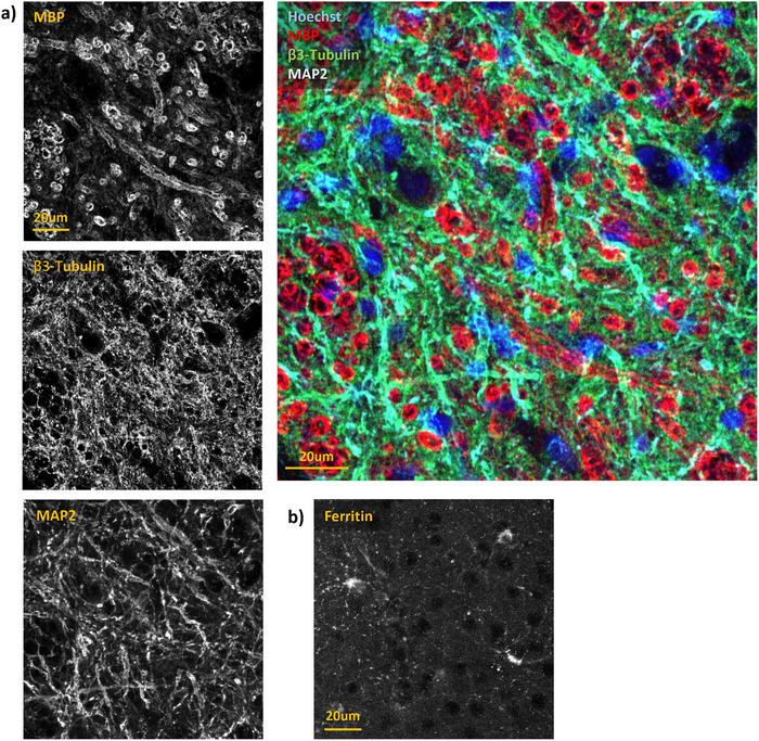
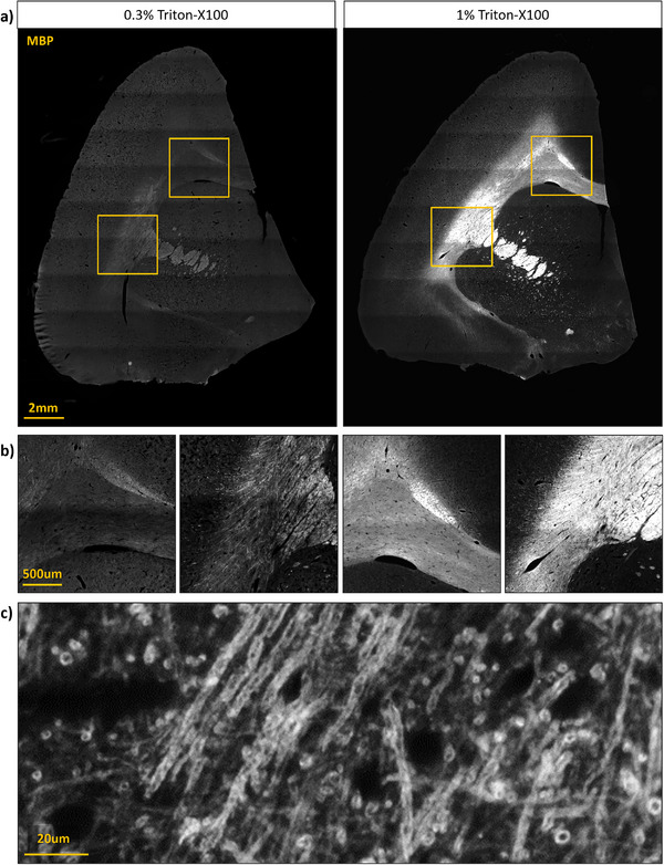
Methods and findings: Here, we have characterized a set of marmoset-compatible fluorescent dyes and antibodies that label myelin, axons, dendrites, and the iron-storage protein ferritin, and developed a batch-style multiplex immunohistochemistry protocol to uniformly process large numbers of tissue slides for multiple cell-type specific markers.

Conclusion: We provide a practical guide for researchers interested in harnessing the potential of marmoset models to advance understanding of brain structure, function, and pathophysiology.

Keywords: high‐throughput; histology; immunofluorescence; marmoset; myelin.


Conflict of interest statement

The authors declare no conflicts of interest.

Figures

FIGURE 1
FIGURE 1
Batch‐style slide immunolabeling protocol. Brain fixation, cryopreservation, and sectioning takes at least 1 week after which slides can be stored until immunolabeling, which requires 4 days. All incubation steps should be carried out on a mechanical shaker. PBS, phosphate‐buffered saline; UV, ultraviolet.
FIGURE 2
FIGURE 2
Photomicrographs of multiplexed immunolabeling on hemisections of marmoset brain. The middle and rightmost columns present magnified images of the boxed regions on the multicolor images. (A) Hoechst dye with immunolabeling for Myelin Basic Protein (MBP), β3‐tubulin, and Microtubule‐Associated Protein 2 (MAP2). (B) Shows α‐ferritin labeling and staining with the dye Fluoromyelin Red.
FIGURE 3
FIGURE 3
Higher magnification confocal microscope images of the distribution of labeling obtained for each of the four antibodies. (A) Multichannel images show myelin sheaths immunolabeled for Myelin Basic Protein (MBP), β3‐tubulin labeling in axons, and Microtubule‐Associated Protein 2 (MAP2) labeling in dendrites, with an enlarged merged image. (B) Ferritin immunolabeling is highly punctate and concentrated in glia. The ferritin image is from a different tissue section from that shown in (A).
FIGURE 4
FIGURE 4
Comparing permeabilization conditions for Myelin Basic Protein (MBP) immunolabeling. (A) Marmoset brain hemisection permeabilized with 0.3% Triton X‐100 on the left, and then after relabeling with 1.0% Triton X‐100 permeabilization. (B) Left and right images present magnified images of the top and bottom boxes, respectively, revealing brighter and more detailed MBP labeling. (C) Higher magnification image of myelinated axons in the corpus callosum, from a separate section permeabilized with 1.0% Triton X‐100, in which individual myelin sheaths are clearly resolved.

Similar articles

References

    1. Alexander, J. E. , Hunt D. F., Lee M. K., et al. 1991. “Characterization of Posttranslational Modifications in Neuron‐Specific Class III Beta‐Tubulin by Mass Spectrometry.” PNAS 88, no. 11: 4685–4689. - PMC - PubMed
    1. Ambasudhan, R. , Talantova M., Coleman R., et al. 2011. “Direct Reprogramming of Adult Human Fibroblasts to Functional Neurons Under Defined Conditions.” Cell Stem Cell 9, no. 2: 113–118. - PMC - PubMed
    1. Atapour, N. , Majka P., Wolkowicz I. H., Malamanova D., Worthy K. H., and Rosa M. G. P.. 2019. “Neuronal Distribution Across the Cerebral Cortex of the Marmoset Monkey (Callithrix jacchus).” Cerebral Cortex (New York, N.Y.: 1991) 29, no. 9: 3836–3863. - PubMed
    1. Friedrich, I. , Reimann K., Jankuhn S., et al. 2021. “Cell Specific Quantitative Iron Mapping on Brain Slices by Immuno‐µPIXE in Healthy Elderly and Parkinson's Disease.” Acta Neuropathologica Communications 9, no. 1: 47. - PMC - PubMed
    1. Fukunaga, M. , Li T. Q., van Gelderen P., et al. 2010. “Layer‐Specific Variation of Iron Content in Cerebral Cortex as a Source of MRI Contrast.” PNAS 107, no. 8: 3834–3839. - PMC - PubMed

LinkOut - more resources