. 2025 Apr;286(4):e70044.
doi: 10.1002/jmor.70044.

Perichordal Vertebral Column Formation in Rana kobai

Affiliations

Perichordal Vertebral Column Formation in Rana kobai

Yu Takahashi et al. J Morphol. 2025 Apr.

Abstract

The vertebral column of anurans exhibits morphological diversity that is often used in phylogenetic studies. The family Ranidae is one of the ecologically most successful groups of anurans, with the genus Rana being distributed broadly in Eurasia. However, there are relatively sparse detailed studies on the development of the vertebral column in Rana species, and images of the entire axial skeleton have seldom been illustrated till date. Here, we provide an illustrated description on the development of the entire vertebral column in Rana kobai, a Japanese small frog from the Amami Islands. Our observation of double-stained skeletal specimens revealed that in R. kobai, the original atlas and the first dorsal are fused into one vertebra, and the ninth neural arch is fused with the tenth arch in half of the examined larvae. Anuran vertebral column development is classified into two modes, perichordal and epichordal. Rana species undergo the typical perichordal mode of centrum formation. Kemp and Hoyt (1969) described that centrum formation in R. pipiens starts from a saddle-shaped bone on the dorsal half of the notochord. Nevertheless, our detailed observations revealed that centrum ossification initially emerges at the base of the paired neural arches and then forms the saddle-shaped bone. In Xenopus, a species with epichordal centra, centrum formation starts from a pair of ovoid bone elements at the base of the neural arches. Overall, our results imply that centrum ossification starts from the base of neural arches in anurans, irrespective of whether it is perichordal or epichordal. Our observations also revealed the presence of the crescent-shaped cartilage domain in the intervertebral region in R. kobai. The location of the crescent-shaped domain in R. kobai is consistent with that of the intercentrum in Ichthyostega and several temnospondyls. Based on our observations, we propose a hypothesis on the difference between perichordal and epichordal modes in light of evolution.

Keywords: Anura; Rana; centrum; vertebral column.


Conflict of interest statement

The authors declare no conflicts of interest.

Figures
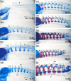
Figure 1
Figure 1
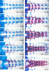
Overview of neural arch development in Rana kobai. Lateral views of (a) St. 27, (b) early St. 29, (c) late St. 29, (d) St. 31, (e) St. 33, (f) St. 35, (g) St. 37, (h) St. 38, and (i) higher magnification of St. 38 larvae. Arrowheads in (a) indicate three pairs of lamina primordia. Arrows in (d), (e), (g), and (h) show the bifurcation of the distal ends of the 1 + 2 arches. Magenta arrowheads show the remains of spinal foramina. lm, lamina; pd, pedicle; poz, postzygapophysis; prz, prezygapophysis; tp, transverse process. The scale bar in (a) is common among (a), (b), (c), and (i). The scale bar in (d) is common among (d) to (h).
Figure 2
Figure 2
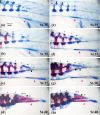
Progression of neural arch ossification and centrum formation in the lateral view of Rana kobai larvae. (a) St. 31, (b–e) St. 33, (f–h) St. 35, (i) St. 37, and (j) St. 40 larvae. Arrows in (a), (b), (c), (e), (g), and (j) show the bifurcation or abnormal shape of the lamina of the 1 + 2 arches. The scale bar in (a) is common among (a) to (j).
Figure 3
Figure 3
Progression of neural arch ossification and centrum formation in the ventral view of Rana kobai larvae. (a) St. 31, (b–e) St. 33, (f–h) St. 35, (i) St. 37, (j) and St. 40 larvae. Arrows in (b) and (c) indicate the cartilaginous cylindrical centrum primordia. The scale bar in (a) is common among (a) to (h). The scale bar in (i) is common between (i) and (j).
Figure 4
Figure 4
Regionalization of the intervertebral region during the larval development of Rana kobai. (a) A St. 40 froglet showing a wedge‐like cartilage domain posteroventral to each centrum (arrowheads). (b) the same froglet as in (a), showing crescent‐shaped domains (white arrowheads) ventral to the notochord in the ventrolateral view. (c) Another St. 40 froglet showing a wedge‐like cartilage domain posteroventral to each centrum (arrowheads). The scale bar in (a) is common among (a) to (c).
Figure 5
Figure 5
Postsacral vertebrae and urostyle development in Rana kobai. (a, b, e) St. 35 larvae with the tenth arches of postsacral vertebrae. (c, f) St. 37 larvae with 10 + 11 arches. (g) a St. 38 larva showing 10 + 11 + 12 fused arches. (d, h) St. 40 froglets exhibiting 10 + 11 + 12 fused arches and regression of the notochord resulting in closure of postsacral arches and the hypochord. Here, two morphotypes are presented; (a–d) Fusion type, in which the ninth arch fuses with the tenth arch. (e–h) Separate type, in which the ninth arch is separated from the tenth arch. The difference in the relationship between the ninth and tenth arches in each type is shown (d, h). Black arrowheads, spinal foramina; magenta arrowheads, hypochord. The scale bar in (a) is common among (a) to (h).
Figure 6
Figure 6
Rib and transverse process development in Rana kobai. (a) St. 31, (b) St. 33, (c) St. 35, (d) St. 37 larvae, and (e) St. 40 froglet in the dorsal view. The fifth to eighth transverse processes elongate as rib‐like processes only at the late stage. The scale bar in (a) is common among (a) to (e).
Figure 7
Figure 7
Difference between perichordal and epichordal modes of centrum formation shown by whole‐mount skeletal specimens slightly before the completion of metamorphosis. (a) Lateral view of a Xenopus laevis larva at St. 61, (b) lateral view of a Rana kobai larva at St. 38. Note that the centrum and intervertebral region are formed around the notochord in (b) and are located dorsally to the thick notochord in (a).
Figure 8
Figure 8
A hypothesis for the derivation of perichordal and epichordal modes in the evolutionary background. The ancestral temnospondyl had mechanisms forming a pair of pleurocentra at the base of neural arches and an intercentrum at the ventral midline, in addition to the cartilage layer around the notochord. The anuran ancestor lost the pleurocentrum and intercentrum as discrete skeletal elements. However, the mechanisms inducing skeletal elements at the base of neural arches are partially maintained, so that centrum ossification precedes at these regions. In addition, a crescent‐shaped cartilage domain is formed at the ventral midline in the intervertebral region. In the epichordal lineage, including Xenopus, the trunk notochord tends to regress, and the mechanisms inducing the cartilage layer around the notochord are severely diminished. This promoted the secondary development of pleurocentrum‐like skeletal elements at the base of neural arches. Furthermore, the Xenopus larva forms a transient cartilage ventral to the notochord. Thus, the difference between perichordal and epichordal modes can be perceived as the difference in the balance between the two mechanisms.

Similar articles

References

    1. Adolphi, H. 1892. “Über Variationen der Spinalnerven und der Wirbelsäule anurer Amphibien. I. (Bufo variabilis Pall.).” Gegenbaurs Morphologisches Jahrbuch 19: 313–375.
    1. Adolphi, H. 1895. “Über Variationen der Spinalnerven und der Wirbelsäule anurer Amphibien. II. (Pelobates fuscus Wagl. und Rana esculenta L.).” Gegenbaurs Morphologisches Jahrbuch 22: 449–490.
    1. Anderson, J. S. , Reisz R. R., Scott D., Fröbisch N. B., and Sumida S. S.. 2008. “A Stem Batrachian From the Early Permian of Texas and the Origin of Frogs and Salamanders.” Nature 453, no. 7194: 515–518. 10.1038/nature06865. - DOI - PubMed
    1. Báez, A. M. , and Nicoli L.. 2004. “A New Look at an Old Frog: The Jurassic Notobatrachus Reig From Patagonia.” Ameghiniana 41: 257–270.
    1. Banbury, B. , and Maglia A. M.. 2006. “Skeletal Development of the Mexican Spadefoot, Spea multiplicata (Anura: Pelobatidae).” Journal of Morphology 267, no. 7: 803–821. 10.1002/jmor.10441. - DOI - PubMed

LinkOut - more resources