. 2025 Apr 25;136(9):1031-1048.
doi: 10.1161/CIRCRESAHA.124.325475. Epub 2025 Apr 4.

Piezo1 in PASMCs: Critical for Hypoxia-Induced Pulmonary Hypertension Development

Affiliations

Piezo1 in PASMCs: Critical for Hypoxia-Induced Pulmonary Hypertension Development

Fenja Knoepp et al. Circ Res. .

Abstract

Background: Pulmonary hypertension (PH) is a life-threatening and progressive yet incurable disease. The hallmarks of PH comprise (1) sustained contraction and (2) excessive proliferation of pulmonary arterial smooth muscle cells (PASMCs). A major stimulus to which PASMCs are exposed during PH development is altered mechanical stress, originating from increased blood pressure, changes in blood flow velocity, and a progressive stiffening of pulmonary arteries. Mechanosensitive ion channels, including Piezo1 (Piezo-type mechanosensitive ion channel component-1), perceive such mechanical stimuli and translate them into a variety of cellular responses, including contractility or proliferation. Thus, the objective of the present study was to elucidate the specific role of Piezo1 in PASMCs for PH development and progression.

Methods: The cell-type specific function of Piezo1 in PH was assessed in (1) PASMCs and lung tissues from patients with PH and (2) 2 mouse strains characterized by smooth muscle cell-specific, conditional Piezo1 knockout. Taking advantage of these strains, the smooth muscle cell-specific role of Piezo1 in PH development and progression was assessed in isolated, perfused, and ventilated mouse lungs, wire myography, and proliferation assays. Finally, in vivo function of smooth muscle cell-specific Piezo1 knockout was evaluated upon induction of chronic hypoxia-induced PH in these mice with insights into pulmonary vascular cell senescence.

Results: Compared with healthy controls, PASMCs from patients with PH featured an elevated Piezo1 expression and increased proliferative phenotype. Smooth muscle cell-specific Piezo1 deletion, as confirmed via quantitative real-time polymerase chain reaction and patch clamp recordings, prevented the hypoxia-induced increase in PASMC proliferation in mice. Moreover, Piezo1 knockout reduced hypoxic pulmonary vasoconstriction in isolated, perfused, and ventilated mouse lungs, endothelial-denuded pulmonary arteries, and hemodynamic measurements in vivo. Consequently, Piezo1-deficient mice were considerably protected against chronic hypoxia-induced PH development with ameliorated right heart hypertrophy and improved hemodynamic function. In addition, distal pulmonary capillaries were preserved in the Piezo1-knockout mice, associated with a lower number of senescent endothelial cells.

Conclusions: This study provides evidence that Piezo1 expressed in PASMCs is critically involved in the pathogenesis of PH by controlling pulmonary vascular tone, arterial remodeling, and associated lung capillary rarefaction due to endothelial cell senescence.

Keywords: blood pressure; cellular senescence; endothelial cells; hypertension, pulmonary; hypoxia; vascular remodeling.


Conflict of interest statement

None.

Figures
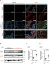
Figure 1.
Figure 1.
Elevated Piezo1 expression in the medial layer of pulmonary vessels in patients with idiopathic pulmonary arterial hypertension (IPAH). A, Representative photomicrographs of confocal immunofluorescence staining of Piezo1 (white), α-SMA (α-smooth muscle actin; red), elastin (green), and 4′,6-diamidino-2-phenylindole (DAPI; blue) in lung tissue of 2 patients with IPAH and respective controls from healthy donors. Remodeled pulmonary vessels of patients with IPAH depict an enhanced expression of Piezo1 in the medial layer of pulmonary vessels, as confirmed by double-staining against SMA. Scale bars, 50 µm. B, Western blot analysis and (C and D) densitometry of Piezo1 expression in pulmonary arterial smooth muscle cells (PASMCs) from pulmonary hypertension (PH; n=6) and control patients (n=6). Differences were assessed via the unpaired t test and displayed as P value.
Figure 2.
Figure 2.
Piezo1 expression and validation of knockdown in pulmonary arterial smooth muscle cells (PASMCs). A, Sections of pulmonary arteries (PAs) from control Piezo1+/+ and reporter Piezo1LacZ/+ mice demonstrating expression of Piezo1 (blue staining) in the smooth muscle layer of PAs. B, Quantitative real-time polymerase chain reaction (qRT-PCR) data illustrating the progressive knockdown of Piezo1 in PAs of Piezo1lox/lox (n=4), Piezo1lox/loxCre (n=3), Piezo1del/lox (n=3), and Piezo1 del/loxCre (n=4) mice. C, No change in the body weight upon Piezo1 deletion was observed (Piezo1lox/lox [n=5], Piezo1lox/loxCre [n=6], Piezo1 del/lox [n=5], and Piezo1 del/loxCre [n=4]). D, Cell-attached patch clamp recordings of Piezo1 currents in response to fast pressure pulse stimulation, recorded at a holding potential of −80 mV (top) and at the predicted reversal potential for Piezo1 (0 mV; bottom). E, Pressure-effect curves for Piezo1 current amplitude in Piezo1lox/lox (n=13) and Piezo1del/loxCre mice (n=18). Differences were assessed via the Mann-Whitney U test and displayed as P values.
Figure 3.
Figure 3.
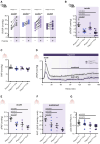
Piezo1 deficiency diminishes the contractility of small intrapulmonary arteries. A, Schematic representation of the procedure of wire myography experiments on endothelial-denuded intrapulmonary arteries (IPAs) of Piezo1-deficient mice and their respective controls. Arteries were first exposed to a wake-up protocol to confirm the viability and assess the general contractility of the vessels. Therefore, the arteries were stimulated 3-fold with exposure to potassium-enriched physiological saline solution (KPSS), separated by wash-out steps. As the final part of the wake-up procedure, phenylephrine (PE, 10 µmol/L) was added to the bath solution. The removal of the endothelium was confirmed by application of acetylcholine (ACh, 10 µmol/L). Only those arteries that exhibit no ACh-mediated vasodilation were regarded as endothelium-denuded and used for further experiments. Hypoxic pulmonary vasoconstriction (HPV) was initiated after pretone with 15-mmol/L potassium chloride (KCl). Finally, arterial stiffness was determined under calcium (Ca2+)-free conditions. B, HPV in endothelial-denuded IPAs of Piezo1-deficient mice (Piezo1del/loxCre, purple; n=11) and their respective controls (Piezo1lox/lox, gray; n=12). Data are normalized to the tension value before the onset of the hypoxic challenge and displayed as mean±SEM (dotted lines). HPV was determined by calculating the area under the curve (AUC, displayed as a shaded area). C, Statistical analysis of the experiments depicted in B (Piezo1lox/lox [n=12], Piezo1lox/loxCre [n=9], Piezo1del/lox [n=9], and Piezo1del/loxCre [n=11]). D, General contractility of the IPAs in response to KPSS and (E) PE was reduced in Piezo1-deficient mice (Piezo1lox/lox [n=12], Piezo1lox/loxCre [n=9], Piezo1del/lox [n=9], and Piezo1del/loxCre [n=11]). F, Stiffness of IPAs remained unaltered by Piezo1 deficiency (Piezo1lox/lox [n=12], Piezo1lox/loxCre [n=9], Piezo1del/lox [n=9], and Piezo1del/loxCre [n=11]). Data were statistically analyzed by the 1-way ANOVA and the uncorrected Fisher least significant difference test for multiple comparisons and displayed as P values. Only differences regarded as significant changes (P<0.05) are displayed as P values.
Figure 4.
Figure 4.
Piezo1 deficiency reduces hypoxic pulmonary vasoconstriction. A, Statistical analysis of changes in right ventricular systolic pressure (RVSP) before (−) and 5 minutes after hypoxic ventilation with 8% O2 (+, shaded in light purple; Piezo1lox/lox [n=6], Piezo1lox/loxCre [n=9], Piezo1del/lox [n=17], and Piezo1del/loxCre [n=11]). Data were statistically analyzed by paired Student t test. B, Pressure differences in RVSP as calculated by ∆RVSP=RVSPhypoxia−RVSPnormoxia revealed a blunted hypoxic pulmonary vasoconstriction (HPV) response in Piezo1-deficient mice (Piezo1lox/lox [n=6], Piezo1lox/loxCre [n=9], Piezo1del/lox [n=17], and Piezo1del/loxCre [n=11]). Data were statistically analyzed by the 1-way ANOVA and the uncorrected Fisher least significant difference test and displayed as P values. C, Baseline pulmonary arterial pressure (PAP) in isolated, ventilated, and perfused mouse lungs (Piezo1lox/lox [n=5], Piezo1lox/loxCre [n=6], Piezo1del/lox [n=5], and Piezo1del/loxCre [n=4]) before the onset of hypoxic ventilation is not affected by Piezo1 deficiency. D, Time course of the strength of HPV during 180 minutes of hypoxic ventilation (1% O2) in isolated, ventilated, and perfused mouse lungs. PAP was normalized to the value before the onset of the hypoxic challenge (∆PAP) and displayed for lungs from control (Piezo1lox/lox, gray; n=5) and Piezo1-deficient mice (Piezo1del/lox, purple; n=4). Strength of HPV was calculated for both, the acute phase (peak within 5–10 minutes) and the sustained phase (from 30 to 180 minutes) by determining the area under the curve (AUC, shaded area). E, Statistical analysis for acute and (F) sustained phase of HPV. G, Piezo1 deficiency was confirmed via quantitative polymerase chain reaction (qPCR); n=8 per genotype. Data were statistically analyzed by the 1-way ANOVA and the uncorrected Fisher least significant difference test. Only differences regarded as significant changes (P<0.05) are displayed as P values.
Figure 5.
Figure 5.
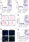
Piezo1 knockout in pulmonary arterial smooth muscle cells (PASMCs) partially confers protection against chronic hypoxia–induced pulmonary hypertension (PH) in mice. A, Right ventricular systolic pressure (RVSP) of mice after 3 weeks of normoxic (white area) or hypoxic (shaded area) exposure (normoxia: Piezo1lox/lox [n=3], Piezo1lox/loxCre [n=7], Piezo1del/lox [n=5], and Piezo1del/loxCre [n=7]; hypoxia: Piezo1lox/lox [n=6], Piezo1lox/loxCre [n=6], Piezo1del/lox [n=6], and Piezo1del/loxCre [n=6]). B, Right heart hypertrophy, depicted as the Fulton index (right ventricle [RV]/[left ventricle (LV)+septum (S)]; normoxia: Piezo1lox/lox [n=5], Piezo1lox/loxCre [n=5], Piezo1del/lox [n=5], and Piezo1del/loxCre [n=5]; hypoxia: Piezo1lox/lox [n=6], Piezo1lox/loxCre [n=6], Piezo1del/lox [n=6], and Piezo1del/loxCre [n=6]). C, Representative photographs and (D) values of mean muscularization of small (20–70 µm) pulmonary vessels from paraffin-embedded lung sections (normoxia: Piezo1lox/lox [n=5], Piezo1lox/loxCre [n=5], Piezo1del/lox [n=5], and Piezo1del/loxCre [n=6]; hypoxia: Piezo1lox/lox [n=6], Piezo1lox/loxCre [n=6], Piezo1del/lox [n=6], and Piezo1del/loxCre [n=6]). Scale bars, 50 µm. E, Representative immunofluorescence staining against PCNA (proliferating cell nuclear antigen, pink) and (F) statistical analysis for evaluation of proliferation in normoxic and chronic hypoxic mice (normoxia: Piezo1lox/lox [n=5], Piezo1lox/loxCre [n=5], Piezo1del/lox [n=5], and Piezo1del/loxCre [n=5]; hypoxia: Piezo1lox/lox [n=5], Piezo1lox/loxCre [n=5], Piezo1del/lox [n=5], and Piezo1del/loxCre [n=6]). Scale bars, 50 µm. Data were statistically analyzed by the 1-way ANOVA and the uncorrected Fisher least significant difference test. Only differences regarded as significant (P<0.05) are displayed as P values.
Figure 6.
Figure 6.
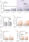
Modulation of Piezo1 activity affects the proliferation of pulmonary arterial smooth muscle cells (PASMCs) from hypoxic mice and patients with pulmonary hypertension (PH). A, Proliferation assays of PASMCs isolated from control (Piezo1lox/lox: normoxia [n=6] and hypoxia [n=5]) and Piezo1-deficient mice (Piezo1lox/loxCre: normoxia [n=4], hypoxia [n=7]; Piezo1del/loxCre: normoxia [n=8] and hypoxia [n=5]) exposed to normoxia or to chronic hypoxia (21 days). Proliferation was assessed in the presence of 0.2 and 10% FCS (fetal calf serum) and after exposure to increasing concentrations of Yoda1 (selective activator of the Piezo1 ion channel) and (B) GsMTx4. Values are expressed as percent of control values in starvation DMEM (Piezo1lox/lox: n=8–9; Piezo1del/loxCre: n=6–8). Data are shown as mean±SEM from at least 3 different experiments. C, Effect of increasing concentrations of Yoda1, (D) margaric acid (MA), and (E) GsMTx4 (GsM toxin 4, a peptide toxin) on the proliferation of PASMCs from patients with PH (n=6–8) and controls (n=8–9) in the presence of 0.2 and 2% FCS. Values are expressed as percent of control values in starvation Dulbecco's Modified Eagle Medium (DMEM). Data are mean±SEM of 8 to 10 values. Data were statistically analyzed by the 2-way ANOVA and the uncorrected Fisher least significant difference test. P values are indicated for comparison among group means.
Figure 7.
Figure 7.
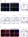
Piezo1 deletion in vascular smooth muscle cells protects Piezo1del/loxCre mice against hypoxia-induced endothelial cell senescence and microvascular rarefaction. A, Representative pictures showing immunofluorescence staining of p16 (white) in normoxic and chronic hypoxic murine lungs. The zoomed areas are indicated by rectangles. Blue: 4′,6-diamidino-2-phenylindole (DAPI) nuclear staining; green: elastin autofluorescence. Scale bars, 200 µm. B, Statistical analysis representing the percentage of p16+ cells in Piezo1lox/lox (normoxia: n=7; hypoxia: n=4) and Piezo1del/loxCre mice (normoxia: n=5; hypoxia: n=6). C, Representative micrographs showing immunofluorescence staining of p21 (brown) in small pulmonary vessels (left) and lung parenchyma (right). Blue: hematoxylin nuclear staining. Scale bars, 50 µm. D, Statistical analysis representing the percentage of p21+ lung cells in Piezo1lox/lox (normoxia: n=7; hypoxia: n=3) and Piezo1del/loxCre mice (n=4). E, Representative micrographs showing immunofluorescence staining of CD31 (red, a marker of endothelial cells). Blue: DAPI nuclear staining. Scale bars, 200 µm. F, Statistical analysis representing the percentage of CD31+ area in lung tissues of Piezo1lox/lox (normoxia: n=5; hypoxia: n=4) and Piezo1del/loxCre mice (normoxia: n=4; hypoxia: n=5). B, D, and F, Data are expressed as individual values per mice and mean±SEM for groups of mice. Data were statistically analyzed by the 2-way ANOVA and the uncorrected Fisher least significant difference test. Only differences regarded as significant changes (P<0.05) are displayed as P values.
Figure 8.
Figure 8.
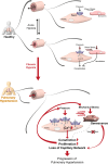
Proposed mechanism for the role of pulmonary arterial smooth muscle cell (PASMC)-Piezo1 in the development and progression of pulmonary hypertension. EC indicates endothelial cell; and HPV, hypoxic pulmonary vasoconstriction.

References

    1. Chin KM, Rubin LJ. Pulmonary arterial hypertension. J Am Coll Cardiol. 2008;51:1527–1538. doi: 10.1016/j.jacc.2008.01.024 - PubMed
    1. Veith C, Varturk-Ozcan I, Wujak M, Hadzic S, Wu CY, Knoepp F, Kraut S, Petrovic A, Gredic M, Pak O, et al. . SPARC, a novel regulator of vascular cell function in pulmonary hypertension. Circulation. 2022;145:916–933. doi: 10.1161/CIRCULATIONAHA.121.057001 - PubMed
    1. Sommer N, Huttemann M, Pak O, Scheibe S, Knoepp F, Sinkler C, Malczyk M, Gierhardt M, Esfandiary A, Kraut S, et al. . Mitochondrial complex IV subunit 4 isoform 2 is essential for acute pulmonary oxygen sensing. Circ Res. 2017;121:424–438. doi: 10.1161/CIRCRESAHA.116.310482 - PMC - PubMed
    1. Sommer N, Alebrahimdehkordi N, Pak O, Knoepp F, Strielkov I, Scheibe S, Dufour E, Andjelkovic A, Sydykov A, Saraji A, et al. . Bypassing mitochondrial complex III using alternative oxidase inhibits acute pulmonary oxygen sensing. Sci Adv. 2020;6:eaba0694. doi: 10.1126/sciadv.aba0694 - PMC - PubMed
    1. Humbert M, Kovacs G, Hoeper MM, Badagliacca R, Berger RMF, Brida M, Carlsen J, Coats AJS, Escribano-Subias P, Ferrari P, et al. ; ESC/ERS Scientific Document Group. 2022 ESC/ERS guidelines for the diagnosis and treatment of pulmonary hypertension. Eur Heart J. 2022;43:3618–3731. doi: 10.1093/eurheartj/ehac237 - PubMed

MeSH terms