. 2025 Apr 2;20(1):20251146.
doi: 10.1515/med-2025-1146. eCollection 2025.

Single-nucleus RNA sequencing reveals ARHGAP28 expression of podocytes as a biomarker in human diabetic nephropathy

Affiliations

Single-nucleus RNA sequencing reveals ARHGAP28 expression of podocytes as a biomarker in human diabetic nephropathy

Fengxia Zhang et al. Open Med (Wars). .

Abstract

Introduction: Diabetic kidney disease (DKD) represents serious diabetes-associated complications, and podocyte loss is an important histologic sign of DKD. The cellular and molecular profiles of podocytes in DKD have yet to be fully elucidated.

Methods: This study analyzed kidney-related single-nucleus RNA-seq datasets (GSE131882, GSE121862, and GSE141115) and human diabetic kidney glomeruli transcriptome profiling (GSE30122). ARHGAP28 expression was validated by western blot and immunohistochemistry.

Results: In human kidney tissues, 154 differentially expressed genes (DEGs) were identified in podocytes, which were enriched in biological processes related to nephron development and extracellular matrix-receptor interactions. Similarly, in the mouse kidney, 344 DEGs were found, clustering in pathways associated with renal development and signaling mechanisms like PI3K/Akt (phosphatidylinositol-3 kinase/protein kinase B) and PPAR (peroxisome proliferator-activated receptor). In diabetic human kidney glomeruli, 438 DEGs were identified, showing significant enrichment in pathways related to diabetic nephropathy. Venn analysis revealed 22 DEGs common across human and mouse podocytes and diabetic glomeruli, with ARHGAP28 being notably overexpressed in podocytes. The diabetic nephropathy model using db/db mice showed that ARHGAP28 expression was significantly upregulated in the kidney cortex and glomeruli. In vitro studies using a high-glucose podocyte model corroborated these findings.

Conclusions: Collectively, this study provides an insight into the function and diagnosis of DKD and indicates that ARHGAP28 in podocytes is a potential biomarker of DKD.

Keywords: ARHGAP28; diabetic kidney disease, DKD; podocyte; single-nucleus RNA sequencing, snRNA-seq.


Conflict of interest statement

Conflict of interest: The authors declare that they have no competing interests.

Figures

Figure 1
Figure 1
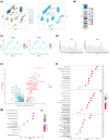
Single-nucleus analysis of the adult human kidney (GSE121862 and GSE131882) and DEGs of podocytes. (a) Cell clusters of adult human kidney and source of the datasets. (b) Cell composition of adult human kidney. (c) Expression of podocyte markers (feature plot). (d) Expression of podocyte markers (violin plot). (e) Volcano plot of DEGs (podocytes vs non-podocytes). (f) GO enrichment of podocyte DEGs (top ten terms). (g) KEGG enrichment of podocyte DEGs.
Figure 2
Figure 2
Single-nucleus analysis of adult mouse kidney (GSE141115) and DEGs of podocytes. (a) Cell clusters of adult mouse kidney and sequencing protocol of the datasets. (b) Cell composition of adult mouse kidney. (c) Expression of podocyte markers (feature plot). (d) Expression of podocyte markers (violin plot). (e) Volcano plot of DEGs (podocytes vs non-podocytes). (f) GO enrichment of podocyte DEGs (top ten terms). (g) KEGG enrichment of podocyte DEGs (top 10 terms).
Figure 3
Figure 3
DEGs of diabetic human kidney glomeruli microarray dataset (GSE30122). (a) Volcano plot of DEGs. (b) GO BP enrichment of DEGs. (c) Heatmap of DEG expression. (d) GSEA enrichment of DEGs.
Figure 4
Figure 4
Narrow down target genes and ARHGAP28 expression in single-cell data. (a) Overlap between DEGs of human podocytes, human orthologs of DEGs of mouse podocytes, and diabetic kidney glomeruli DEGs. (b) Expression of 22 overlap genes in human single-cell data (violin plot); all cells are grouped into podocytes and non-podocytes. (c) Expression of 22 overlap genes in mouse single-cell data (violin plot); all cells are grouped into podocytes and non-podocytes. (d) Expression of ARHGP28 in human single cell data (feature plot). (e) Expression of ARHGAP28 in mouse single cell data (feature plot). (f) Sub-clusters of human podocytes. (g) Expression of ARHGAP28 in human podocyte sub-clusters (feature plot). (h) Sub-clusters of mouse podocytes. (i) Expression of ARHGAP28 in mouse podocyte sub-clusters (feature plot).
Figure 5
Figure 5
DKD modeling. (a) Body weight, (b) blood glucose, (c) serum creatinine, (d) urine albumin/creatinine, (e) semi-quantitative analysis of mesangial matrix, (f) representative photomicrographs of PAS staining of glomeruli of db/db mice and nondiabetic mice. *P < 0.05. Scale bar = 25 µm.
Figure 6
Figure 6
Validation of ARHGAP28 in DKD. (a) Western blot gel showing ARHGAP28 protein expression in the mouse kidney cortex. (b) Semiquantitative analysis of ARHGAP28 protein expression in the mouse kidney cortex. (c) Images of glomerular ARHGAP28 immunohistochemical staining from mouse kidney sections. (d) Semi-quantitative analysis of ARHGAP28 immunohistochemical staining from mouse kidney sections. *P < 0.05. Scale bar = 25 µm. Red arrows indicate the positive staining of ARHGAP28. (e) Podocytes were cultured in vitro and stimulated by high glucose. Expression of mRNA of ARHGAP28 in the treated group and normal control group was tested by qPCR. *P < 0.05.

References

    1. Mahtal N, Lenoir O, Tharaux P. Glomerular endothelial cell crosstalk with podocytes in diabetic kidney disease. Front Med. 2021;8:659013. - PMC - PubMed
    1. Jha R, Lopez-Trevino S, Kankanamalage HR, Jha JC. Diabetes and renal complications: An overview on pathophysiology, biomarkers and therapeutic interventions. Biomedicines. 2024;12(5):1098. - PMC - PubMed
    1. Fang Z, Liu R, Xie J, He JC. Molecular mechanism of renal lipid accumulation in diabetic kidney disease. J Cell Mol Med. 2024;28(11):e18364. - PMC - PubMed
    1. Deng L, Shi C, Li R, Zhang Y, Wangc X, Cai G, et al. The mechanisms underlying Chinese medicines to treat inflammation in diabetic kidney disease. J Ethnopharmacol. 2024;7:118424. - PubMed
    1. Ruiz-Ortega M, Rodrigues-Diez RR, Lavoz C, Rayego-Mateos S. Special issue “Diabetic nephropathy: Diagnosis, prevention and treatment”. J Clin Med. 2020;9(3):813. - PMC - PubMed

LinkOut - more resources