. 2025 Mar 13:16:1549107.
doi: 10.3389/fimmu.2025.1549107. eCollection 2025.

Remodeling of immune system functions by extracellular vesicles

Affiliations

Remodeling of immune system functions by extracellular vesicles

Deborah Neyrinck-Leglantier et al. Front Immunol. .

Abstract

Introduction: The treatment of chronic viral infections can often bring viral replication under control. However, chronic immune activation persists and can lead to the development of comorbid conditions, such as cardiovascular disease and cancer. This is particularly true for people living with HIV (PLWH), who have significantly more extracellular vesicles from membrane budding, also called plasma microparticles (MPs), than healthy individuals (HDs), and a much more immunomodulatory phenotype. We hypothesized that the number and phenotypic heterogeneity of MPs can trigger a functional remodeling of immune responses in PLWH, preventing full immune restoration.

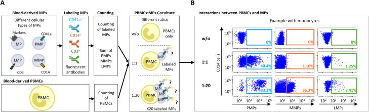
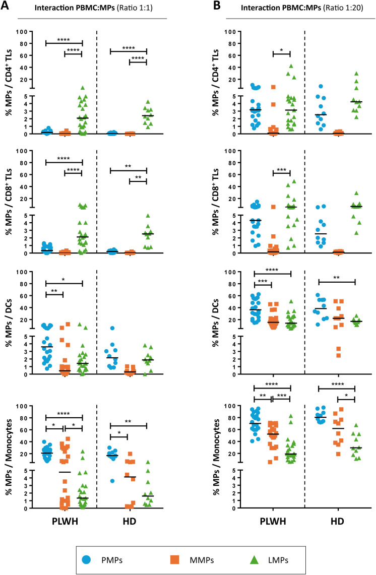
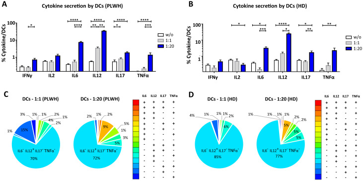
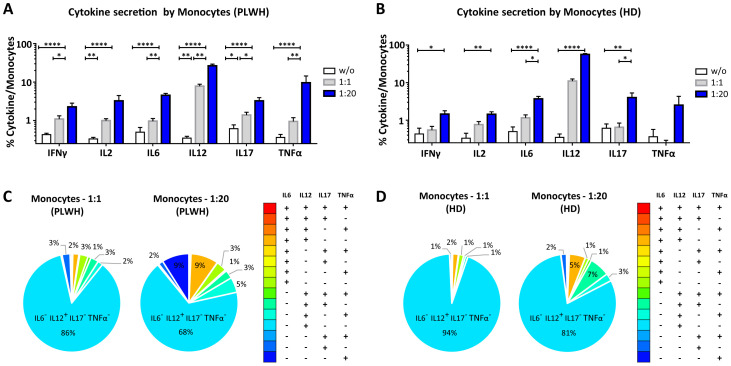
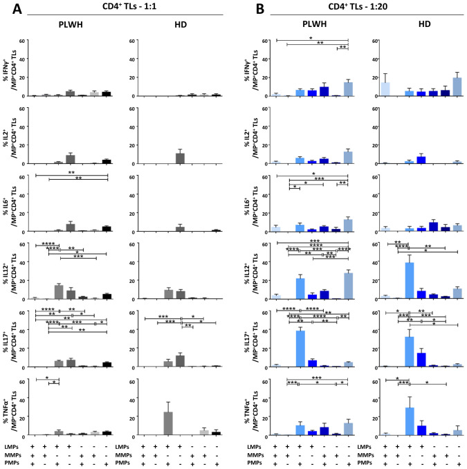
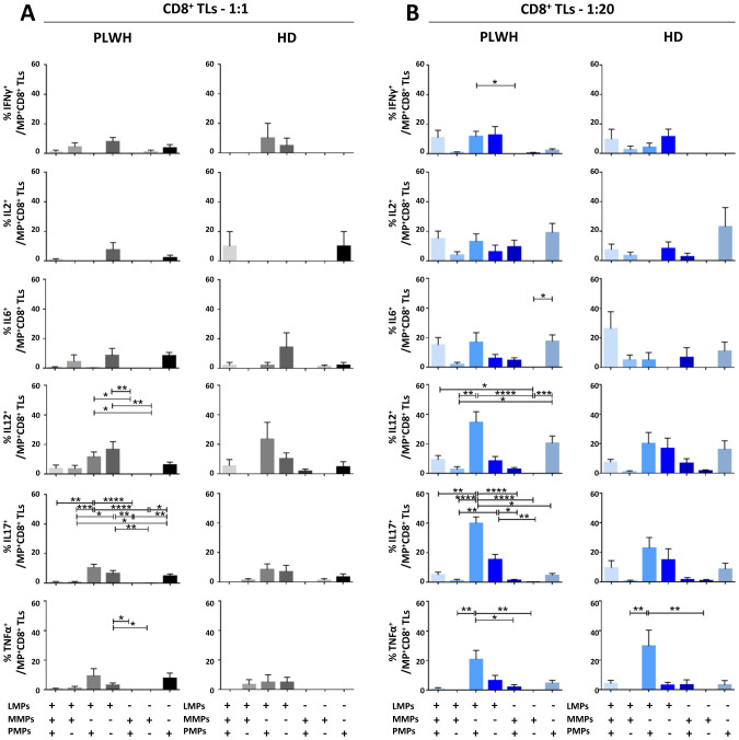
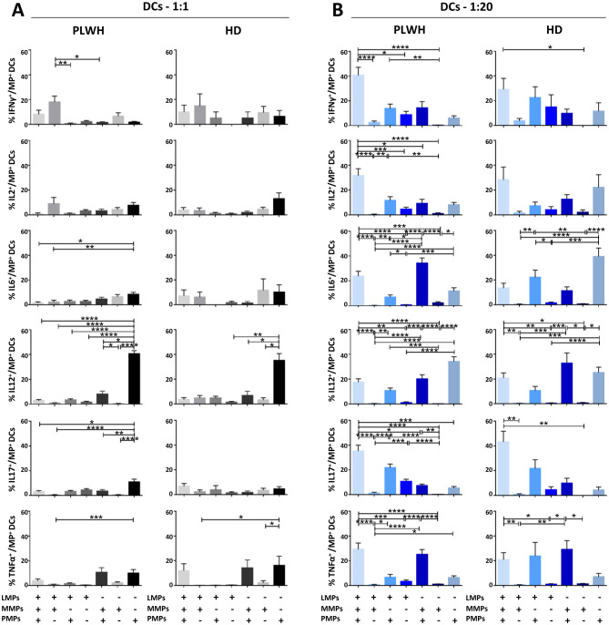
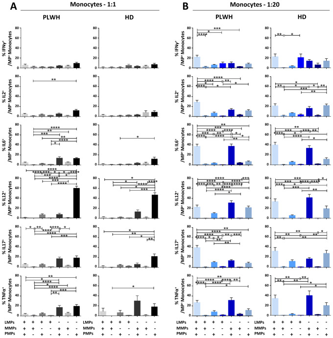
Methods: We investigated the rapid impact of three types of MPs - derived from membrane budding in platelets (CD41a+ PMPs), monocytes (CD14+ MMPs) and lymphocytes (CD3+ LMPs) in the plasma of PLWH or HDs-on four cell types (CD4+ and CD8+T lymphocytes, monocytes and DCs).

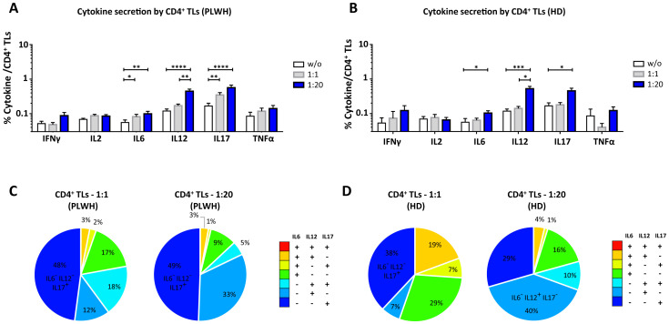
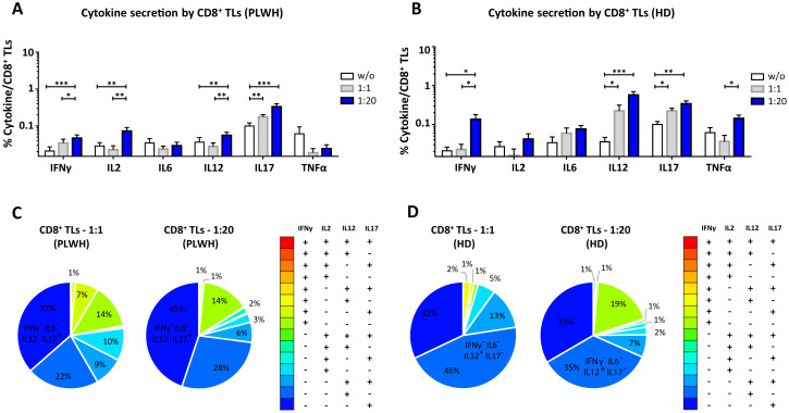
Results: These investigations of the short multiple interactions and functions of MPs with these cells revealed an increase in the secretion of cytokines such as IFNg, IL2, IL6, IL12, IL17 and TNFa by the immune cells studied following interactions with MPs. We show that this functional remodeling of immune cells depends not only on the number, but also on the phenotype of MPs.

Conclusion: These data suggest that the large numbers of MPs and their impact on functional remodeling in PLWH may be incompatible with the effective control of chronic infections, potentially leading to chronic immune activation and the onset of comorbid diseases.

Keywords: PLWH; cellular remodeling; cytokine secretion; extracellular vesicles (EV); immunomodulation.


Conflict of interest statement

The authors declare that the research was conducted in the absence of any commercial or financial relationships that could be construed as a potential conflict of interest.

Figures

Figure 1
Figure 1
Assays of MP coculture with PBMCs to study interactions between MPs and immune cells. (A) Schematic representation of MP coculture assays with PBMCs at different ratios for investigations of the interaction of CD41a+ PMPs, CD14+ MMPs and CD3+ LMPs with immune cells. MPs were isolated and labeled with an anti-CD41a antibody for PMPs, an anti-CD14 antibody for MMPs and an anti-CD3 antibody for LMPs. The total number of labeled MPs (CD41a+ PMPs + CD14+ MMPs + CD3+ LMPs) was calculated by flow cytometry with Trucount beads. The MPs were then cocultured with PBMCs at ratios of 1:1 and 1:20 (PBMCs: MPs). As a control, PBMCs were cultured without MPs (w/o). After coculture, cells that had and had not interacted with MPs were harvested and labeled to study the co-expression of cell markers with CD41a from PMPs, CD14 from MMPs or CD3 from LMPs. (B) Example of the gating strategy used for flow cytometric assessment of the interaction of CD41a+ PMPs, CD14+ MMPs and CD3+ LMPs with monocytes (CD14+) for one representative experiment.
Figure 2
Figure 2
Interaction of CD41a+ PMPs, CD14+ MMPs and CD3+ LMPs with immune cells. (A, B) Percentage of cells expressing CD41a from PMPs (blue circle), CD14 from MMPs (orange square) or CD3 from LMPs (green triangle) of patients (PLWH, n=22) or healthy donors (HD, n=10) at coculture ratios of 1:1 (A) or 1:20 (B) (PBMCs: MPs). Horizontal bars indicate the median value. P values (P<0.05 considered significant) were obtained in ANOVA and Kruskal-Wallis post hoc tests. ****P<0.0001, ***P<0.001, **P<0.01, *P<0.05.
Figure 3
Figure 3
Functional impact of MPs on cytokine secretion by CD4+ T lymphocytes. PBMCs were cocultured for 18 hours with known numbers of CD41a+ PMPs and CD14+ MMPs and CD3+ LMPs at ratios of 1:1 and 1:20 (PBMCs: MPs). As a control, PBMCs were also cultured without MPs (w/o). The cells were then harvested, fixed, permeabilized and labeled to investigate cytokine secretion. (A, B) The percentages (mean ± SD) of CD4+ T lymphocytes secreting IFNγ, IL2, IL6, IL12, IL17 and TNFα were determined without (white bars) or with coculture with MPs from PLWH [(A), n=22, except for TNF, n=16, 10 independent coculture experiments] or HDs [(B), n=10, except for TNF n=6, three independent coculture experiments] at ratios of 1:1 (gray bars) and 1:20 (blue bars) (PBMCs: MPs). (C, D) Co-expression data for IL6, IL12 and IL17 are presented for CD4+ T lymphocytes cocultured with MPs from PLWH [(C), n=16, eight independent coculture experiments] or HDs [(D), n=6, two independent coculture experiments] at ratios of 1:1 and 1:20 (PBMCs: MPs). P values (P<0.05 considered significant) were obtained in ANOVA and Friedman’s post hoc tests. ****P<0.0001, ***P<0.001, **P<0.01, *P<0.05.
Figure 4
Figure 4
Functional impact of MPs on cytokine secretion by CD8+ T lymphocytes. PBMCs were cocultured for 18 hours with known numbers of CD41a+ PMPs and CD14+ MMPs and CD3+ LMPs at ratios of 1:1 and 1:20 (PBMCs: MPs). As a control, PBMCs were also cultured without MPs (w/o). The cells were then harvested, fixed, permeabilized and labeled to investigate cytokine secretion. (A, B) The percentages (mean ± SD) of CD8+ T lymphocytes secreting IFNγ, IL2, IL6, IL12, IL17 and TNFα were determined without (white bars) or with coculture with MPs from PLWH ((A), n=22, except for TNF, n=16, 10 independent coculture experiments) or HDs [(B), n=10, except for TNF n=6, three independent coculture experiments] at ratios of 1:1 (gray bars) and 1:20 (blue bars) (PBMCs: MPs). (C, D) Co-expression data for IFNγ, IL2, IL12 and IL17 are presented for CD8+ T lymphocytes cocultured with MPs from PLWH [(C), n=16, eight independent coculture experiments] or HDs [(D), n=6, two independent coculture experiments] at ratios of 1:1 and 1:20 (PBMCs: MPs). P values (P<0.05 considered significant) were obtained in ANOVA and Friedman’s post hoc tests. ***P<0.001, **P<0.01, *P<0.05.
Figure 5
Figure 5
Functional impact of MPs on cytokine secretion by DCs. PBMCs were cocultured for 18 hours with known numbers of CD41a+ PMPs and CD14+ MMPs and CD3+ LMPs at ratios of 1:1 and 1:20 (PBMCs: MPs). As a control, PBMCs were also cultured without MPs (w/o). The cells were then harvested, fixed, permeabilized and labeled to investigate cytokine secretion. (A, B) The percentages (mean ± SD) of DCs secreting IFNγ, IL2, IL6, IL12, IL17 and TNFα were determined without (white bars) or with coculture with MPs from PLWH [(A), n=22, except for TNF, n=16, 10 independent coculture experiments] or HDs [(B), n=10, except for TNF n=6, three independent coculture experiments] at ratios of 1:1 (gray bars) and 1:20 (blue bars) (PBMCs: MPs). (C, D) Co-expression data for IL6, IL12, IL17 and TNFα are presented for DCs cocultured with MPs from PLWH [(C), n=16, eight independent coculture experiments] or HDs [(D), n=6, two independent coculture experiments] at ratios of 1:1 and 1:20 (PBMCs: MPs). P values (P<0.05 considered significant) were obtained in ANOVA and Friedman’s post hoc tests. ****P<0.0001, ***P<0.001, **P<0.01, *P<0.05.
Figure 6
Figure 6
Functional impact of MPs on cytokine secretion by monocytes. PBMCs were cocultured for 18 hours with known numbers of CD41a+ PMPs and CD14+ MMPs and CD3+ LMPs at ratios of 1:1 and 1:20 (PBMCs: MPs). As a control, PBMCs were also cultured without MPs (w/o). The cells were then harvested, fixed, permeabilized and labeled to investigate cytokine secretion. (A, B) The percentages (mean ± SD) of monocytes secreting IFNγ, IL2, IL6, IL12, IL17 and TNFα were determined without (white bars) or with coculture with MPs from PLWH [(A), n=22, except for TNF, n=16, 10 independent coculture experiments] or HDs [(B), n=10, except for TNF n=6, three independent coculture experiments] at ratios of 1:1 (gray bars) and 1:20 (blue bars) (PBMCs: MPs). (C, D) Co-expression data for IL6, IL12, IL17 and TNFα are presented for monocytes cocultured with MPs from PLWH [(C), n=16, eight independent coculture experiments] or HDs [(D), n=6, two independent coculture experiments] at ratios of 1:1 and 1:20 (PBMCs: MPs). P values (P<0.05 considered significant) were obtained in ANOVA and Friedman’s post hoc tests. ****P<0.0001, **P<0.01, *P<0.05.
Figure 7
Figure 7
Identification of the MP subpopulations involved in cytokine secretion by CD4+ T lymphocytes. (A, B) Co-expression data for CD41a+ PMPs, CD14+ MMPs and CD3+ LMPs are presented for each cytokine secreted by CD4+ T lymphocytes cocultured with MPs from PLWH (n=22, except for TNF, n=16, 10 independent coculture experiments) or HDs (n=10, except for TNF n=6, three independent coculture experiments) at ratios of 1:1 (A) or 1:20 (B) (PBMCs: MPs). P values (P<0.05 considered significant) were obtained in ANOVA and Friedman’s post hoc tests. ****P<0.0001, ***P<0.001, **P<0.01, *P<0.05.
Figure 8
Figure 8
Identification of the MP subpopulations involved in cytokine secretion by CD8+ T lymphocytes. (A, B) Co-expression data for CD41a+ PMPs, CD14+ MMPs and CD3+ LMPs are presented for each cytokine secreted by CD8+ T lymphocytes cocultured with MPs from PLWH (n=22, except for TNF, n=16, 10 independent coculture experiments) or HDs (n=10, except for TNF n=6, three independent coculture experiments) at ratios of 1:1 (A) or 1:20 (B) (PBMCs: MPs). P values (P<0.05 considered significant) were obtained in ANOVA and Friedman’s post hoc tests. ****P<0.0001, ***P<0.001, **P<0.01, *P<0.05.
Figure 9
Figure 9
Identification of the MP subpopulations involved in cytokine secretion by DCs. (A, B) Co-expression data for CD41a+ PMPs, CD14+ MMPs and CD3+ LMPs are presented for each cytokine secreted by DCs cocultured with MPs from PLWH (n=22, except for TNF, n=16, 10 independent coculture experiments) or HDs (n=10, except for TNF n=6, three independent coculture experiments) at ratios of 1:1 (A) or 1:20 (B) (PBMCs: MPs). P values (P<0.05 considered significant) were obtained in ANOVA and Friedman’s post hoc tests. ****P<0.0001, ***P<0.001, **P<0.01, *P<0.05.
Figure 10
Figure 10
Identification of the MP subpopulations involved in cytokine secretion by monocytes. (A, B) Co-expression data for CD41a+ PMPs, CD14+ MMPs and CD3+ LMPs are presented for each cytokine secreted by monocytes cocultured with MPs from PLWH (n=22, except for TNF, n=16, 10 independent coculture experiments) or HDs (n=10, except for TNF n=6, three independent coculture experiments) at ratios of 1:1 (A) or 1:20 (B) (PBMCs: MPs). P values (P<0.05 considered significant) were obtained in ANOVA and Friedman’s post hoc tests. ****P<0.0001, ***P<0.001, **P<0.01, *P<0.05.

Similar articles

References

    1. Kaminski H, Lemoine M, Pradeu T. Immunological exhaustion: How to make a disparate concept operational? PloS Pathog. (2021) 17:e1009892. doi: 10.1371/journal.ppat.1009892 - DOI - PMC - PubMed
    1. Vingert B, Benati D, Lambotte O, de Truchis P, Slama L, Jeannin P, et al. . HIV controllers maintain a population of highly efficient Th1 effector cells in contrast to patients treated in the long term. J virology. (2012) 86:10661–74. doi: 10.1128/JVI.00056-12 - DOI - PMC - PubMed
    1. Ackerman M, Mikhailova A, Brown E, Dowell K, Walker B, Bailey-Kellogg C, et al. . Polyfunctional HIV-specific antibody responses are associated with spontaneous HIV control. PloS pathogens. (2016) 12:e1005315. doi: 10.1371/journal.ppat.1005315 - DOI - PMC - PubMed
    1. Zicari S, Sessa L, Cotugno N, Ruggiero A, Morrocchi E, Concato C, et al. . Immune activation, inflammation, and non-AIDS co-morbidities in HIV-infected patients under long-term ART. Viruses. (2019) 11:200. doi: 10.3390/v11030200 - DOI - PMC - PubMed
    1. Appay V, Almeida J, Sauce D, Autran B, Papagno L. Accelerated immune senescence and HIV-1 infection. Exp gerontology. (2007) 42:432–7. doi: 10.1016/j.exger.2006.12.003 - DOI - PubMed

MeSH terms