. 2025 Mar 20:16:1542191.
doi: 10.3389/fimmu.2025.1542191. eCollection 2025.

Neutrophils negatively control IL-17A-producing γδ T cell frequencies in a contact-dependent manner under physiological conditions

Affiliations

Neutrophils negatively control IL-17A-producing γδ T cell frequencies in a contact-dependent manner under physiological conditions

Xinhua Yu et al. Front Immunol. .

Abstract

Background: In addition to serving as the primary effector cells against infections, neutrophils have been implicated in the regulation of both innate and adaptive immunity. In this study, we aimed to investigate the role of neutrophils in the regulation of the immune system under physiological conditions.

Methods: The in vivo effect of neutrophils on the immune system was examined using neutropenic mice. The interaction between neutrophils and γδ T cells was investigated using an in vitro co-culture system.

Findings: Unexpectedly, we observed an accumulation of γδ T cells in the cervical lymph nodes of neutropenic mice. Transcriptomic analysis revealed that these γδ T cells exhibited unique expression profiles of cell surface molecules and genes involved in defense responses. Further characterization indicated that the accumulated γδ T cells were IL-17 producing CD44+CD62L-CD27- memory cells. Additionally, in vitro experiments demonstrated that neutrophils could inhibit the function of IL-17A producing γδ T cells by inducing cell death in a contact-dependent manner.

Conclusion: This present study demonstrates that neutrophils negatively regulate IL-17 producing γδ T cells under physiological conditions. Given that IL-17A is a critical cytokine for the recruitment of neutrophils to peripheral tissues, our study suggests that the crosstalk between neutrophils and IL-17A producing γδ T cells is a crucial mechanism for maintaining immune homeostasis under physiological conditions.

Keywords: IL-17A; cell death; innate immunity; neutrophils; physiological condition; γδ T cells.


Conflict of interest statement

The authors declare that the research was conducted in the absence of any commercial or financial relationships that could be construed as a potential conflict of interest. The author(s) declared that they were an editorial board member of Frontiers, at the time of submission. This had no impact on the peer review process and the final decision.

Figures
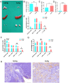
Figure 1
Figure 1
Accumulation of γδT cells in neutropenic mice. (A) Representative picture of thymus, spleen and cervical lymph nodes (LNs) from Mcl-1 f/f; LysMCre neutropenic male mice (ho/tg) and their littermate Mcl-1 f/wt; LysMCre (het/tg) control male mice at the age of 10-12 weeks old. Single cell suspensions were prepared from thymus, spleen and cervical LNs and counted and stained with anti-CD19, anti-CD4, anti-CD8, anti-CD3, and anti-γδTCR IgGs to defined B cells, CD4+ T cells, CD8+ T cells, and γδT cells. Total number of cells in thymus (B), spleen (C) and cervical LNs (D) were compared between neutropenic mice (n=6) and control mice (n=6). Percentages of CD19+ B cells, CD4+ T cells, CD8+ T cells and γδT cells in thymus (E) spleen (F) and cervical LNs (G) were compared between neutropenic (n=6) and control (n=6) mice. (H) Representative picture of immunohistochemistry staining for γδT cell in cervial LNs of neutropenic and control mice. Bar=50 µm. Data are presented as mean ± SEM, ns, not significant; *p<0.05; **p<0.01; ****p<0.0001.
Figure 2
Figure 2
Unique features of γδT cells accumulated in neutropenic mice. Hierarchic cluster of differentially expressed genes between γδT cells from neutropenic and control mice. Blue color represents a low expression and yellow color stand for a high expression. GO term enrichment analysis was performed using DAVID 6.8 software for both upregulated and downregulated genes, and top enriched GO terms are indicated. Upregulated (B) and downregulated (C) gene encoding cluster of differentiation (CD) molecules. (D-F) Characterization of γδT cells isolated from neutropenic mice and littermate controls. Single cell suspension was prepared from cervical LNs from neutropenic and control mice characterized either by staining the freshly prepared cells with CD3, γδTCR, CD44 and CD62L. Expression of CD3 and γδTCR on γδT cells in neutropenic (n=5) and control (n=5) mice are shown as representative sample (D) and quantitative values of percentage of CD3 bright γδT cells (E). Expression of CD62L and CD44 on γδT cells in neutropenic (n=5) and control (n=5) mice are shown as representative sample (F) and quantitative values of percentage of CD44+CD62L- γδT cells (G). Data are presented as mean ± SEM, ****p<0.0001.
Figure 3
Figure 3
Production of IL-17A in γδT cells of neutropenic mice and littermate controls. Single-cell suspensions were prepared from cervical LNs from neutropenic and control mice characterized either by staining the freshly prepared cells with surface markers or by staining the cultured cells with anti-IL-17A IgG. Representative sample (A) and quantitative values of percentage of CD27- γδT cells (B) show the expression of CD27 on γδT cells in neutropenic (n=5) and control (n=5) mice. Freshly prepared cells from cervical LNs from neutropenic mice (n=6) and littermate controls (n=6) were cultured for 5 hours with/without stimulation of phorbol myristate acetate and calcium ionophore (PMA/I) and stained with anti-IL-17A IgG to determine the IL-17A production in γδT cells. Representative samples (C) and quantitative values (D) of percentage of IL-17A-producing γδT cells are shown. Data are presented as mean ± SEM, ns, not significant; ***p<0.001; ****p<0.0001.
Figure 4
Figure 4
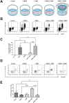
Neutrophils regulate IL-17A production and cell death of γδT cells in vitro. (A) Schematic diagram of the detailed experimental conditions. Single-cell suspensions were prepared from the cervical LNs of neutropenic mice (n=6) and cultured for 5 hours with or without stimulation of phorbol myristate acetate and calcium ionophore (PMA/I), in presence or absence of neutrophils (PMN) isolated from littermate control mice at 1:1 ratio, or presence of neutrophils in a transwell co-culture manner (PMNt). In the transwell co-culture system, which employed transwell inserts, single-cell suspensions prepared from the cervical LNs were seeded into the wells of the culture plate, while neutrophils were seeded onto the transwell inserts. After the culture, cells were collected and stained with surface markers to define γδT cells and with anti-IL-17A IgG to determine the expression of IL-17A. (B) Representative samples show the expression of IL-17A in gated living CD3 T cells, where living γδT cells are further defined as γδTCR positive cells. Quantitative values of percentage of IL-17A+ γδT cells in CD3 cells are shown in (C) Cell death was evaluated by staining the cultures cells with Annexin V and propidium iodide (PI). (D) Representative samples show the cell death of total γδT cells which are defined as CD3+γδT+ cells. Quantitative values of percentage of Annxin V+PI+ γδT cells (dead cells) are shown in (E) Data are presented as mean ± SEM, ns, not significant; **p<0.01; ***p<0.001; ****p<0.0001.
Figure 5
Figure 5
Contact-dependent induction of γδT cell death by living neutrophils. Sorted murine γδT cells (green) from neutropenic mice were co-cultured with in vitro-differentiated neutrophils (red) in the presence of PMA (100 ng/ml) for three hours. Dead cells are indicated by DAPI-positive staining (blue). The interaction between γδT cells and neutrophils was monitored using a confocal microscope every 2.5 minutes. Rectangular boxes highlight γδT cells and neutrophils in direct contact, with white boxes indicating interactions with living neutrophils and green boxes indicating interactions with dead neutrophils. Representative images are shown at three distinct time points corresponding to different interaction stages: before contact [110 min, (A)], during contact [118 min, (B)], and after contact [137 min, (C)].

Similar articles

References

    1. Mocsai A. Diverse novel functions of neutrophils in immunity, inflammation, and beyond. J Exp Med. (2013) 210:1283–99. doi: 10.1084/jem.20122220 - DOI - PMC - PubMed
    1. Sutherland TE, Logan N, Ruckerl D, Humbles AA, Allan SM, Papayannopoulos V, et al. . Chitinase-like proteins promote IL-17-mediated neutrophilia in a tradeoff between nematode killing and host damage. Nat Immunol. (2014) 15:1116–25. doi: 10.1038/ni.3023 - DOI - PMC - PubMed
    1. Rosales C. Neutrophils at the crossroads of innate and adaptive immunity. J Leukoc Biol. (2020) 108:377–96. doi: 10.1002/JLB.4MIR0220-574RR - DOI - PubMed
    1. Schuster S, Hurrell B, Tacchini-Cottier F. Crosstalk between neutrophils and dendritic cells: a context-dependent process. J Leukoc Biol. (2013) 94:671–5. doi: 10.1189/jlb.1012540 - DOI - PubMed
    1. Valayer A, Brea D, Lajoie L, Avezard L, Combes-Soia L, Labas V, et al. . Neutrophils can disarm NK cell response through cleavage of NKp46. J Leukoc Biol. (2017) 101:253–9. doi: 10.1189/jlb.3AB0316-140RR - DOI - PubMed

MeSH terms

Substances

LinkOut - more resources