

Static and temporal dynamic changes in brain activity in patients with post-stroke balance dysfunction: a pilot resting state fMRI
- PMID: 40182145
- PMCID: PMC11965596
- DOI: 10.3389/fnins.2025.1558069
Static and temporal dynamic changes in brain activity in patients with post-stroke balance dysfunction: a pilot resting state fMRI
Abstract
Objective: The aim of this study was to investigate the characteristics of brain activity changes in patients with post-stroke balance dysfunction and their relationship with clinical assessment, and to construct a classification model based on the extreme Gradient Boosting (XGBoost) algorithm to discriminate between stroke patients and healthy controls (HCs).
Methods: In the current study, twenty-six patients with post-stroke balance dysfunction and twenty-four HCs were examined by resting-state functional magnetic resonance imaging (rs-fMRI). Static amplitude of low frequency fluctuation (sALFF), static fractional ALFF (sfALFF), static regional homogeneity (sReHo), dynamic ALFF (dALFF), dynamic fALFF (dfALFF) and dynamic ReHo (dReHo) values were calculated and compared between the two groups. The values of the imaging metrics for the brain regions with significant differences were used in Pearson correlation analyses with the Berg Balance Scale (BBS) scores and as features in the construction of the XGBoost model.
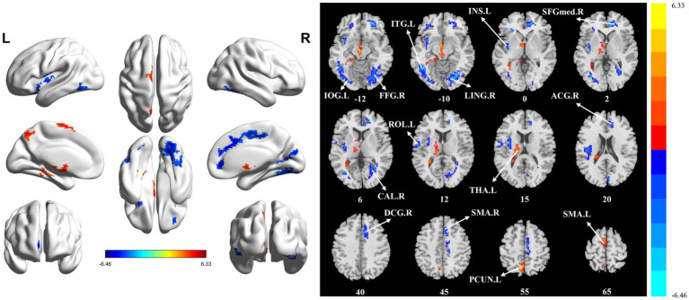
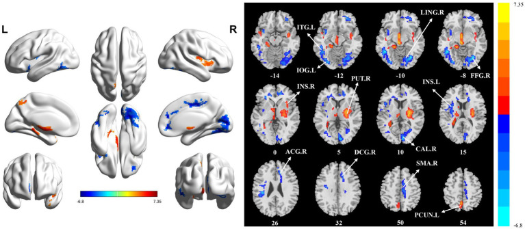
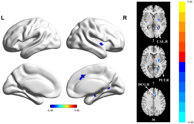
Results: Compared to HCs, the brain regions with significant functional abnormalities in patients with post-stroke balance dysfunction were mainly involved bilateral insula, right fusiform gyrus, right lingual gyrus, left thalamus, left inferior occipital gyrus, left inferior temporal gyrus, right calcarine fissure and surrounding cortex, left precuneus, right median cingulate and paracingulate gyri, right anterior cingulate and paracingulate gyri, bilateral supplementary motor area, right putamen, and left cerebellar crus II. XGBoost results show that the model constructed based on static imaging features has the best classification prediction performance.
Conclusion: In conclusion, this study provided evidence of functional abnormalities in local brain regions in patients with post-stroke balance dysfunction. The results suggested that the abnormal brain regions were mainly related to visual processing, motor execution, motor coordination, sensorimotor control and cognitive function, which contributed to our understanding of the neuropathological mechanisms of post-stroke balance dysfunction. XGBoost is a promising machine learning method to explore these changes.
Keywords: amplitude of low frequency fluctuation; balance dysfunction; extreme gradient boosting; fractional amplitude of low frequency fluctuation; regional homogeneity; resting-state functional magnetic resonance imaging; stroke.
Copyright © 2025 Tang, Liu, Long, Ren, Liu, Li, Han, Liao, Zhang, Lu and Zhang.
Conflict of interest statement
The authors declare that the research was conducted in the absence of any commercial or financial relationships that could be construed as a potential conflict of interest.
Figures


Comment in
-
Commentary: Static and temporal dynamic changes in brain activity in patients with post-stroke balance dysfunction: a pilot resting state fMRI.Front Neurosci. 2025 May 30;19:1607090. doi: 10.3389/fnins.2025.1607090. eCollection 2025. Front Neurosci. 2025. PMID: 40520503 Free PMC article. No abstract available.
Similar articles
-
Alterations of static and dynamic changes in intrinsic brain activity and its relation to behavioral outcomes in subcortical ischemic stroke after one-month intervention.Sci Rep. 2025 Apr 8;15(1):11961. doi: 10.1038/s41598-025-96163-8. Sci Rep. 2025. PMID: 40200032 Free PMC article.
-
Regional brain dysfunction in insomnia after ischemic stroke: A resting-state fMRI study.Front Neurol. 2022 Nov 25;13:1025174. doi: 10.3389/fneur.2022.1025174. eCollection 2022. Front Neurol. 2022. PMID: 36504641 Free PMC article.
-
A Resting State Functional Magnetic Resonance Imaging Study of Unmedicated Adolescents With Non-suicidal Self-Injury Behaviors: Evidence From the Amplitude of Low-Frequency Fluctuation and Regional Homogeneity Indicator.Front Psychiatry. 2022 Jun 17;13:925672. doi: 10.3389/fpsyt.2022.925672. eCollection 2022. Front Psychiatry. 2022. PMID: 35782416 Free PMC article.
-
Alterations of regional spontaneous brain activity in obsessive-compulsive disorders: A meta-analysis.J Psychiatr Res. 2023 Sep;165:325-335. doi: 10.1016/j.jpsychires.2023.07.036. Epub 2023 Aug 2. J Psychiatr Res. 2023. PMID: 37573797 Review.
-
Common and distinct patterns of intrinsic brain activity alterations in major depression and bipolar disorder: voxel-based meta-analysis.Transl Psychiatry. 2020 Oct 19;10(1):353. doi: 10.1038/s41398-020-01036-5. Transl Psychiatry. 2020. PMID: 33077728 Free PMC article. Review.
Cited by
-
Commentary: Static and temporal dynamic changes in brain activity in patients with post-stroke balance dysfunction: a pilot resting state fMRI.Front Neurosci. 2025 May 30;19:1607090. doi: 10.3389/fnins.2025.1607090. eCollection 2025. Front Neurosci. 2025. PMID: 40520503 Free PMC article. No abstract available.
References
-
- Berg K. O., Maki B. E., Williams J. I., Holliday P. J., Wood-Dauphinee S. L. (1992). Clinical and laboratory measures of postural balance in an elderly population. Arch. Phys. Med. Rehabil. 73, 1073–1080, PMID: - PubMed
LinkOut - more resources
Full Text Sources