

Rapidly Growing Malignant Peripheral Nerve Sheath Tumors Arising From Neurofibromatosis Type 1: A Case Report by Rehabilitation Physicians
- PMID: 40182390
- PMCID: PMC11965776
- DOI: 10.7759/cureus.79995
Rapidly Growing Malignant Peripheral Nerve Sheath Tumors Arising From Neurofibromatosis Type 1: A Case Report by Rehabilitation Physicians
Abstract
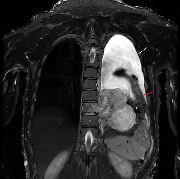
We describe a case of a 40-year-old Japanese woman with rapidly growing malignant peripheral nerve sheath tumors (MPNSTs) arising from neurofibromatosis type 1 (NF-1). The patient presented numbness in both legs, back pain, and gait disturbances. Magnetic resonance imaging (MRI) revealed a spinal tumor at the thoracic level. To resolve her symptoms, a laminectomy and intradural tumor resection were performed. The tumor was diagnosed as a neurofibroma with no malignant characteristics. After the surgery, she participated in a rehabilitation program aimed at promoting independence in daily activities and enhancing muscle strength. Initially, her walking ability showed improvement; however, she soon experienced complications, including challenges with bowel movements and a gradual decline in her walking function. A follow-up MRI on the 67th day post-surgery showed tumor regrowth that necessitated reoperation. After the surgery, the neurological symptoms improved temporarily, but they worsened again, ultimately leading to a shift to palliative care and her demise several days later. This case underscores the challenges in pathological diagnosis and the aggressive nature of MPNSTs, emphasizing the need for vigilant monitoring and timely intervention. Despite initial surgical success, rapid tumor growth can occur during rehabilitation, highlighting the importance of a multidisciplinary approach for accurate diagnosis and treatment. Early detection of tumor progression through meticulous neurological monitoring and prompt surgical consultation are critical for optimal outcomes. Further research into more definitive diagnostic tools and effective treatment strategies for MPNSTs is crucial to improve patient care.
Keywords: malignant peripheral nerve sheath tumor (mpnst); malignant spinal tumor; nerve schwannoma; neurofibromatosis type 1 (nf-1); spinal cord tumor.
Copyright © 2025, Hayashi et al.
Conflict of interest statement
Human subjects: Consent for treatment and open access publication was obtained or waived by all participants in this study. Conflicts of interest: In compliance with the ICMJE uniform disclosure form, all authors declare the following: Payment/services info: All authors have declared that no financial support was received from any organization for the submitted work. Financial relationships: All authors have declared that they have no financial relationships at present or within the previous three years with any organizations that might have an interest in the submitted work. Other relationships: All authors have declared that there are no other relationships or activities that could appear to have influenced the submitted work.
Figures


References
-
- Pediatric and adult malignant peripheral nerve sheath tumors: an analysis of data from the surveillance, epidemiology, and end results program. Amirian ES, Goodman JC, New P, Scheurer ME. J Neurooncol. 2014;116:609–616. - PubMed
-
- Extremity malignant peripheral nerve sheath tumors (neurogenic sarcomas): a 10-year experience. Vauthey JN, Woodruff JM, Brennan MF. Ann Surg Oncol. 1995;2:126–131. - PubMed
-
- Malignant peripheral nerve sheath tumors (MPNST): the Mayo Clinic experience. Stucky CC, Johnson KN, Gray RJ, Pockaj BA, Ocal IT, Rose PS, Wasif N. Ann Surg Oncol. 2012;19:878–885. - PubMed
-
- Malignant peripheral nerve sheath tumour (MPNST): the clinical implications of cellular signalling pathways. Katz D, Lazar A, Lev D. Expert Rev Mol Med. 2009;11:0. - PubMed
Publication types
LinkOut - more resources
Full Text Sources
Research Materials
Miscellaneous