Expression of Tim-3 on neutrophils as a novel indicator to assess disease activity and severity in ankylosing spondylitis
- PMID: 40182847
- PMCID: PMC11966500
- DOI: 10.3389/fmed.2025.1530077
Expression of Tim-3 on neutrophils as a novel indicator to assess disease activity and severity in ankylosing spondylitis
Abstract
Objective: To investigate the expression of Tim-3 on neutrophils in ankylosing spondylitis (AS) patients and its correlation with disease activity, severity, and inflammatory markers.
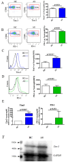
Methods: Sixty-two AS patients from Guangdong Second Provincial General Hospital and 38 healthy controls (HC) were enrolled. Clinical data, physical exams, and laboratory measurements were recorded. Flow cytometry measured Tim-3 and PD-1 expression on neutrophils, real-time PCR quantified mRNA levels and protein expression of Tim-3 was determined by Western blot. We analyzed the correlation between Tim-3 mean fluorescence intensity (MFI) on neutrophils, inflammatory markers, and AS disease activity and severity.
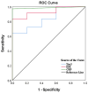
Results: Tim-3 expression on neutrophils was higher in AS patients than in HC, showing a positive correlation with erythrocyte sedimentation rate (ESR), c-reactive protein (CRP), and Ankylosing Spondylitis Disease Activity Score (ASDAS). Active AS patients (ASDAS ≥ 1.3) had increased Tim-3 MFI compared to inactive ones (ASDAS < 1.3). Regular treatment with non-steroidal anti-inflammatory drugs (NSAIDs), biological disease-modifying anti-rheumatic drugs (bDMARDs), and conventional synthetic disease-modifying anti-rheumatic drugs (csDMARDs) over a month significantly reduced Tim-3 MFI in AS patients.
Conclusion: Elevated Tim-3 expression on neutrophils correlates with increased inflammatory markers and AS activity. Treatment lowered Tim-3 MFI, suggesting its potential as an indicator for assessing AS disease activity and severity and as a feedback mechanism to reduce tissue damage from inflammation.
Keywords: Tim-3; ankylosing spondylitis; disease activity; immunomodulation; neutrophils.
Copyright © 2025 Huang, He, Yi, Zheng, Deng, Chen, Zhu, Wang, Chen, Zheng, Huang and Li.
Conflict of interest statement
The authors declare that the research was conducted in the absence of any commercial or financial relationships that could be construed as a potential conflict of interest.
Figures


Similar articles
-
Add-On Effects of Conventional Synthetic Disease-Modifying Anti-Rheumatic Drugs in Ankylosing Spondylitis: Data from a Real-World Registered Study in China.Med Sci Monit. 2020 Jan 21;26:e921055. doi: 10.12659/MSM.921055. Med Sci Monit. 2020. PMID: 31959738 Free PMC article.
-
Platelet to albumin ratio is an independent indicator for disease activity in ankylosing spondylitis.Clin Rheumatol. 2023 Feb;42(2):407-413. doi: 10.1007/s10067-022-06439-x. Epub 2022 Nov 21. Clin Rheumatol. 2023. PMID: 36414863
-
Procollagen I N-terminal peptide correlates with inflammation on sacroiliac joint magnetic resonance imaging in ankylosing spondylitis but not in non-radiographic axial spondyloarthritis: A cross-sectional study.Mod Rheumatol. 2022 Jul 1;32(4):770-775. doi: 10.1093/mr/roab044. Mod Rheumatol. 2022. PMID: 34897520
-
The Discriminative Values of the Bath Ankylosing Spondylitis Disease Activity Index, Ankylosing Spondylitis Disease Activity Score, C-Reactive Protein, and Erythrocyte Sedimentation Rate in Spondyloarthritis-Related Axial Arthritis.J Clin Rheumatol. 2017 Aug;23(5):267-272. doi: 10.1097/RHU.0000000000000522. J Clin Rheumatol. 2017. PMID: 28661926
-
Comparison of Ankylosing Spondylitis Disease Activity Score and Bath Ankylosing Spondylitis Disease Activity Index tools in assessment of axial spondyloarthritis activity.Reumatologia. 2024;62(1):64-69. doi: 10.5114/reum/185429. Epub 2024 Mar 18. Reumatologia. 2024. PMID: 38558891 Free PMC article. Review.
References
LinkOut - more resources
Full Text Sources
Research Materials
Miscellaneous

