

The Effect of Arm Abduction and Forearm Muscle Activation on Kinematics During Elbow Flexion
- PMID: 40182878
- PMCID: PMC11963008
- DOI: 10.1016/j.jhsg.2024.11.006
The Effect of Arm Abduction and Forearm Muscle Activation on Kinematics During Elbow Flexion
Abstract
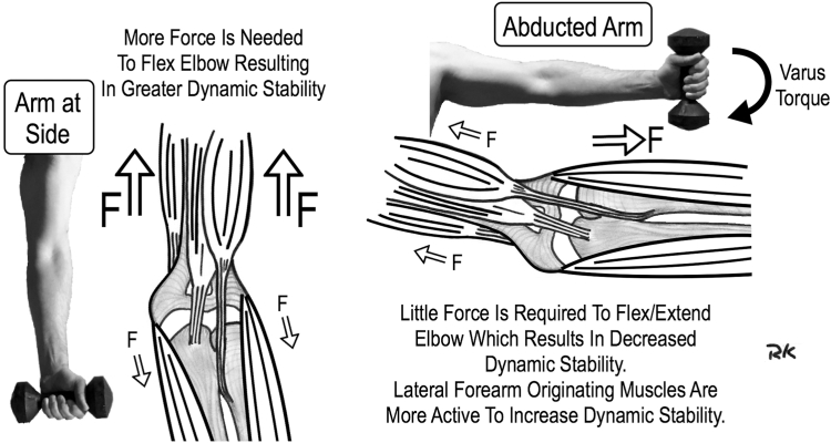
Purpose: As the elbow flexes with the arm at the side (0° humerothoracic abduction, HTA), it loses its valgus carrying angle. When the arm is abducted to 90° HTA, a varus torque tensions the lateral ligaments. Our purpose was to quantify the effect of abduction on elbow kinematics during active motion and the effect of lateral forearm muscle activation. We hypothesized that arm abduction would increase elbow varus angulation throughout flexion, and lateral forearm muscle activation would decrease varus angulation.
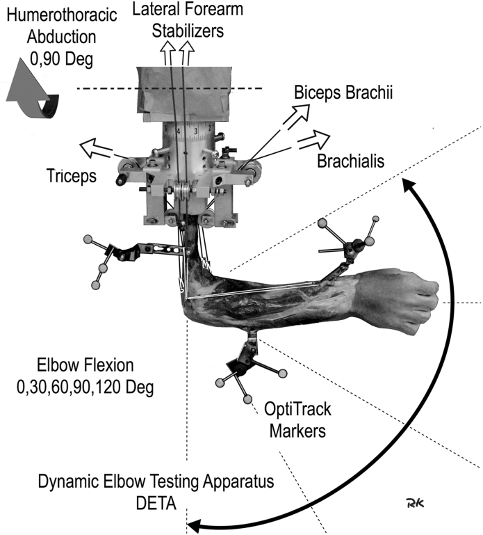
Methods: A dynamic elbow testing apparatus was employed in six human cadaver arms at two levels of arm abduction, 0° and 90° HTA. Six electromechanical actuators simulated muscle action, whereas joint position was measured to quantify the relationship between the forearm and humerus as the elbow was actively flexed.
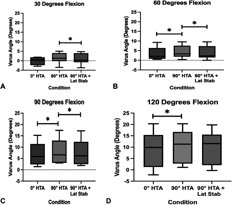
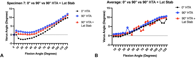
Results: All elbows maintained greater varus angle with the arm at 90° HTA compared with 0° HTA, significant at 60° flexion, 4.3° versus 3.4°, 90° flexion, 8.0° versus 6.8°, and 120° flexion, 10.5° versus 8.9°. The abducted elbow demonstrated less varus angle when the lateral stabilizers were activated. A significant difference was found at 30° flexion, 0.9 versus 1.5, 60° flexion, 3.8 versus 4.3, and 90° flexion, 7.6 versus 8.0.
Conclusions: Elbow joint coronal plane kinematics were influenced by abduction of the arm to 90° HTA, and greater elbow varus angles were found throughout flexion when compared with the arm at side position (0° HTA). In addition, activation of lateral forearm muscles (90° HTA + Lat Stab) decreased elbow varus angulation throughout flexion.
Clinical relevance: Understanding the effect of varus torque on elbow biomechanics and the degree to which these effects are countered through dynamic stabilization may assist in arthroplasty and ligamentous reconstruction designs.
Keywords: Dynamic stabilizers; Elbow biomechanics; Elbow kinematics; Elbow valgus.
© 2024 The Authors.
Conflict of interest statement
No benefits in any form have been received or will be received related directly to this article.
Figures
Similar articles
-
The Effect of Elbow Flexion On Valgus Carrying Angle.J Hand Surg Am. 2025 Mar;50(3):373.e1-373.e6. doi: 10.1016/j.jhsa.2023.07.010. Epub 2023 Aug 17. J Hand Surg Am. 2025. PMID: 37589618
-
A Dynamic Elbow Testing Apparatus for Simulating Elbow Joint Motion in Varying Shoulder Positions.J Hand Surg Glob Online. 2023 Aug 31;5(6):823-827. doi: 10.1016/j.jhsg.2023.07.017. eCollection 2023 Nov. J Hand Surg Glob Online. 2023. PMID: 38106931 Free PMC article.
-
Effect of the posterior bundle of the medial collateral ligament on elbow stability.J Hand Surg Am. 2009 Jan;34(1):116-23. doi: 10.1016/j.jhsa.2008.09.016. J Hand Surg Am. 2009. PMID: 19121737
-
The role of the elbow musculature, forearm rotation, and elbow flexion in elbow stability: an in vitro study.J Shoulder Elbow Surg. 2009 Mar-Apr;18(2):260-8. doi: 10.1016/j.jse.2008.08.004. Epub 2008 Nov 30. J Shoulder Elbow Surg. 2009. PMID: 19046641
-
Elbow Biomechanics: Bony and Dynamic Stabilizers.J Hand Surg Am. 2020 Jun;45(6):528-535. doi: 10.1016/j.jhsa.2020.01.016. Epub 2020 Apr 13. J Hand Surg Am. 2020. PMID: 32299691 Review.
References
-
- Shiba R., Sorbie C., Siu D.W., Bryant J.T., Cooke T.D., Wevers H.W. Geometry of the humeroulnar joint. J Orthop Res. 1988;6(6):897–906. - PubMed
-
- Van Roy P., Baeyens J.P., Fauvart D., Lanssiers R., Clarijs J.P. Arthro-kinematics of the elbow: study of the carrying angle. Ergonomics. 2005;48(11e14):1645e1656. - PubMed
-
- Kincaid B.L., An K.-N. Elbow joint biomechanics for preclinical evaluation of total elbow prostheses. J Biomech. 2013;46:2331–2341. - PubMed
-
- Nicol A.C., Berme N., Paul J.P. Proceeding of the IMechE Conference of Joint Replacement of the Upper Extremity. Institute of Mechanical Engineers; 1977. A Biomechanical analysis of elbow joint function; pp. 45–51.
LinkOut - more resources
Full Text Sources
Research Materials
