. 2025 Mar 20:13:1547044.
doi: 10.3389/fbioe.2025.1547044. eCollection 2025.

Towards propagation of epidermal cells for wound repair: glass, as cell culture substrate, enhances proliferation and migration of human keratinocytes

Affiliations

Towards propagation of epidermal cells for wound repair: glass, as cell culture substrate, enhances proliferation and migration of human keratinocytes

Hady Shahin et al. Front Bioeng Biotechnol. .

Abstract

Introduction: Human keratinocytes require relatively long propagation time which impedes their availability as autologous cell transplantation within a clinically reasonable timeframe. There is an unmet need for efficient xeno-free cell expansion approaches to propagate human keratinocytes as regenerative therapy.

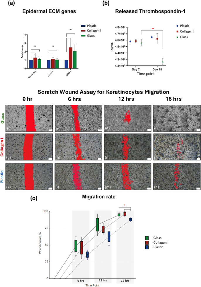
Methods: Primary human keratinocytes and HaCaT cells were cultured on glass, plastic, and animal-derived collagen I matrix for 10 days. Proliferation, migration, DNA methylation, as well as gene and protein expression were assessed to characterize the effect of the tested culture substrates on keratinocytes at the molecular and functional levels.

Results: Keratinocytes cultured on glass exhibited faster proliferation, global DNA demethylation and upregulation of epidermal differentiation markers. Scratch wound assay revealed that keratinocytes cultured on glass demonstrated enhanced cell migration compared to those on plastic or collagen I. Multiplex immunoassays identified temporal and substrate-dependent variations in a panel of keratinocyte-specific secreted factors, encompassing immunomodulatory cytokines, growth factors, and angiogenic factors.

Discussion: Glass, as a culture substrate, promotes epidermal differentiation and enhances keratinocyte migration. The latter is a critical factor in re-epithelialization and wound healing. Functional properties suggest that glass may optimize the inflammatory response and promote efficient wound repair, making it a promising candidate for the short-term expansion of keratinocytes for transplantation purposes. Further in-vivo validation is required to definitively establish the efficacy of keratinocytes cultured on glass for clinical applications.

Keywords: ATMP; culture substrate; epidermal differentiation; glass; keratinocytes; migration; wound healing.


Conflict of interest statement

The authors declare that the research was conducted in the absence of any commercial or financial relationships that could be construed as a potential conflict of interest.

Figures

FIGURE 1
FIGURE 1
Live images showed HaCaT’s polygonal ‘cobblestone’ shape and colony formatiom (a–c) at 48 h and (d–f) at 72 h; scale bar = 100 µm. Images are representatives of four independent experiments. (g) HaCaT cultured on three substrates maintaining an exponential growth curve over 24, 48 and 72 h. Cells cultured on glass showed superior proliferation rate. (h) Gene expression of basal epidermal genes after 10 days in culture on the three substrates. p63 showed no significant changes between the three groups, K5 showed slight downregulation at FC of 0.6 and 0.7 in cells grown on collagen I and glass respectively, K14 showed upregulation in cells grown on glass by 1.7 FC compared to plastic. Gene expression data is shown as the mean of fold change of four independent experiments and the standard error of mean, expression was normalized to the plastic control FC of 1. *Statistically significant *p < 0.05, **p < 0.01, ***p < 0.001, & ****p < 0.0001 vs. the control.
FIGURE 2
FIGURE 2
(a) Global DNA methylation level. Quantification of 5-mC content of DNA samples from keratinocytes cultured on studied substrates. Glass and collagen I decreased the methylation level significantly to almost 50% (0.9 ± 0.3 ng and 1 ± 0.1 ng, respectively) of the plastic control (2.3 ± 0.5 ng). (b) Gene expression analysis of epidermal differentiation markers shows upregulation in cells grown on glass of K1, K10, involucrin, stratifin and loricrin; whereas filaggrin shows an upregulation trend in glass compared to plastic at FC of 2.9. Gene expression data was presented as the mean of fold change of four independent experiments and the standard error of mean, expression was normalized to the plastic control FC of 1. *Statistically significant *p < 0.05, **p < 0.01 & ***p < 0.001 vs. the control.
FIGURE 3
FIGURE 3
(a) Gene expression of ECM markers, shows comparable expression of fibronectin, Col IV and MMP1 after 10 days of culture on the three substrates. Gene expression data is shown as the mean of fold change of four independent experiments and the standard error of mean. (b) Comparable amounts of thrombospondin-1 released by keratinocytes cultured on all three substrates at ∼ 4.6 × 10³ ng/mL at day 7, then at day 10 thrombospondin-1 released by keratinocytes cultured on glass declined to 4.2 × 10³ ng/mL compared to day 7 (p-value = 0.008). (c–f) SWA images from glass group, showing faster cell migration in the glass group at 6, 12 & 18 h (g–j) SWA images from collagen I group, (k–n) SWA images from plastic group. Scale bars = 200 µm. (o) After 6 h the scratch wounds in glass and collagen I groups showing 48.4% and 45.8% healing respectively compared to 35.5% in the plastic group, at 12 h healing% shows 80.2%, 73.4% in glass and collagen I respectively compared to plastic 62.4%, at 18 h the scratch wounds almost entirely healed in the glass and collagen I groups with 94.8% and 96% respectively compared to plastic 87.9% closure (p-value = 0.02). SWA data is presented as Box: mean ± SE; Whisker: mean ± SD from three independent biological replicates. *Statistically significant *p < 0.05 & **p < 0.01 vs. the control.
FIGURE 4
FIGURE 4
(a) Quantification of total protein content in the culture supernatant of keratinocytes. The data are presented as a violin plot, showing all individual data points. The borders of the violin represent the distribution, while the solid lines indicate the median of each dataset. The dotted lines denote the upper and lower quartiles, and the mean is shown as a separate marker. (b–i) Luminex® multiplex immunoassays quantifying the levels of analytes secreted by cultured keratinocytes on days 3, 7 and 10. The analytes measured are (b) VEGF-A, (c) EGF, (d) TIMP-1, (e) TNF-α, (f) IL-1α, (g) IL-8, (H) KLK-5, and (i) KLK-6. Data from four independent experimental replicates are presented as superimposed symbols representing the mean ± standard error of the mean, with connecting lines illustrating trends. The data are normalized to the total protein content of each corresponding sample. Statistical significance was considered when p < 0.05 using Welch’s t-test. Statistical differences between each surface are detailed in Table 2. * Signifies statistical significance in the temporal differences between time points within each group.

Similar articles

References

    1. Alam H., Sehgal L., Kundu S. T., Dalal S. N., Vaidya M. M. (2011). Novel function of keratins 5 and 14 in proliferation and differentiation of stratified epithelial cells. Mol. Biol. Cell 22, 4068–4078. 10.1091/mbc.e10-08-0703 - DOI - PMC - PubMed
    1. Alghfeli L., Parambath D., Manzoor S., Roach H. I., Oreffo R. O. C., El-Serafi A. T. (2021). Synthesis of scaffold-free, three dimensional, osteogenic constructs following culture of skeletal osteoprogenitor cells on glass surfaces. Bone Rep. 15, 101143. 10.1016/j.bonr.2021.101143 - DOI - PMC - PubMed
    1. Alghfeli L., Parambath D., Tag Eldeen L. A., El-Serafi I., El-Serafi A. T. (2022). Non-additive effect of the DNA methylation inhibitor, 5-Aza-dC, and glass as a culture surface on osteogenic differentiation. Heliyon 8, e12433. 10.1016/j.heliyon.2022.e12433 - DOI - PMC - PubMed
    1. Bae O. N., Noh M., Chun Y. J., Jeong T. C. (2015). Keratinocytic vascular endothelial growth factor as a novel biomarker for pathological skin condition. Biomol. Ther. Seoul. 23, 12–18. 10.4062/biomolther.2014.102 - DOI - PMC - PubMed
    1. Banno T., Gazel A., Blumenberg M. (2004). Effects of tumor necrosis factor-alpha (TNF alpha) in epidermal keratinocytes revealed using global transcriptional profiling. J. Biol. Chem. 279, 32633–32642. 10.1074/jbc.m400642200 - DOI - PubMed