. 2025 Mar 20:16:1549183.
doi: 10.3389/fphar.2025.1549183. eCollection 2025.

Pharmacological evaluation of drug therapies in Aicardi-Goutières syndrome: insights from patient-derived neural stem cells

Affiliations

Pharmacological evaluation of drug therapies in Aicardi-Goutières syndrome: insights from patient-derived neural stem cells

Stefania Braidotti et al. Front Pharmacol. .

Abstract

Aicardi-Goutières syndrome (AGS) is a rare genetic disorder classified among type I interferonopathies. Current pharmacological management of AGS is symptomatic and supportive, with recent clinical applications of JAK inhibitors (JAKi) and antiretroviral therapies (RTIs). To investigate the effects of these therapies, patient-specific induced pluripotent stem cells (iPSCs) were generated by reprogramming fibroblasts from three AGS patients with distinct genetic mutations (AGS1, AGS2, AGS7) and differentiated into neural stem cells (NSCs). iPSCs and NSCs derived from commercial BJ fibroblasts of a healthy donor served as control. The cytotoxic effects of glucocorticoids, thiopurines, JAK inhibitors (ruxolitinib, baricitinib, tofacitinib, pacritinib), and RTIs (abacavir, lamivudine, zidovudine) were evaluated using the MTT assay. Results showed that glucocorticoids did not compromise NSC viability. Among thiopurines, thioguanine, but not mercaptopurine, exhibited cytotoxicity in NSCs. All tested JAK inhibitors, except pacritinib, were non-toxic to iPSCs and NSCs. Interestingly, high concentrations of certain JAK inhibitors (ruxolitinib, baricitinib, tofacitinib) led to an unexpected increase in cell viability in AGS patient-derived cells compared to control, suggesting potential alterations in cell proliferation or stress responses. RTIs demonstrated no cytotoxicity, except for zidovudine, which showed selective toxicity in AGS2-derived iPSCs compared to controls. These findings suggest that glucocorticoids, JAK inhibitors (excluding pacritinib), and RTIs are likely safe for NSCs of AGS patients, while caution is warranted with thioguanine and pacritinib. Further studies are needed to explore the mechanisms underlying increased cell viability at high JAK inhibitor concentrations and the selective sensitivity to zidovudine.

Keywords: Aicardi-Goutières syndrome; JAK inhibitors; antiretrovirals; drug sensitivity; patient-derived stem cell.


Conflict of interest statement

The authors declare that the research was conducted in the absence of any commercial or financial relationships that could be construed as a potential conflict of interest.

Figures

FIGURE 1
FIGURE 1
Expression levels of iPSCs (OCT4, SOX2) and NSCs stemness specific markers (SOX2, Nestin, SOX1, PAX6) in iPSC and NSCs. Gene expression was normalized to housekeeping actin β gene expression, and relative expression was calculated as 2−ΔCt. P-value according to t-test analysis, *P < 0.05, **P < 0.01, ***P < 0.001, ****P < 0.0001. The data are reported as means ± standard error (SE) of 3 independent experiments performed in triplicate.
FIGURE 2
FIGURE 2
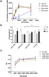
Stem cell viability assays. (A) iPSCs proliferation assay; [3H]-thymidine incorporation into DNA was expressed counts per minute (or CPM). (B) iPSCs cell cycle assay. (C) NSCs MTT assay; NSCs metabolic activity was evaluated under basal conditions. Cell densities referred to initial cell/well seeding in a 96-wells plate. OD: optical density. Data are reported as means ± SE of at least 3 in-dependent experiments performed in triplicate. Statistical analysis vs control BJ-stem cells: *P < 0.05, ***P < 0.001, Two-way ANOVA, and Bonferroni’s post-test.
FIGURE 3
FIGURE 3
Drug safety of immunosuppressant drugs. (A) Dose-response curve (by MTT assay) of dexamethasone on NSCs. Immortalized lymphoblastoid cell line NALM6 was used as control. (B) Relative NR3C1 gene expression. (C) Dose-response curve (by MTT assay) of thiopurines on NSCs. (D) Relative HPRT1 gene expression. Drug cytotoxic effects were analyzed by MTT assay after 72 h; results are presented as mean ± standard error (SE) from at least 3 independent experiments for each cell line; P-value were calculated according to Two-way ANOVA, Bonferroni’s post-test. Gene expression was calculated with respect the housekeeping gene actin β (relative expression of ΔCT (2−ΔCT)); P-value were calculated according to one-way ANOVA analysis. *, p < 0.05, **, p < 0.01, ***, p < 0.001,****, p < 0.0001.
FIGURE 4
FIGURE 4
Dose-response curve (by MTT assay) after 72 h exposure of ruxolitinib, baricitinib, tofacitinib, pacritinib in iPSCs (A–D) and NSCs (E–H). Seeding density: AGS2-iPSCs: 3.0 × 10⁴ cells/well, all other iPSCs and NSCs: 1.0 × 10⁴ cells/well in 96-well plate. The data are reported as percentage of cell viability as means ± standard error (SE) of 3 independent experiments performed in triplicate. Statistical analysis vs control BJ stem cells: *P < 0.05, **P < 0.01, ***P < 0.001, ****P < 0.0001, Two-way ANOVA and Bonferroni’s post-test. Green asterisks referred to AGS7- versus BJ-stem cells; blue asterisks to AGS2- versus BJ-stem cells; red asterisks to AGS1- versus BJ-stem cells.
FIGURE 5
FIGURE 5
Dose-response curve (by MTT assay) after 72 h exposure with abacavir, lamivudine, zidovudine, in iPSCs (A–C) and NSCs (D–F), respectively. The data are reported as percentage of cell viability as means ± standard error (SE) of 3 independent experiments performed in triplicate. Statistical analysis vs control BJ: *P < 0.05, ***P < 0.001, Two-way ANOVA and Bonferroni’s post-test. Blue asterisks reffered to AGS2- versus BJ-stem cells.

References

    1. Aicardi J., Goutières F. (1984). A Progressive familial encephalopathy in infancy with calcifications of the basal ganglia and chronic cerebrospinal fluid lymphocytosis. Ann. Neurology 15 (1), 49–54. 10.1002/ana.410150109 - DOI - PubMed
    1. Akwa Y., Hassett D. E., Eloranta M. L., Sandberg K., Masliah E., Powell H., et al. (1998). Transgenic expression of IFN-α in the central nervous system of mice Protects against Lethal Neurotropic viral Infection but Induces inflammation and Neurodegeneration. J. Immunol. 161 (9), 5016–5026. 10.4049/jimmunol.161.9.5016 - DOI - PubMed
    1. Ali E., Ferraro R. M., Lanzi G., Masneri S., Piovani G., Mazzoldi E. L., et al. (2020). Generation of induced pluripotent stem cell (iPSC) lines from a Joubert syndrome patient with compound heterozygous mutations in C5orf42 gene. Stem Cell Res. 49, 102007. 10.1016/j.scr.2020.102007 - DOI - PubMed
    1. Al Mutairi F., Alfadhel M., Nashabat M., El-Hattab A. W., Ben-Omran T., Hertecant J., et al. (2018). Phenotypic and molecular Spectrum of Aicardi-Goutières syndrome: a study of 24 patients. Pediatr. Neurol. 78, 35–40. 10.1016/j.pediatrneurol.2017.09.002 - DOI - PubMed
    1. Aquaro S., Borrajo A., Pellegrino M., Svicher V. (2020). Mechanisms underlying of antiretroviral drugs in different cellular reservoirs with a focus on macrophages. Virulence 11 (1), 400–413. 10.1080/21505594.2020.1760443 - DOI - PMC - PubMed

LinkOut - more resources