. 2025 Jun;31(6):146.
doi: 10.3892/mmr.2025.13511. Epub 2025 Apr 4.

α‑ketoglutarate protects against septic cardiomyopathy by improving mitochondrial mitophagy and fission

Affiliations

α‑ketoglutarate protects against septic cardiomyopathy by improving mitochondrial mitophagy and fission

Wei Wu et al. Mol Med Rep. 2025 Jun.

Abstract

Septic cardiomyopathy is a considerable complication in sepsis, which has high mortality rates and an incompletely understood pathophysiology, which hinders the development of effective treatments. α‑ketoglutarate (AKG), a component of the tricarboxylic acid cycle, serves a role in cellular metabolic regulation. The present study delved into the therapeutic potential and underlying mechanisms of AKG in ameliorating septic cardiomyopathy. A mouse model of sepsis was generated and treated with AKG via the drinking water. Cardiac function was assessed using echocardiography, while the mitochondrial ultrastructure was examined using transmission electron microscopy. Additionally, in vitro, rat neonatal ventricular myocytes were treated with lipopolysaccharide (LPS) as a model of sepsis and then treated with AKG. Mitochondrial function was evaluated via ATP production and Seahorse assays. Additionally, the levels of reactive oxygen species were determined using dihydroethidium and chloromethyl derivative CM‑H2DCFDA staining, apoptosis was assessed using a TUNEL assay, and the expression levels of mitochondria‑associated proteins were analyzed by western blotting. Mice subjected to LPS treatment exhibited compromised cardiac function, reflected by elevated levels of atrial natriuretic peptide, B‑type natriuretic peptide and β‑myosin heavy chain. These mice also exhibited pronounced mitochondrial morphological disruptions and dysfunction in myocardial tissues; treatment with AKG ameliorated these changes. AKG restored cardiac function, reduced mitochondrial damage and corrected mitochondrial dysfunction. This was achieved primarily through increasing mitophagy and mitochondrial fission. In vitro, AKG reversed LPS‑induced cardiomyocyte apoptosis and dysregulation of mitochondrial energy metabolism by increasing mitophagy and fission. These results revealed that AKG administration mitigated cardiac dysfunction in septic cardiomyopathy by promoting the clearance of damaged mitochondria by increasing mitophagy and fission, underscoring its therapeutic potential in this context.

Keywords: AKG; fission; mitochondria; mitophagy; septic cardiomyopathy.


Conflict of interest statement

The authors declare that they have no competing interests.

Figures

Figure 1.
Figure 1.
AKG improves cardiac dysfunction in an LPS-induced mouse model of septic cardiomyopathy. (A) Representative 2D echocardiographic images of the LV short-axis from mice treated with control, AKG, LPS and AKG + LPS. (C) LVEF, LVFS, LVEDD and LVESD based on the LV ultrasound images (n=8/group). (B) Protein and mRNA expression levels of ANP, BNP and β-MHC in LV myocardium (n=6/group), GAPDH was used as the loading control for ANP and BNP, while HSP90 was used as the loading control for β-MHC. Data are presented as the mean ± SEM. *P<0.05, P<0.01, #P<0.001. AKG, α-ketoglutarate; LPS, lipopolysaccharide; LV, left ventricle; LVEF, LV ejection fraction; LVFS, LV fractional shortening; LVEDD, LV end-diastolic dimension; LVESD, LV end-systolic dimension; ANP, atrial natriuretic peptide; BNP, brain natriuretic peptide; β-MHC, β-major histocompatibility complex; HSP90, heat shock protein 90.
Figure 2.
Figure 2.
AKG alleviates myocardial mitochondrial morphological damage induced by LPS. (A) Electron microscopy images of left ventricular myocardium. Magnification: Top, ×10,000; bottom, ×30,000. *Mitochondria with dissolved cristae; the arrow indicates mitochondria with incomplete outer membranes (n=6/group). (B) Representative immunoblotting images and semi-quantitative analysis of protein expression of markers of mitochondrial mitophagy (Bnip3 and LC3), fission (DRP1 and Fis1) and mitochondrial content (Ndufab1 and Ndufa12; n=6/group). (C) Quantitative analysis of mitochondria. The percentage of mitochondria with an incomplete outer membrane and dissolved cristae, and the number of mitochondria per unit area (n=6/group). Data are presented as the mean ± SEM. *P<0.05, P<0.01, #P<0.001. AKG, α-ketoglutarate; m, mitochondria; Z, Z line; Mito number, the number of mitochondria per unit area; Bnip3, BCL2 interacting protein 3; DRP1, dynamin-related protein 1; Nduf, NADH:ubiquinone oxidoreductase subunits; LPS, lipopolysaccharide.
Figure 3.
Figure 3.
AKG recovers myocardial mitochondrial energy metabolism in an LPS-induced mouse model of septic cardiomyopathy. Western blotting was carried out using protein extracted from LV tissues, and protein expression was semi-quantified and normalized to GAPDH (n=6/group). (A) Representative blots of marker proteins for mitochondrial energy metabolism: Mitochondrial complex I (MT-ND1), complex II (SDHA), COX IV, tricarboxylic acid cycle (OGDH), a key enzyme for acetyl-CoA synthesis (PDH) and anaerobic glycolysis (HIF-1α). (B) Semi-quantitative analysis of mitochondrial energy metabolism marker proteins. (C) ATP content in the LV tissues, and lactate content in the plasma and LV tissues, as determined by an ELISA (n=6/group). Data are presented as the mean ± SEM. *P<0.05, P<0.01, #P<0.001. LV, left ventricle; MT-ND1, mitochondrial NADH-ubiquinone oxidoreductase chain 1; HIF-1α, hypoxia inducible factor-1α; OGDH, ketoglutarate dehydrogenase; PDH, pyruvate dehydrogenase; SDHA, succinate dehydrogenase; AKG, α-ketoglutarate; LPS, lipopolysaccharide; COX IV, complex IV; prot, protein.
Figure 4.
Figure 4.
AKG reduces myocardial ROS and apoptosis in an LPS-induced mouse model of septic cardiomyopathy. ROS levels were detected using various methods, including DCF staining, DHE staining, MDA content determination using an ELISA, and examination of NOX2/4 protein expression. Apoptosis was assessed via TUNEL staining, and examination of the protein expression levels of Bax and Bcl-2. (A) From top to bottom: Representative images of DCF, DHE, TUNEL and H&E staining (n=6/group). (B) Changes in myocardial and plasma levels of MDA (n=6/group). (C) Western blot bands and analysis of marker proteins for NOX2 and NOX4 (n=6/group). (D) Quantitative analysis of DCF, DHE and TUNEL staining (n=6/group). (E) Representative blots and analysis of marker proteins for Bax and Bcl-2 (n=6/group). Data are presented as the mean ± SEM. *P<0.05, P<0.01, #P<0.001. AKG, α-ketoglutarate; LPS, lipopolysaccharide; ROS, reactive oxygen species; DCF, chloromethyl derivative CM-H2DCFDA; DHE, dihydroethidium; MDA, malondialdehyde; NOX, NADPH oxidase; prot, protein.
Figure 5.
Figure 5.
Effect of AKG on apoptosis and mitochondrial metabolism and dynamics in vitro. Neonatal rat ventricular myocytes were treated with vehicle, AKG, LPS or AKG + LPS. (A) Images and quantitative analysis of the TUNEL staining (n=6/group). (B) Representative western blot images and (C) semi-quantification of markers for mitochondrial metabolism (MT-ND1, SDHA, COX IV, OGDH and PDH) (n=6/group). (D) Protein expression levels of selected markers for apoptosis (Bcl-2), mitochondrial quality control (Bnip3 and DRP1) and mitochondrial number (Ndufa12). (E) Semi-quantitative analysis of protein expression of Bcl-2, Bnip3, DRP1 and Ndufa12) (n=6/group). Data are presented as the mean ± SEM. *P<0.05, P<0.01, #P<0.001. AKG, α-ketoglutarate; LPS, lipopolysaccharide; MT-ND1, mitochondrial NADH-ubiquinone oxidoreductase chain 1; SDHA, succinate dehydrogenase; COX IV, complex IV; OGDH, ketoglutarate dehydrogenase; PDH, pyruvate dehydrogenase; Bnip3, BCL2 interacting protein 3; DRP1, dynamin-related protein 1; Nduf, NADH:ubiquinone oxidoreductase subunits.
Figure 6.
Figure 6.
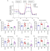
Effect of AKG on mitochondrial respiratory capacity in vitro. (A) OCR curve of the control, AKG, LPS and AKG + LPS groups (n=5/group). (B) Non-mitochondrial respiration, (C) basal respiration, (D) maximal respiration, (E) ATP production, (F) proton leak and (G) spare respiration capacity were calculated. Data are presented as the mean ± SEM. *P<0.05, P<0.01, #P<0.001. AKG, α-ketoglutarate; LPS, lipopolysaccharide; OCR, oxygen consumption rate; FCCP, trifluoromethoxy carbonylcyanide phenylhydrazone.

Similar articles

References

    1. Singer M, Deutschman CS, Seymour CW, Shankar-Hari M, Annane D, Bauer M, Bellomo R, Bernard GR, Chiche JD, Coopersmith CM, et al. The third international consensus definitions for sepsis and septic shock (sepsis-3) JAMA. 2016;315:801–810. doi: 10.1001/jama.2016.0287. - DOI - PMC - PubMed
    1. Fleischmann C, Scherag A, Adhikari NK, Hartog CS, Tsaganos T, Schlattmann P, Angus DC, Reinhart K, International Forum of Acute Care Trialists Assessment of global incidence and mortality of hospital-treated sepsis. Current estimates and limitations. Am J Respir Crit Care Med. 2016;193:259–272. doi: 10.1164/rccm.201504-0781OC. - DOI - PubMed
    1. Prescott HC, Angus DC. Enhancing recovery from sepsis: A review. JAMA. 2018;319:62–75. doi: 10.1001/jama.2017.17687. - DOI - PMC - PubMed
    1. Sato R, Nasu M. A review of sepsis-induced cardiomyopathy. J Intensive Care. 2015;3:48. doi: 10.1186/s40560-015-0112-5. - DOI - PMC - PubMed
    1. Hollenberg SM, Singer M. Pathophysiology of sepsis-induced cardiomyopathy. Nat Rev Cardiol. 2021;18:424–434. doi: 10.1038/s41569-020-00492-2. - DOI - PubMed

MeSH terms