. 2025 Mar 20;53(6):gkaf241.
doi: 10.1093/nar/gkaf241.

AOC 1044 induces exon 44 skipping and restores dystrophin protein in preclinical models of Duchenne muscular dystrophy

Affiliations

AOC 1044 induces exon 44 skipping and restores dystrophin protein in preclinical models of Duchenne muscular dystrophy

Usue Etxaniz et al. Nucleic Acids Res. .

Abstract

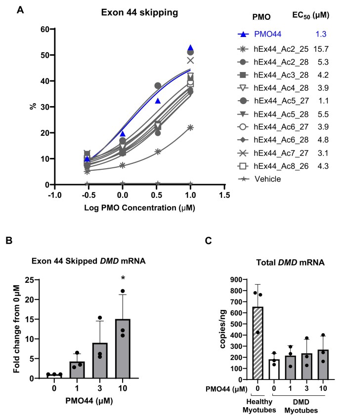
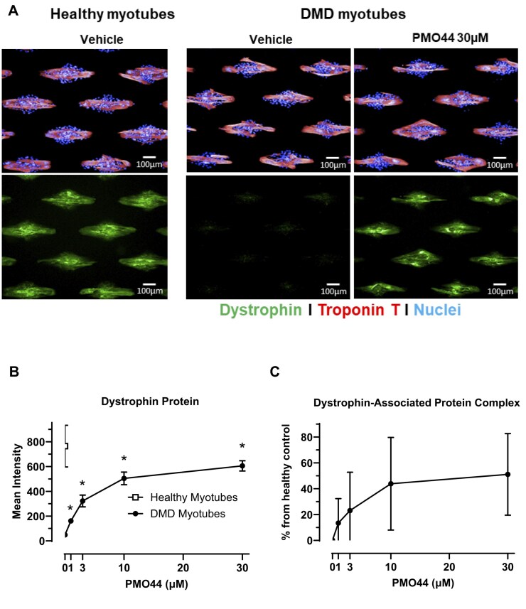
Duchenne muscular dystrophy (DMD) is a severe disorder caused by mutations in the dystrophin gene, resulting in loss of functional dystrophin protein in muscle. While phosphorodiamidate morpholino oligomers (PMOs) are promising exon-skipping therapeutics aimed at restoring dystrophin expression, their effectiveness is often limited by poor muscle delivery. We developed AOC 1044, an antibody-oligonucleotide conjugate (AOC) that combines a PMO-targeting exon 44 with an antibody against the transferrin receptor (TfR1), enhancing delivery to muscle tissues for patients with DMD amenable to exon 44 skipping (DMD44). AOC 1044 induces dose-dependent exon 44 skipping and its mouse-active variant elicited dose-dependent dystrophin restoration in skeletal and cardiac muscle in a DMD mouse model. This treatment also reduced muscle damage, as evidenced by decreases in serum creatine kinase and key liver enzymes, suggesting that restored dystrophin is functionally active. In nonhuman primates, single or repeated AOC 1044 doses resulted in dose-dependent increases in PMO concentration and exon 44 skipping across a range of muscle tissues, including the heart. Collectively, these findings highlight AOC 1044 as a promising therapeutic candidate for patients with DMD44, offering improved muscle targeting and meaningful dystrophin restoration, with potential clinical benefits in reducing muscle degeneration.


Conflict of interest statement

Usue Etxaniz, Isaac Marks, Michael Cochran, and Venkata R. Doppalapudi are employees of Avidity Biosciences, Inc. who receive stock and stock options and have patent applications or pending or awarded patents. Matthew Diaz, Aaron Anderson, Olecya Tyaglo, Tiffany Hoang, Maria Azzurra Missinato, Kristoffer Svensson, Ben Badillo, Philip R. Kovach, Laura Leung, Hae Won Kwon, Husam S. Younis, W. Michael Flanagan, Arthur A. Levin, Hanhua Huang, and Georgios Karamanlidis are employees of Avidity Biosciences, Inc. who receives stock and stock options. Tyler Albin, Raghav Bhardwaj, Maria Azzurra Missinato, and Beatrice Darimont are former employees of Avidity Biosciences, Inc and may hold stock and/or stock options (and Tyler Albin and Beatrice Darimont have patent applications). Toshifumi Yokota and Rika Maruyama are co-founders and shareholders of OligomicsTX, Inc.

Figures

Graphical Abstract
Graphical Abstract
Figure 1.
Figure 1.
PMO44-mediated exon 44 skipping in DMD-patient-derived myotubes. (A) Primary human skeletal myotubes were treated with PMOs (0.3–10 μM) using Endoporter as a transfection reagent. Total and exon 44-skipped DMD mRNA transcripts were quantified by reverse-transcription quantitative polymerase chain reaction (RT-qPCR) at 48 h post-treatment. Data represent means from a single experiment performed in duplicate. (B, C) Myotubes derived from myoblast cell lines from DMD patients were treated with PMO44 (0–10 μM). Healthy myotubes were also included as a control in (C). Exon-44-skipped mRNA and total DMD (exon 73–74) levels were determined by ddPCR 48 h post-treatment. Data are presented as mean ± standard error of the mean (SEM), n = 3/group. * Statistical difference relative to vehicle control (P < .05) using one-way ANOVA with Dunnett’s post-hoc test.
Figure 2.
Figure 2.
PMO44-mediated dystrophin protein restoration in DMD-patient-derived myotubes. (A) Dystrophin protein (green staining) was evaluated by immunofluorescence after 6 days of PMO treatment. (B) Quantitative microscopy of dystrophin staining was performed after 6 days of PMO treatment. (C) DAPC was evaluated after 6 days of PMO treatment using quantitative immunofluorescence using CellProfiler to evaluate restoration. Data were acquired using Operetta HCS imaging system and analyzed using Acapella software (PerkinElmer) and are plotted as means ± SEM. DMD myotubes were derived from three different patients, and n = 4 for healthy myotubes, >50 myotubes per replicate were analyzed. *P < .05 relative to 0 μM using one-way ANOVA with Dunnett’s post-hoc test.
Figure 3.
Figure 3.
mAOC 1044 exon 44 skipping in mice expressing WT hDMD or hDMDdel45/mdx. (A) PMO44 concentration, (B) exon 44 skipping, and (C) total DMD mRNA in mice administered a single IV dose of mAOC 1044 (30 mg/kg, per PMO44 component) from which muscle tissue was harvested at 2 weeks post-dose. Data presented as mean ± SEM, n = 3–4 per group. *P < .05 compared to WT, using unpaired t-test.
Figure 4.
Figure 4.
mAOC 1044 produced increased PMO44 muscle tissue concentration in hDMDdel45/mdx mice. hDMDdel45/mdx mice were administered a single IV dose of mAOC 1044 (10 or 30 mg/kg, per PMO44 component) or unconjugated PMO44 (30 mg/kg) and muscle tissue was collected 2 or 4 weeks after dosing (n = 4/group). (AC) PMO concentrations, (DF) exon-44-skipped DMD mRNA, and (G–I) dystrophin protein was evaluated in gastrocnemius, diaphragm, and heart muscles. All PMO44 dose group samples had values below the limit of quantification (5.4 nM). Data presented as mean ± SEM. All mAOC 1044 treatment groups at all time points were statistically significant relative to vehicle control *(P < .05) using two-way ANOVA with Tukey’s post-hoc test. This figure displays data from an experiment that utilized the same set of animals, n = 4 per group.
Figure 5.
Figure 5.
PK/PD profile of mAOC 1044 in gastrocnemius muscle of hDMDdel45/mdx mice. hDMDdel45/mdx mice were administered a single IV dose of mAOC 1044 (10 or 30 mg/kg, PMO44 component). The PK/PD model was developed based on the PMO44 concentrations, exon 44 skipping, and dystrophin protein production in muscle at 4 weeks post-dose, n = 4/group.
Figure 6.
Figure 6.
mAOC 1044 produced reductions in serum markers of muscle damage in hDMDdel45/mdx mice (4–6 months old). (A) CK (B) ALT and (C) AST in serum collected from hDMDdel45/mdx mice treated with a single IV dose of mAOC 1044 (0, 10, or 30 mg/kg, PMO44 component) or unconjugated PMO44 (30 mg/kg) for 4 weeks. Data presented as mean ± SEM, n = 4/group. *P < .05 relative to 0 mg/kg group using one-way ANOVA with Dunnett’s post-hoc test. The data shown in this figure were obtained from the same set of animals shown in Fig. 4, collected at 4 weeks post-dose.
Figure 7.
Figure 7.
Pharmacological and functional activity of mAOC 23 in the mdx mouse model of DMD. Mdx mice were administered a single IV dose of vehicle or mAOC 23 at 30 mg/kg (per PMO23 component). WT animals were administered a single dose of vehicle. (A) Exon 23 skipping, and dystrophin protein were evaluated in gastrocnemius tissue 4 weeks after dosing. (B) Representative micrograph of dystrophin protein staining in quadriceps muscle. (C) Distance traveled in open field arena was quantified. Data are presented as means ± SEM, n = 4–8/group, *P < .05 compared to mdx-vehicle group using an unpaired t-test. (D) A representative image of the open field arena test for a single mouse in each group is shown for illustration purposes. In this figure, panels (A) and (C)/(D) reflect data from different animal sets within the same experiment, while panel (B) presents data from different animals in an independent study.
Figure 8.
Figure 8.
AOC 1044 produced dose-dependent increases in PMO44 concentration and exon 44 skipping in muscle of NHPs. (A) PMO44 tissue concentration and (B) exon 44 skipping in NHPs administered IV AOC 1044 (0, 5, 15 or 45 mg/kg PMO44 component) every 4 weeks for 3 months. Data are presented as mean ± SEM, n = 3–5/group/time point. (C) Exon 44 skipping in a range of skeletal muscles in monkeys 6 weeks after receiving a single injection of AOC 1044 at 30 mg/kg (based on the PMO44 component). Data are presented as mean ± SEM. *P < .05 compared to vehicle group, n = 4 group using unpaired t-test. In this figure, the same animal set was used for the tissue concentration and exon skipping data. Panel (C) presents data from an independent NHP study.
Figure 9.
Figure 9.
AOC 1044 was well-tolerated in NHPs following 9 months of treatment. (A) Body weight (B), heart rate (C), PR interval (D), and QRS interval in NHPs administered IV AOC 1044 (vehicle or 5, 15, or 45 mg/kg PMO44 component) every 4 weeks for up to 9 months, followed by a 4-month treatment-free period wherever indicated. Data are presented as mean ± SD, n = 3–13 for body weight and n = 5–13 for electrocardiogram. The same animals were analyzed over time.

References

    1. Juan-Mateu J, Gonzalez-Quereda L, Rodriguez MJ et al. . DMD mutations in 576 dystrophinopathy families: a step forward in genotype-phenotype correlations. PLoS One. 2015; 10:e0135189.10.1371/journal.pone.0135189. - DOI - PMC - PubMed
    1. Hoffman EP, Brown RH Jr., Kunkel LM Dystrophin: the protein product of the Duchenne muscular dystrophy locus. Cell. 1987; 51:919–28.10.1016/0092-8674(87)90579-4. - DOI - PubMed
    1. Ryder S, Leadley RM, Armstrong N et al. . The burden, epidemiology, costs and treatment for Duchenne muscular dystrophy: an evidence review. Orphanet J Rare Dis. 2017; 12:79.10.1186/s13023-017-0631-3. - DOI - PMC - PubMed
    1. Duan D, Goemans N, Takeda S et al. . Duchenne muscular dystrophy. Nat Rev Dis Primers. 2021; 7:13.10.1038/s41572-021-00248-3. - DOI - PMC - PubMed
    1. Findlay AR, Wein N, Kaminoh Y et al. . Clinical phenotypes as predictors of the outcome of skipping around DMD exon 45. Ann Neurol. 2015; 77:668–74.10.1002/ana.24365. - DOI - PMC - PubMed

MeSH terms

Grants and funding

LinkOut - more resources