. 2025 May;14(13):e2405257.
doi: 10.1002/adhm.202405257. Epub 2025 Apr 4.

Development of Itaconate Polymers Microparticles for Intracellular Regulation of Pro-Inflammatory Macrophage Activation

Affiliations

Development of Itaconate Polymers Microparticles for Intracellular Regulation of Pro-Inflammatory Macrophage Activation

Kaitlyn E Woodworth et al. Adv Healthc Mater. 2025 May.

Abstract

Itaconate (IA) is an endogenous metabolite and a potent regulator of the innate immune system. It's use in immunomodulatory therapies has faced limitations due to challenges in controlled delivery and requirements of high extracellular concentrations for internalization of the highly polar small molecule to achieve its intracellular therapeutic activity. Microparticle (MP)-based delivery strategies are a promising approach for intracellular delivery of small molecule metabolites through macrophage phagocytosis and subsequent intracellular polymer degradation-based delivery. Toward the goal of intracellular delivery of IA, degradable polyester polymer- (poly(dodecyl itaconate)) based IA polymer microparticles (IA-MPs) are generated using an emulsion method, forming micron-scale (≈1.5 µm) degradable microspheres. IA-MPs are characterized with respect to their material properties and IA release kinetics to inform particle fabrication. Treatment of murine bone marrow-derived macrophages with an optimized particle concentration of 0.1 mg million-1 cells enables phagocytosis-mediated internalization and low levels of cytotoxicity. Flow cytometry demonstrates IA-MP-specific regulation of IA-sensitive inflammatory targets. Metabolic analyses demonstrate that IA-MP internalization inhibits oxidative metabolism and induced glycolytic reliance, consistent with the established mechanism of IA-associated inhibition of succinate dehydrogenase. This development of IA-based polymer microparticles provides a basis for additional innovative metabolite-based microparticle drug delivery systems for the treatment of inflammatory disease.

Keywords: biomaterials; immunometabolism; inflammation; microparticles; polyester.


Conflict of interest statement

K.W., N.C., and L.D.H. hold intellectual property rights related to poly(dodecyl itaconate) degradable polyester particles for intracellular itaconate delivery.

Figures

Figure 1
Figure 1
Development of degradable itaconate‐containing polymer microparticles. A) Fabrication of polymer microparticles provides an intracellular delivery method of itaconate to modulate macrophage metabolic function and regulate inflammatory pathways. B) Bulk polycondensation of DMI and DoD in the presence of a radical inhibitor and a tin‐based catalyst generates an IA polymer. C) Polymer purity and structural characterization using 1H NMR. D) Scanning electron microscope image of IA‐MPs generated using increasing homogenization speeds (scale bar = 20 µm), and E) corresponding violin plot of IA‐MP size distributions. F) Quantified soluble IA released from hydrolytically degraded poly (IA‐DoD) polymer in Milli‐Q water and pH 4.5 solution over 48 h (n = 2). G) Confocal image of internalized IA‐MP stained with DAPI (blue; cell nuclei); Rhodamine 6G (magenta; labeled IA‐MPs), and Phalloidin 647 (green, F‐actin) (scale bar = 10 µm).
Figure 2
Figure 2
IA‐MPs are internalized and minimally toxic in LPS‐treated bone marrow‐derived monocytes. Cell death (%, A) and total cell count (%, B) after 24 h IA‐MP treatment (IA‐MP concentrations: 0, 0.001, 0.025, 0.05, 0.1, 0.25, and 0.5 mg million−1 cells). C) Representative BMDM uptake (DAPI (blue; cell nuclei); Rhodamine 6G (red; labeled IA‐MPs)) after 24 h of IA‐MP treatment of 0.5, 0.1, 0.01, and 0 mg million−1 cells (scale bar = 100 µm). Data are expressed as mean ± SD, n = 6. Statistical significance is indicated as * p < 0.05, ** p < 0.01, *** p < 0.001, **** p < 0.0001.
Figure 3
Figure 3
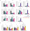
IA‐MPs modulate inflammatory macrophage signaling. Comparative BMDM expression of pro‐inflammatory macrophage markers. A) Relative transcript expression (2−∆∆CT) of IL‐6, IL‐1β, Ccl5, IL‐12p40, Nos2, Hif‐1α, Gclm, Nqo1, Hmox1, Gsr, and Taldo. B,C) Expression of inflammatory B) IL‐6 (6, 12, and 24 h) and C) TNF‐α protein expression. Expression is normalized to LPS media. Data are expressed as mean ± SD, n = 3 (q‐PCR media condition), n = 5 (ELISA 4OI treatment condition), or 6 (all other experimental treatments). Statistical significance is indicated by * < 0.05 ** < 0.01, *** p < 0.001, **** < 0.0001.
Figure 4
Figure 4
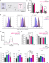
IA‐MPs modulate inflammatory macrophage signaling in an uptake‐specific manner. A–C) Expression of pro‐inflammatory markers by IA‐MP‐treated BMDMs is altered by IA‐MP uptake. A) IA‐MP‐treated BMDMs were gated according to PE (Rhodamine 6G) and SSC to generate low‐ and high‐uptake groups for analysis. B) Representative histograms of IL‐6, IL‐12p40, and MHC‐II expression in IA‐MP low‐ and high‐uptake subpopulations. C) Quantitative expression of IL‐6, IL‐12p40, and MHC‐II in macrophage‐based treatment with IA (5 mmol L−1), 4OI (0.125 mmol L−1), or IA‐MPs (low and high relative uptake). Data are expressed as mean ± SD, = 6. Statistical significance is indicated by * p < 0.05 ** p < 0.01, *** p < 0.001, **** p < 0.0001.
Figure 5
Figure 5
IA‐MPs alter macrophage metabolic function. A) Graphic overview of flow cytometric SCENITH method used to assess reliance on oxidative or glycolytic metabolism through quantification of protein synthesis (corresponding to the level of puromycin incorporation). B) Modal histogram representation of SCENITH‐quantified puromycin levels differentiated by control (black), harringtonine (grey), oligomycin A (pink), and 2DG (purple) treatment conditions. C) SCENITH‐quantified puromycin mean fluorescence intensity in control, harringtonine, oligomycin, and 2DG conditions of macrophages treated with IA‐MPs with respect to media ± LPS. D–F) Respirometric outputs of human macrophage cultures in control, low‐ or high‐dose IA‐MPs, or bulk IA treatments, including D) oxygen consumption rate, E) non‐mitochondrial oxygen consumption, and F) uncoupled: control ratio. G) Relative transcript expression (2−∆∆CT) of Glut‐1, HK‐1, SDHA, and PKM‐1. Data are expressed as mean ± SD, = 3 (media) or 6 (experimental treatments). Statistical significance is indicated as *p < 0.05 ** p < 0.01, *** p < 0.001, **** p < 0.0001.

Update of

References

    1. Phillips C. M., Chen L‐W., Heude B., Bernard J. Y., Harvey N. C., Duijts L., Mensink‐Bout S. M., Polanska K., Mancano G., Suderman M., Shivappa N., Hébert J. R., Nutrients 2019, 11, 1873. - PMC - PubMed
    1. Hasturk H., Kantarci A., Van Dyke T. E., Front. Immunol. 2012, 3, 118. - PMC - PubMed
    1. Fujiwara N., Kobayashi K., Curr. Drug Target: Inflammation Allergy 2005, 4, 281. - PubMed
    1. Zhang C., Yang M., Ericsson A. C., Front. Immunol. 2021, 12, 620510. - PMC - PubMed
    1. Wofford K. L., Singh B. S., Cullen D. K., Spiller K. L., Acta Biomater. 2020, 101, 237. - PMC - PubMed

LinkOut - more resources