. 2025 Apr 4;149(1):31.
doi: 10.1007/s00401-025-02869-4.

Seeding-competent early tau multimers are associated with cell type-specific transcriptional signatures

Affiliations

Seeding-competent early tau multimers are associated with cell type-specific transcriptional signatures

Rahel Feleke et al. Acta Neuropathol. .

Abstract

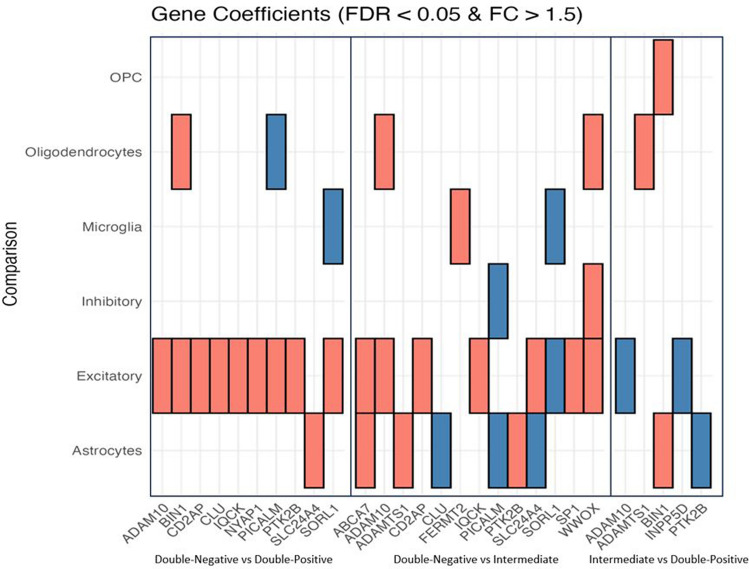
The initial molecular alterations of Alzheimer's disease (AD) are unknown. Established AD is characterized by profound structural and transcriptional alterations in the human brain, with the hallmark neuropathological features being beta-amyloid (Aβ) accumulation in senile plaques and hyperphosphorylated fibrillar tau in neurofibrillary tangles (NFTs). Previous evidence indicates that tau multimerization into small aggregates is one of the earliest molecular alterations, anticipating the accumulation of hyperphosphorylated tau in NFTs. In this study, we investigated the seeding capacity of these early small tau multimers and the transcriptional changes associated with them, aiming to unveil early pathogenic processes in AD-type tau pathology. Early tau multimers visualized with tau proximity ligation assay (tau-PLA) in the post-mortem temporal cortex demonstrated high seeding activity detected by real-time quaking-induced conversion (RT-QuIC) assay and induction of aggregates in a tau biosensor cell line. Using single-nucleus transcriptomics, we showed that brain tissue harboring seeding-competent early tau multimers, but without significant NFT pathology, is associated with substantial gene expression alterations across diverse cell types when compared to control tissue lacking either multimers or NFTs. Differentially expressed genes, such as APP, MAPT, and PSEN1, exhibited significant enrichment of AD heritability in up-regulated genes within excitatory neurons, astrocytes, and oligodendrocytes. Pseudotime analysis exposed a positive correlation between the progression of tau pathology and the expression of genes marking reactive astrocytes. In summary, our results support the hypothesis that seeding-competent tau multimerization may initiate AD-type tau pathology cascades before the accumulation of tau in NFTs. This research contributes valuable insights into the early molecular events associated with AD, with implications for future diagnostic and therapeutic strategies.

Keywords: Alzheimer's disease; Early pathology; Proximity-ligation assay; Seeding; Single-nucleus RNA sequencing; Tau multimers.


Conflict of interest statement

Declarations. Conflict of interest: The authors declare no competing interests.

Figures

Fig. 1
Fig. 1
Early tau multimerization detection across the different Braak Stages as defined by BrainNet Europe diagnostic protocol. A Scanned tau-PLA labeled post-mortem human brain tissue sections. Sections from the hippocampal region, temporal cortex (MTC), and occipital cortex were stained for tau-PLA. B FFPE sections from postmortem human brain tissue from the brain regions of the hippocampus, temporal (MTC), and occipital cortices were stained for AT8-IHC and tau-PLA. The Braak stage of each case was determined according to the modified Braak staging system based on AT8-immunoreactive NTs across the brain tissue. Scale bar 50 μm. EC entorhinal cortex, CA1 hippocampus, TC temporal cortex, OC occipital cortex, MTC middle temporal cortex
Fig. 2
Fig. 2
Diffused tau pathology labeled with different phospho-tau and conformational tau antibodies across the different Braak stages as defined by BrainNet Europe diagnostic protocol. A FFPE sections from post-mortem human brain tissue from the middle temporal cortex stained for AT180-, T217-, AT8-, PHF1-, S422-, MC1-, and Alz50-IHC. Scale bar 50 μm. B Quantification of diffuse tau pathology labeled with tau-PLA and various phospho-tau and conformational tau antibodies across Braak stages in EC, TC, and OC. The y-axis represents a semi-quantitative score from 0 to 6 (Supplementary Fig. 1a), while the x-axis corresponds to Braak stages (B0–BVI). Each antibody at each Braak stage was compared to Braak 0 through a One-way ANOVA (Dunnett); N = 11/12/12/9/7/8/8. Each bar represents the mean ± standard error of the mean (SEM). Full statistical details can be found in Supplementary Table 2. EC entorhinal cortex, TC temporal cortex, OC occipital cortex
Fig. 3
Fig. 3
Overview of samples used for tau RT-QuIC, cell-based seeding (FRET), and snRNA-seq assays. 6 subjects were designated to the Double-Negative group, exhibiting no detectable tau aggregates, implying an absence of tau pathology (A); 5 subjects were designated to the Intermediate group, demonstrating early signs of tau multimerization with a minimal or negligible number of large fibrils (B); 5 subjects were designated to the Double-Positive group, characterized by both diffuse tau pathology and pronounced large lesions in the temporal region (C). RT-QuIC Real-Time Quaking-Induced Conversion, FRET fluorescence resonance energy transfer, SnRNA-seq single-nucleus RNA sequencing
Fig. 4
Fig. 4
Tau RT-QuIC analysis. A RT-QuIC analysis of negative control (tau MAPT KO mouse brain homogenate), positive control (τ306 aggregates), Double-Negative, Intermediate, and Double-Positive brain homogenates. Each curve represents a single case, run in triplicate. B Comparison of tau seeding activity of negative control (tau MAPT KO mouse brain homogenate), positive control (τ306 aggregates), Double-Negative, Intermediate, and Double-Positive brain homogenates with RT-QuIC. Fmax (maximum ThT fluorescence), lag time (reaction time to exceed a ThT fluorescence threshold of the average baseline fluorescence + 5 SD), time to reach maximum ThT fluorescence, and Vmax (maximum slope) were analyzed. The assay cut-off was determined to be 52 h as a reproducible endpoint before the spontaneous amyloid aggregation in the negative control wells. The endpoint of 52 h was used for any data points with ThT fluorescence values at or greater than 52 h. Groups with dilution 1 × 10–1 were assessed through a One-way ANOVA (Bonferroni); N = 6/5/5. Each bar represents the mean ± standard deviation (SD). *p < 0.05, **p < 0.01, ***p < 0.001, ****p < 0.0001
Fig. 5
Fig. 5
Seeding amplification assay using tau biosensor cell line. Comparison of tau seeding activity of Double-Negative, Intermediate, and Double-Positive brain homogenates of temporal with FRET-based biosensor cell line. τ306 monomers were used as a control. A Fluorescent imaging showcases cells post 48-h treatment with respective brain homogenates; scale bar = 50 µm. B Quantitative analysis displaying the percentage of cells with intracellular tau inclusions, assessed through one-way ANOVA (Bonferroni); N = 6/5/5. Each bar represents the mean ± standard deviation (SD). *p < 0.05, **p < 0.01, ***p < 0.001, ****p < 0.0001
Fig. 6
Fig. 6
Seeding activity of non-immunodepleted and immunodepleted brain homogenates assessed by tau RT-QuIC. A Fluorescence curves of non-immunodepleted brain homogenates (pre-BH, visualized in red), immunodepleted brain homogenates with AT8 antibody (post-BH—AT8; visualized in blue), and immunodepleted brain homogenates with tau5 antibody (post-BH—tau5; visualized in green) across Double-Negative, Intermediate, and Double-Positive groups. B Comparative analysis of seeding kinetics, including maximum fluorescence (Fmax), lag time (reaction time to exceed a ThT fluorescence threshold of the average baseline fluorescence + 5 standard deviations), time to maximum fluorescence, and maximum aggregation rate (Vmax), revealing differences in tau aggregation potential between groups and immunodepletion conditions. One sample from each group was analyzed in triplicates. Data was evaluated using Two-way ANOVA comparing to the control (pre-BH) followed by Dunnett's Multiple Comparison Test. Each bar represents the mean ± standard error of the mean (SEM). *p < 0.05, **p < 0.01, ***p < 0.001
Fig. 7
Fig. 7
Cell-type-specific gene expression changes across all comparisons. A Total number of DEGs across all pairwise comparisons (FDR < 0.05, log2(fold change) > log2(1.5)). B, C A yes/no plot indicating if a gene (column) is expressed in a given comparison. The total number of DEGs (unique) in each comparison is indicated on the x-axis. The full list of DEGs across all pairwise comparisons in the panel is available in Supplementary Table 5
Fig. 8
Fig. 8
Expression levels of previously identified Alzheimer’s Disease-relevant genes in the current datasets. Red color denotes up-regulated genes, while blue color denotes down-regulated
Fig. 9
Fig. 9
Significantly (FDR < 0.05) enriched pathways identified using DEG sets from each pairwise comparison. The fill of each tile indicates the − log10FDR value of the most significant child term associated with the parent term. The full list of child GO terms assigned to each parent term across all pairwise comparisons in the panel is available in Supplementary Table 6
Fig. 10
Fig. 10
Genetic association with cell-type specific differentially expressed genes across all pairwise comparisons. LDSC was used to identify associations. The x-axis indicates enrichment p-values. The black line indicates Bonferroni significance threshold (p-values adjusted for the number of cell types tested; FDR < 0.05). The color of the bars indicates if the DEGs were up- or down-regulated. Full results can be found in Supplementary Table 7

References

    1. Aissani B (2014) Confounding by linkage disequilibrium. J Hum Genet 59(2):110–115 - PMC - PubMed
    1. Alafuzoff I et al (2008) Staging of neurofibrillary pathology in Alzheimer’s disease: a study of the BrainNet Europe Consortium. Brain Pathol 18(4):484–496 - PMC - PubMed
    1. Ando K et al (2022) PICALM and Alzheimer’s disease: an update and perspectives. Cells 11(24):3994–3994 - PMC - PubMed
    1. Aran KR, Singh S (2023) Mitochondrial dysfunction and oxidative stress in Alzheimer’s disease—a step towards mitochondria based therapeutic strategies. Aging Health Res 3(4):100169
    1. Arbaciauskaite M, Lei Y, Cho YK (2021) High-specificity antibodies and detection methods for quantifying phosphorylated tau from clinical samples. Antib Ther 4(1):34–44 - PMC - PubMed

Publication types

LinkOut - more resources