. 2025 Jul;12(25):e2414587.
doi: 10.1002/advs.202414587. Epub 2025 Apr 4.

Isoquercitrin Alleviates Diabetic Nephropathy by Inhibiting STAT3 Phosphorylation and Dimerization

Affiliations

Isoquercitrin Alleviates Diabetic Nephropathy by Inhibiting STAT3 Phosphorylation and Dimerization

Chen Xuan et al. Adv Sci (Weinh). 2025 Jul.

Abstract

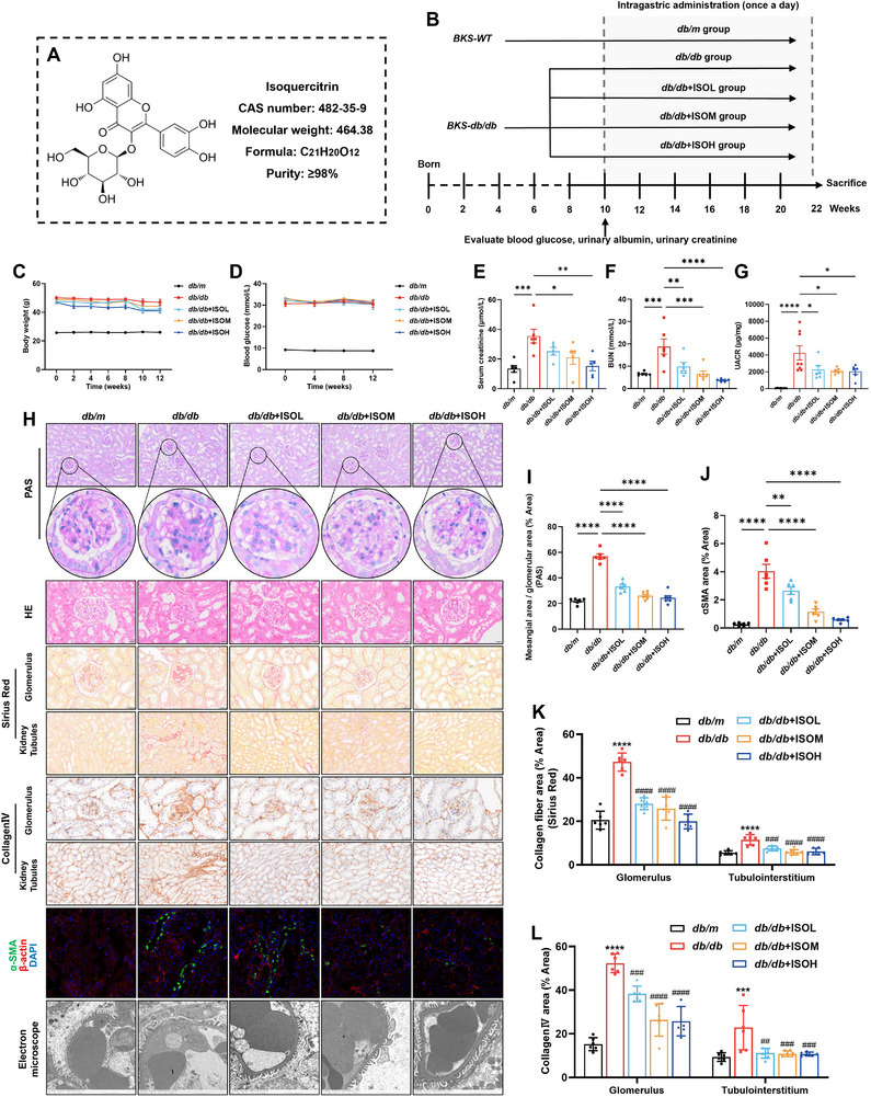
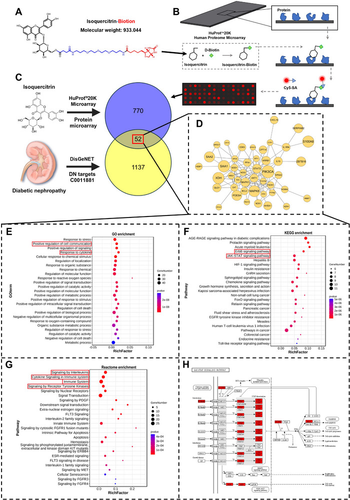
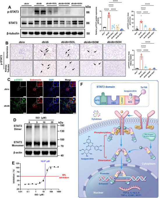
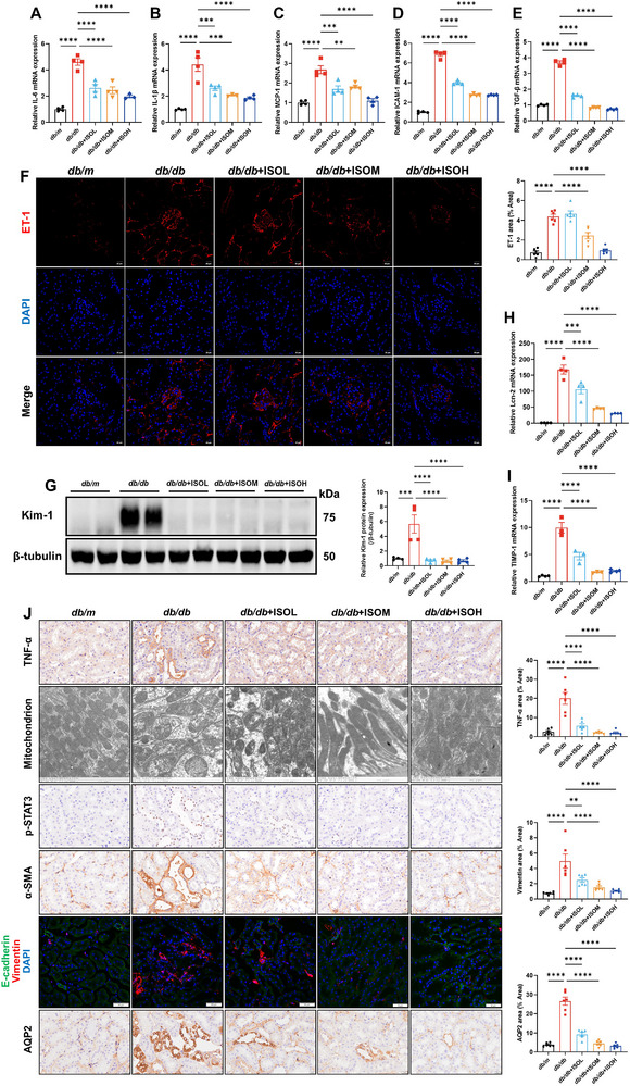
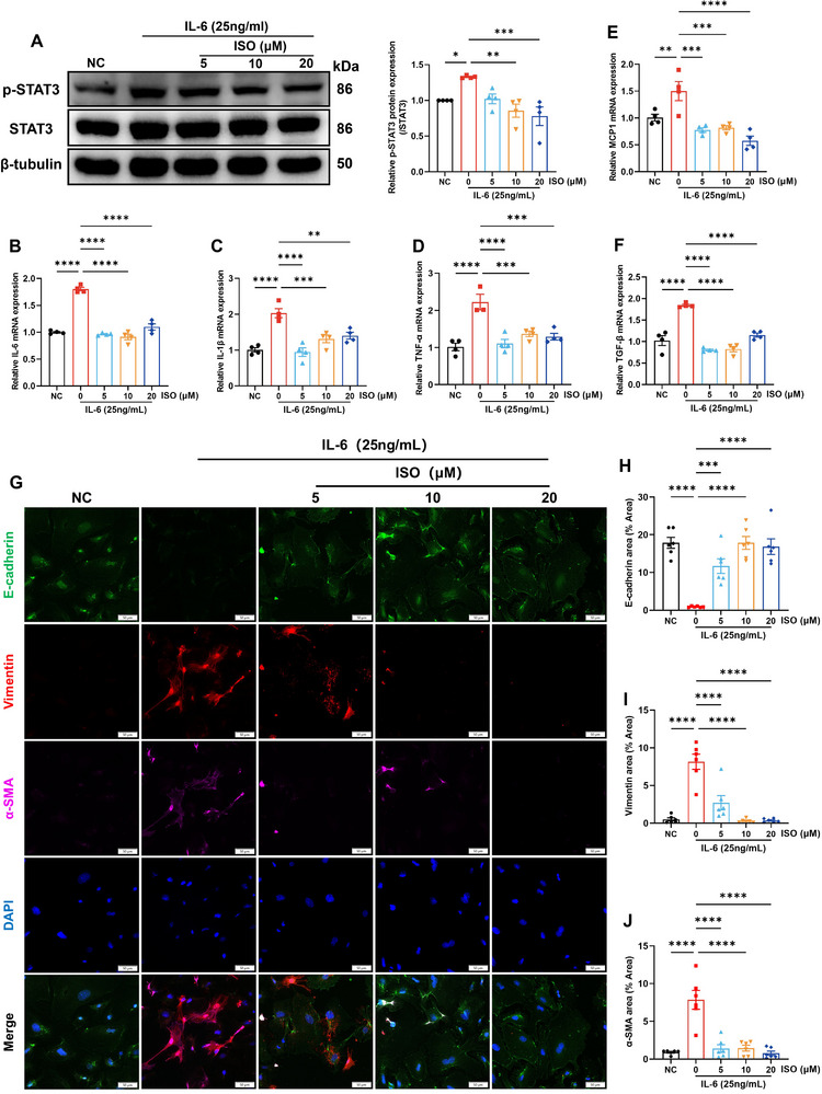
At the convergence point of multiple cytokine signals, signal transducer and activator of transcription 3 (STAT3) is a highly promising therapeutic target for diabetic nephropathy. Isoquercitrin, a natural small-molecule inhibitor of STAT3, may have beneficial effects on diabetic nephropathy; however, the underlying mechanism remains unclear. Isoquercitrin significantly mitigated renal inflammation and fibrosis by inhibiting STAT3 activity in mice with diabetic nephropathy. Moreover, STAT3 is a direct molecular target of isoquercitrin, which as corroborated by tight and stable noncovalent binding between them. This interaction is mechanistically supported by the affinity of isoquercitrin for the Ser668-Gln635-Gln633 region within the pY+1 binding pocket of the SH2 domain. This binding obstructs pivotal processes like STAT3 phosphorylation and dimerization, thereby suppressing its transcriptional function. Finally, a kidney-targeted nanocarrier, Iso@PEG-GK, is developed to load isoquercitrin, thus enhancing its therapeutic precision for diabetic nephropathy. Iso@PEG-GK significantly improved the absorption and renal distribution of isoquercitrin. This study is the first to demonstrate that isoquercitrin exerts a significant protective effect against diabetic nephropathy and may provide a novel therapeutic drug for this disease.

Keywords: Iso@PEG‐GK; SH2 domain; STAT3; diabetic nephropathy; dimerization; isoquercitrin; phosphorylation.


Conflict of interest statement

The authors declare no conflict of interest.

Figures

Figure 1
Figure 1
Isoquercitrin alleviates renal pathological damage and protects renal function in db/db mice. A) Molecular structure and information of isoquercitrin. B) Schematic overview of the experimental design of the mice model. The body weight (C) and blood glucose levels (D) of the mice were recorded during treatment with isoquercitrin (n = 6). Blood and urine samples were collected to detect serum creatinine (E), blood urea nitrogen (BUN) (F), and the urinary albumin/creatinine ratio (UACR) (G) at the end of the 12 weeks of isoquercitrin treatment (n = 6). Kidney tissues were prepared into paraffin sections and subjected to periodic acid–Schiff (PAS), hematoxylin and eosin (HE), and Sirius Red staining (H). The mesangial area (I) and collagen fiber area (K) were calculated. Immunohistochemistry and immunofluorescence detection of the expression of collagen IV (H, L) and alpha‐smooth muscle actin (α‐SMA) (H, J) in kidney tissues (n = 6). H) Electron microscopy observation of pathological changes of podocytes and the basement membrane (n = 3). Data are presented as the mean ± standard error of the mean (SEM). *p < 0.05, **/## p < 0.01, ***/### p < 0.001, ****/#### p < 0.0001. In (K) and (L), “*” indicates comparison with db/m mice, and “#” indicates comparison with db/db mice. One‐way ANOVA followed by the Dunnett's post hoc test.
Figure 2
Figure 2
Isoquercitrin targets the JAK‐STAT pathway, thereby regulating the immune‐inflammatory response. Isoquercitrin labeled with D‐biotin (A) was incubated with the HuProt™20K human proteome microarray. B) Workflow of the proteome microarray. C) The significantly specific binding proteins of isoquercitrin screened by the proteome microarray were intersected with diabetic nephropathy disease targets. The common targets (D) were extracted and subjected to Gene Ontology (E), Kyoto Encyclopedia of Genes and Genomes (F), and Reactome (G) enrichment analysis. H) The common targets were mapped onto the JAK‐STAT signaling pathway.
Figure 3
Figure 3
STAT3 is a direct binding target for isoquercitrin, with isoquercitrin specifically binding to the Ser668–Gln635–Gln633 site of STAT3. A) STAT3 in proteome microarray results. B) Pull‐down experiments to detect the binding of isoquercitrin to STAT3 (n = 3). C) Surface plasmon resonance precisely measures the binding affinity and mode of isoquercitrin to STAT3. D) Cellular thermal shift assay (CETSA) confirms whether isoquercitrin can enter cells and bind to STAT3 (n = 3). E) Molecular docking and dynamics simulations assessing the binding mode of isoquercitrin with STAT3. F) The MM/GBSA method calculates the contributions of different amino acid residues in the isoquercitrin–STAT3 binding. The stability of the isoquercitrin–STAT3 binding was assessed through root‐mean‐square deviation (G), radius of gyration (H), and root‐mean‐square fluctuation (I). J) Alanine scan calculates the changes in affinity between isoquercitrin and STAT3 after mutating amino acid residues to alanine. The changes in the binding of isoquercitrin to STAT3 were detected using CETSA after mutating specific amino acids to alanine (K, L, M) (n = 3).
Figure 4
Figure 4
Isoquercitrin occupies the SH2 domain, inhibiting STAT3 phosphorylation, dimerization, and downregulating its transcriptional activity. A) Western blot (WB) detection of isoquercitrin effects on phosphorylated STAT3 (p‐STAT3) and total STAT3 in kidneys (n = 4). B) Immunohistochemistry detection of p‐STAT3 in glomerulus and kidney tubules (n = 6). Black arrows indicate p‐STAT3 positive regions. C) Immunofluorescence co‐staining of p‐STAT3 and the endothelial cell marker Endomucin. White arrows indicate p‐STAT3 positive regions. D) After crosslinking protein–protein interactions with DSS, the effect of isoquercitrin on STAT3 dimers in 293T cells was detected using WB. E) The determination of the half‐maximal inhibitory concentration for the inhibition of STAT3 transcriptional activity by isoquercitrin. F) Isoquercitrin inhibits STAT3 phosphorylation and dimerization. Data are presented as the mean ± SEM. ****p < 0.0001. One‐way ANOVA followed by the Dunnett's post hoc test.
Figure 5
Figure 5
Isoquercitrin inhibits STAT3 to improve endothelial and renal tubular epithelial cell injury. A–E) RT‐qPCR detection of mRNA expression levels of interleukin (IL)‐6, IL‐1β, monocyte chemoattractant protein‐1 (MCP‐1), intercellular adhesion molecule‐1 (ICAM‐1), and tumor growth factor beta (TGF‐β) in kidneys (n = 4). F) Immunofluorescence detection of endothelial cell injury marker ET‐1 in kidneys (n = 6). G) WB detection of Kim‐1 expression, a marker of renal tubular epithelial cell injury, in mouse kidneys (n = 4). RT‐qPCR detection of lipocalin‐2 (Lcn‐2) (H) and tissue inhibitor of metalloproteinase 1 (TIMP‐1) (I) mRNA expression levels in kidneys (n = 4). J) Immunohistochemistry and immunofluorescence detection of TNF‐α, p‐STAT3, α‐SMA, E‐cadherin, vimentin, and aquaporin 2 (AQP2) expression in kidney tubules (n = 6). Electron microscopy observation of the morphology of mitochondria in renal tubular epithelial cells of mice (J) (n = 3). Data are presented as the mean ± SEM. **p < 0.01, ***p < 0.001, ****p < 0.0001. One‐way ANOVA followed by the Dunnett's post hoc test.
Figure 6
Figure 6
Isoquercitrin alleviates high glucose or IL‐6‐induced endothelial cell injury by inhibiting STAT3 activity. RPMI Medium 1640 containing 45 mM glucose was used as a high glucose (HG) culture medium to induce human umbilical vein endothelial cells (HUVECs). RPMI Medium 1640 with a glucose concentration of 11 mM was used to culture HUVECs in the normal glucose (NG) control group. Subsequently, isoquercitrin (ISO) at concentrations of 5, 10, and 20 µM was added to the culture medium for intervention. A) WB detection of the p‐STAT3, STAT3, and TGF‐β expression levels in HG‐stimulated HUVECs (n = 4). B) The mRNA expression changes of cytokines IL‐6, TNF‐α, ICAM‐1, and MCP‐1 in HUVECs after HG stimulation and ISO intervention (n = 3). C) WB detection of the expression of endothelial dysfunction marker inducible nitric oxide synthase (iNOS) and endothelial glycocalyx components syndecan‐1 and glypican‐1 in HUVECs (n = 4). D) WB detection of endothelial cell marker VE‐cadherin and mesenchymal cell markers vimentin and α‐SMA in HUVECs (n = 4). HUVECs were stimulated with 25 ng mL−1 IL‐6, whereas HUVECs without IL‐6 stimulation were the normal control (NC) group. E) WB detection of p‐STAT3, STAT3, IL‐6, and IL‐1β expression in HUVECs stimulated with IL‐6 (n = 3). F) The mRNA expression changes of cytokines IL‐1β, TNF‐α, and MCP‐1 in HUVECs after IL‐6 stimulation and ISO intervention (n = 6). STAT3 overexpression in HUVECs via plasmid transfection, with WB detection of transfection efficiency (G) (n = 3). H) WB detection of the effects of IL‐6 and ISO on p‐STAT3 and STAT3 expressions in HUVEC cells under transfected or nontransfected conditions (n = 6). I–K) RT‐qPCR detection of the effects of IL‐6 and ISO on the transcription levels of cytokines IL‐6, IL‐1β, and TNF‐α in HUVEC cells with or without transfection (n = 4). Data are presented as the mean ± SEM. */# p < 0.05, **/## p < 0.01, ***/### p < 0.001, ****/#### p < 0.0001. In (C), (D), and (F), * indicates comparison with db/m, and # indicates comparison with db/db. One‐way ANOVA followed by the Dunnett's post hoc test.
Figure 7
Figure 7
Isoquercitrin inhibits STAT3 in renal tubular epithelial cells, reducing IL‐6‐induced pro‐inflammatory and profibrotic cytokines. Human kidney 2 cells (HK2) cells were stimulated with 25 ng mL−1 IL‐6, whereas HK2 cells without IL‐6 stimulation were the normal control (NC) group. Subsequently, interventions were conducted with 5, 10, and 20 µM of isoquercitrin. A) WB detection of p‐STAT3 and STAT3 expression in HK2 cells (n = 4). RT‐qPCR detection of the mRNA expression of pro‐inflammatory cytokines IL‐6 (B), IL‐1β (C), and TNF‐α (D), macrophage chemoattractant protein MCP‐1 (E), and profibrotic cytokine TGF‐β (F) (n = 4). G–J) Isoquercitrin inhibits EMT in renal tubular epithelial cells. Isoquercitrin downregulates the abnormally high expression of mesenchymal markers vimentin and α‐SMA, while restoring the expression of the epithelial marker E‐cadherin (n = 6). Data are presented as the mean ± SEM. *p < 0.05, **p < 0.01, ***p < 0.001, ****p < 0.0001. One‐way ANOVA followed by the Dunnett's post hoc test.
Figure 8
Figure 8
Nanocarrier Iso@PEG‐GK optimizes the pharmacokinetic properties of isoquercitrin, enhances renal targeting, and is safe. A) Synthesis process of Iso@PEG‐GK. The hydrodynamic diameter (B), polydispersity index (PDI) (C) (n = 3), and zeta potentials (D) (n = 5) of nanocarriers synthesized by the nanocoprecipitated (blue and purple) and thin film hydration (orange and green) method. E) Transmission electron microscopy observation of the shape and distribution of Iso@PEG‐GK and its control groups Iso@PEG and Iso@PEG‐G. F) Dynamic light scattering detection of the hydrodynamic size of Iso@PEG‐GK, Iso@PEG, and Iso@PEG‐G. G) The zeta potentials of nanocarriers (n = 3). H) The isoquercitrin release curve of Iso@PEG‐GK. I) Changes in blood drug concentration of nanocarriers within 72 h (n = 3). J) The distribution of Iso@PEG‐GK, Iso@PEG, and Iso@PEG‐G in the heart, liver, spleen, lungs, and kidneys at different time points (n = 3). K) Hemolysis assay was used to detect the red blood cell biocompatibility of nanocarriers (n = 3). L) HE staining were used to detect the tissue morphology of major organs after nanocarrier intervention (n = 3).
Figure 9
Figure 9
Schematic diagram showing the mechanism of isoquercitrin inhibiting STAT3 to ameliorate diabetic nephropathy. Isoquercitrin alleviates the injury of endothelial and renal tubular epithelial cells in diabetic nephropathy by inhibiting STAT3 phosphorylation and dimerization. Based on these actions, isoquercitrin reduces the expression of pro‐inflammatory and profibrotic cytokines such as IL‐1β, IL‐6, TNF‐α, ICAM‐1, MCP‐1, and TGF‐β, thereby alleviating renal inflammation and extracellular matrix accumulation.

Similar articles

Cited by

References

    1. Alicic R. Z., Rooney M. T., Tuttle K. R., Clin. J. Am. Soc. Nephrol. 2017, 12, 2032. - PMC - PubMed
    1. Gupta S., Dominguez M., Golestaneh L., Med. Clin. North Am. 2023, 107, 689. - PubMed
    1. Schena F. P., Gesualdo L., J. Am. Soc. Nephrol. 2005, 16, S30. - PubMed
    1. Navarro‐González J. F., Mora‐Fernández C., J. Am. Soc. Nephrol. 2008, 19, 433. - PubMed
    1. Shikata K., Makino H., J. Diabetes Investig. 2013, 4, 142. - PMC - PubMed

MeSH terms

LinkOut - more resources