. 2025 Apr 4;16(1):249.
doi: 10.1038/s41419-025-07580-6.

Cancer-associated fibroblasts secrete CSF3 to promote TNBC progression via enhancing PGM2L1-dependent glycolysis reprogramming

Affiliations

Cancer-associated fibroblasts secrete CSF3 to promote TNBC progression via enhancing PGM2L1-dependent glycolysis reprogramming

Wenqi Qin et al. Cell Death Dis. .

Abstract

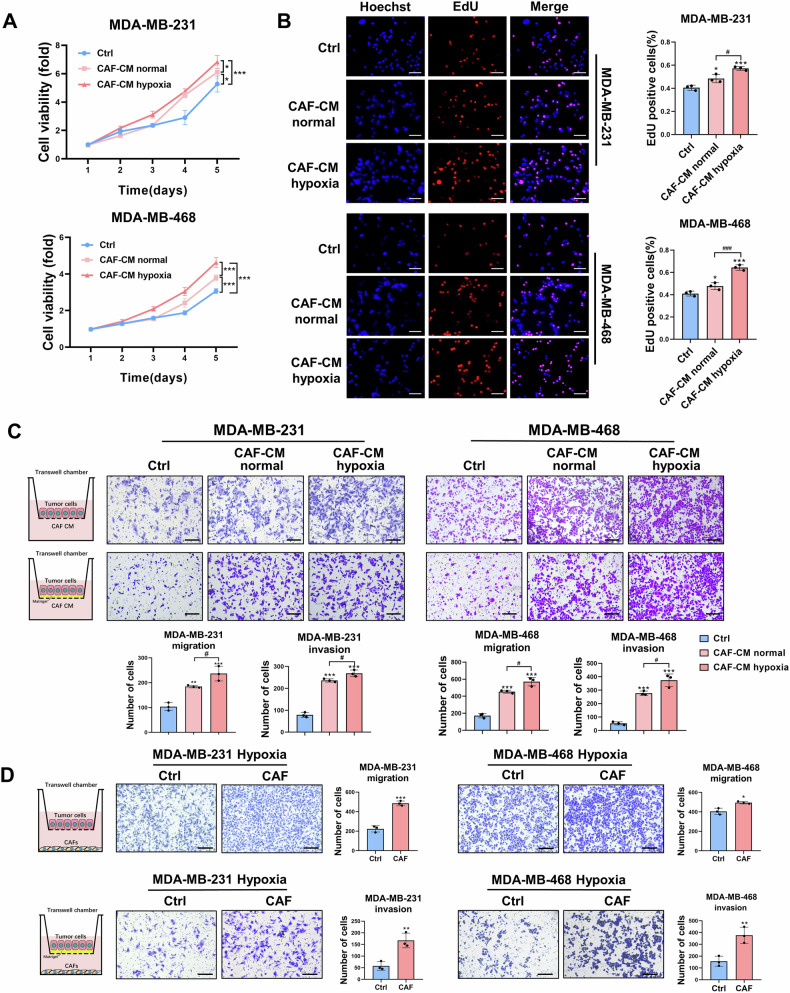
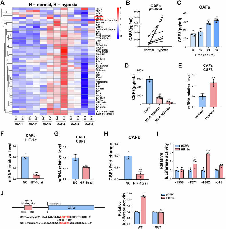
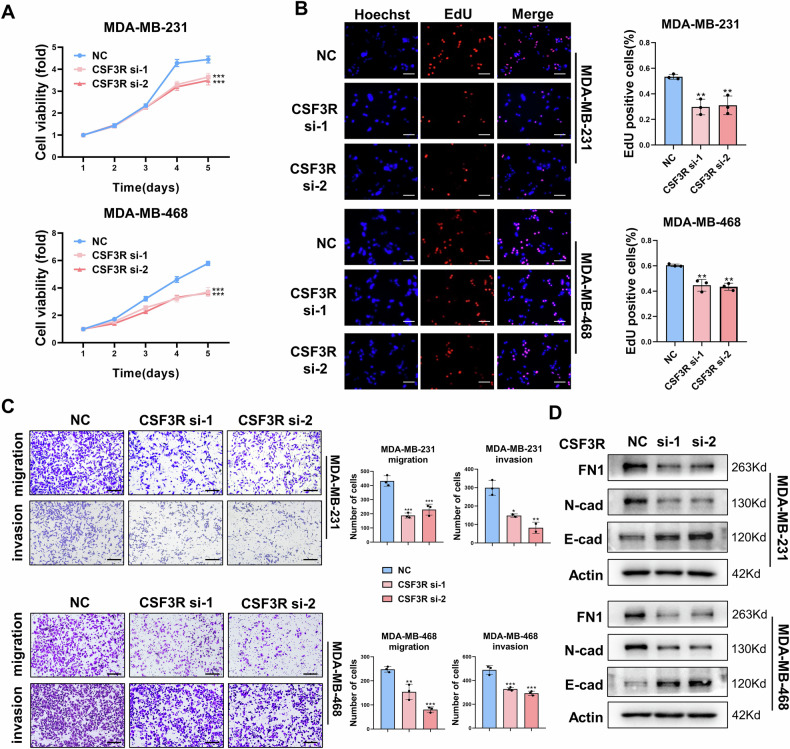
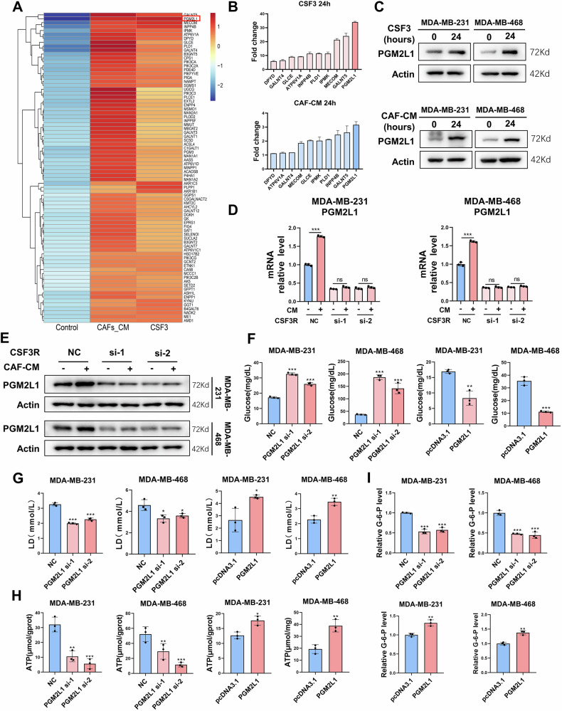
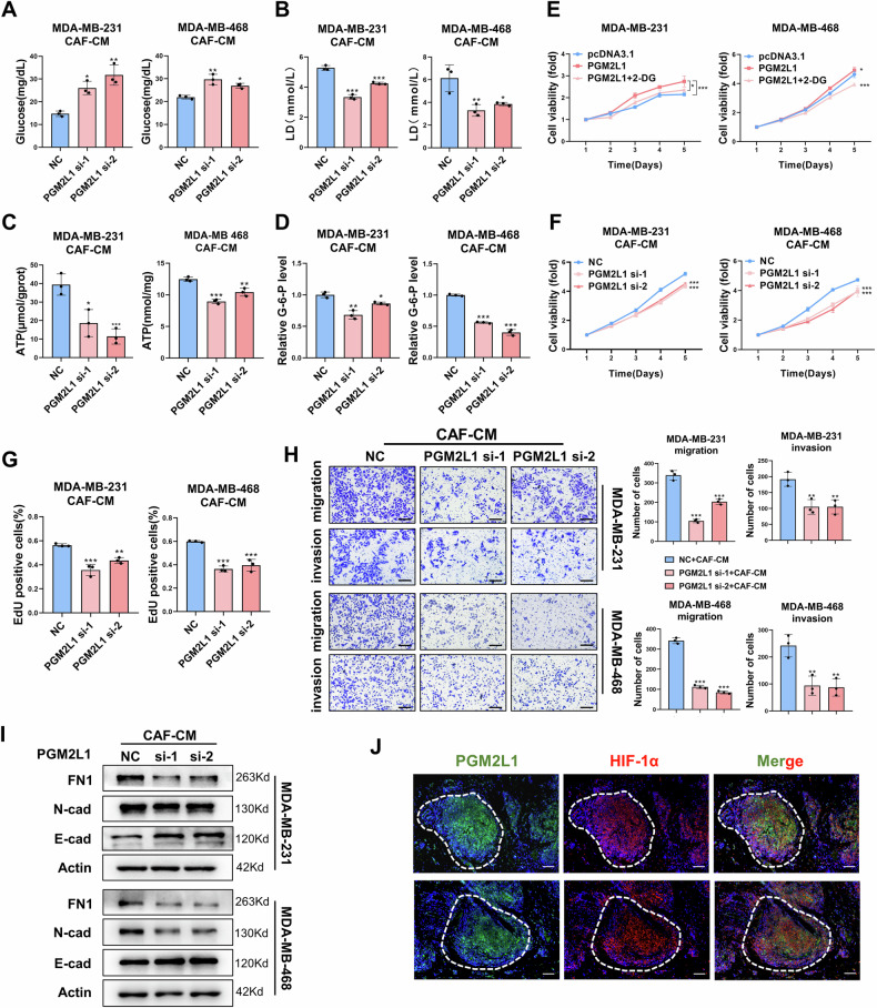
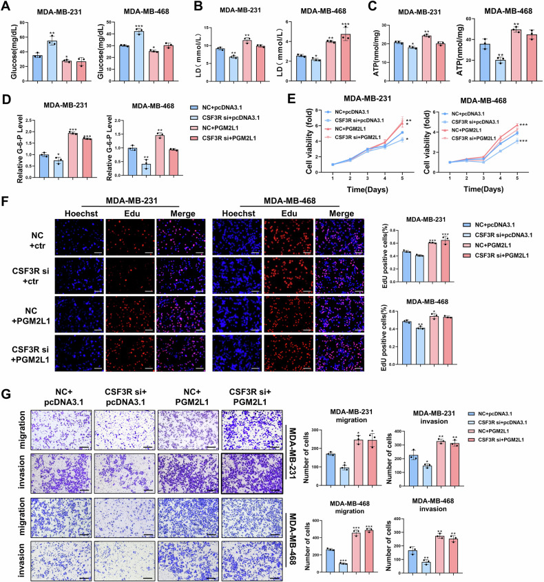
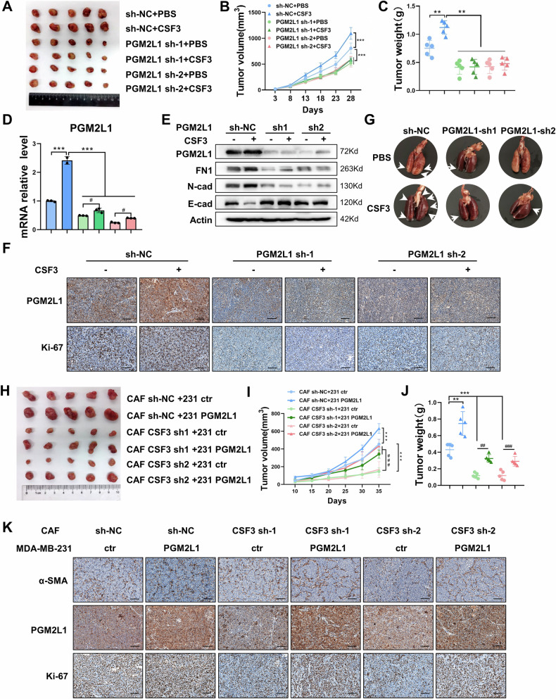
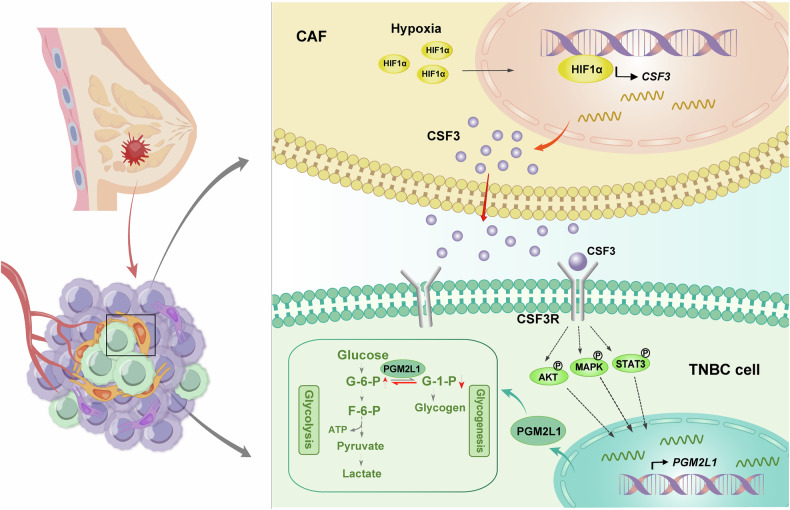
Triple-negative breast cancer (TNBC) is characterized by a pronounced hypoxic tumor microenvironment, with cancer-associated fibroblasts (CAFs) serving as the predominant cellular component and playing crucial roles in regulating tumor progression. However, the mechanism by which CAFs affect the biological behavior of tumor cells in hypoxic environment remain elusive. This study employed a bead-based multiplex immunoassay to analyze a panel of cytokines/chemokines and identified colony stimulating factor 3 (CSF3) as a significantly elevated component in the secretome of hypoxic CAFs. We found that CSF3 promoted the invasive behavior of TNBC cells by activating the downstream signaling pathway of its receptor, CSF3R. RNA sequencing analysis further revealed that phosphoglucomutase 2-like 1 (PGM2L1) is a downstream target of the CSF3/CSF3R signaling, enhancing the glycolysis pathway and providing energy to support the malignant phenotype of breast cancer. In vivo, we further confirmed that CSF3 promotes TNBC progression by targeting PGM2L1. These findings suggest that targeting CSF3/CSF3R may represent a potential therapeutic approach for TNBC.


Conflict of interest statement

Competing interests: The authors declare no competing interests. Ethics: Human BC tissues and corresponding normal breast tissues were obtained from patients undergoing surgery in Qilu Hospital, Shandong University (Jinan, China). All experimental procedures were approved by the Ethics Committee of Qilu Hospital, Shandong University. The number of ethics approval is KYLL-2022(ZM)-1215. All patients written informed consent for the use of their clinical samples in research. All animal experiments were approved by the Shandong University Animal Care and Use Committee. The diameters of the tumors did not exceed 2 cm, which is the maximum diameter as specified by the committee.

Figures

Fig. 1
Fig. 1. CAFs promote the proliferation, migration, and invasion of TNBC cells under hypoxic conditions.
The proliferation ability of TNBC cells treated with CAF-CM was detected by MTT assay (A, n = 4) and EdU assay (B, n = 3). Scale bars, 100 μm. C Transwell assay was performed to evaluate the effect of normoxic or hypoxic CAF-CM on the migration and invasion abilities of TNBC cells (n = 3). Scale bars, 200 μm. D Transwell assay was performed to evaluate the effect of CAFs on the migration and invasion abilities of TNBC cells under hypoxia (n = 3). Scale bars, 200 μm. The data are presented as the mean ± SD. *,#P < 0.05, **P < 0.01, ***,###P < 0.001.
Fig. 2
Fig. 2. Hypoxia induces the secretion of CSF3 from CAFs.
A The bead-based multiplex immunoassay assay to test cytokines secreted by CAFs under normoxic and hypoxic conditions (n = 6). B ELISA assay to quantify CSF3 secreted from paired CAFs under normoxic and hypoxic conditions (n = 10). C The secretion level of CSF3 increases with the prolonged hypoxic exposure (n = 3). D The comparation of CSF3 level from CAFs and TNBC cells (n = 3). E The expression of CSF3 under normoxic and hypoxic conditions was assessed by qRT-PCR (n = 3). The HIF-1α silencing efficiency (F) and CSF3 expression level (G) were verified using qRT-PCR (n = 3). H ELISA assay to test CSF3 level after interfering HIF-1α (n = 3). I The luciferase activity of different targeted plasmids containing potential binding region (n = 3). J Dual-luciferase reporter assay to verify CSF3’ binding region for HIF-1α (n = 3). The data are presented as the mean ± SD. ns, no significance; *P < 0.05, **P < 0.01, ***P < 0.001.
Fig. 3
Fig. 3. CSF3/CSF3R promotes the proliferation, migration, and invasion of TNBC cells.
The proliferation ability of TNBC cells silencing CSF3R was determined by MTT assay(A, n = 4) and EdU assay (B, n = 3). Scale bars, 100 μm. C Transwell assay was implemented to assessed the migration and invasion abilities of TNBC cells silencing CSF3R (n = 3). Scale bars, 200 μm. D The expression of EMT-related markers was examined via western blot. The data are presented as the mean ± SD. *P < 0.05, **P < 0.01, ***P < 0.001.
Fig. 4
Fig. 4. PGM2L1 is a downstream regulatory target of CSF3/CSF3R signaling.
A Heat map of top 80 metabolic pathways-related DEGs in both CSF3 and CAF-CM groups. B The expression level of top differential genes by qRT-PCR (n = 3). C The protein level of PGM2L1 was upregulated by CSF3 and CAF-CM. The expression level of PGM2L1 was decreased by silencing CSF3R in mRNA level (D, n = 3) and protein level (E). Colorimetry assays to measure glycolysis of MDA-MB-231 and MDA-MB-468 cells under hypoxia, including glucose (F), lactic acid (G), ATP (H), and G-6-P (I) (n = 3). The data are presented as the mean ± SD. ns, no significance; *P < 0.05, **P < 0.01, ***P <0.001.
Fig. 5
Fig. 5. PGM2L1 promotes the glycolysis and progression of TNBC cells under hypoxia.
Colorimetry assays to measure glycolysis level of MDA-MB-231 and MDA-MB-468 cells treated with hypoxic CAF-CM, including glucose (A), lactic acid (B), ATP (C), and G-6-P(D) (n = 3). E MTT assay to assess cellular proliferation with or without 2-DG (n = 4). MTT (F, n = 4) and EdU (G, n = 3) assays to evaluate the proliferative ability of TNBC cells treated with hypoxic CAF-CM following PGM2L1 silencing. H Transwell assay to assess the migration and invasion abilities of TNBC cells treated with hypoxic CAF-CM following PGM2L1 silencing (n = 3). Scale bars, 200 μm. I The EMT-makers were examined by western blot. J Fluorescent immunohistochemical staining of the expression of PGM2L1 and HIF-1α in TNBC tissues. Scale bars, 100 μm. The data are presented as the mean ± SD. *P < 0.05, **P < 0.01, ***P < 0.001.
Fig. 6
Fig. 6. Down-regulation CSF3R inhibits PGM2L1-mediated TNBC progression and glycolytic reprogramming.
Following CSF3R-siRNA and PGM2L1-ove plasmid transfection, TNBC cells were tested by glycolysis level, including glucose (A), lactic acid (B), ATP (C), and G-6-P (D) (n = 3). MTT(E, n = 4) and EdU (F, n = 3) assays were performed to assessed cellular proliferation ability. Scale bars, 100 μm. G Transwell assay was employed to appraise cells migration and invasion abilities (n = 3). Scale bars, 200 μm. The data are presented as the mean ± SD. *P < 0.05, **P < 0.01, ***P < 0.001.
Fig. 7
Fig. 7. CSF3 and PGM2L1 promote TNBC progression in vivo.
A Control or PGM2L1-knockdown MDA-MB-231 cells were transplanted into nude mice, followed by PBS or CSF3 treatment. Images of tumors were harvested (n = 5 mice). B Tumor volumes were measured once 5 days beginning 3rd day after implantation (n = 5 mice). C Tumor weight was measured in each group (n = 5 mice). D The expression of PGM2L1 of xenograft tumors were examined by qRT-PCR (n = 3). E The protein level of PGM2L1 and EMT-related markers of xenograft tumors were examined by western blot. F Ki-67 and PGM2L1 in each group were tested by IHC staining. Scale bars, 100 μm. G Representative lung metastatic nodules in mice from different groups were counted (n = 5 mice). H Images of xenograft tumors of MDA-MB-231 cells mixed CAFs (n = 5 mice). I Tumor volumes were measured once 5 days beginning 10th day after implantation (n = 5 mice). J Tumor weight was measured in each group (n = 5 mice). K IHC staining of α-SMA, Ki-67, and PGM2L1 in each group (n = 5). Scale bars, 100 μm. The data are presented as the mean ± SD. #P < 0.05, **,##P < 0.01, ***,###P < 0.001.
Fig. 8
Fig. 8. A schematic graph of the mechanism.
CSF3 acts as a key mediator integrating CAFs and TNBC cells, promoting TNBC proliferation and metastasis via enhancing PGM2L1-dependent glycolysis reprogramming.

Similar articles

References

    1. Langley RR, Fidler IJ. The seed and soil hypothesis revisited-the role of tumor-stroma interactions in metastasis to different organs. Int J Cancer. 2011;128:2527–35. - PMC - PubMed
    1. Hu M, Huang L. Strategies targeting tumor immune and stromal microenvironment and their clinical relevance. Adv Drug Deliv Rev. 2022;183:114137. - PubMed
    1. Sahai E, Astsaturov I, Cukierman E, DeNardo DG, Egeblad M, Evans RM, et al. A framework for advancing our understanding of cancer-associated fibroblasts. Nat Rev Cancer. 2020;20:174–86. - PMC - PubMed
    1. Chhabra Y, Weeraratna AT. Fibroblasts in cancer: unity in heterogeneity. Cell. 2023;186:1580–609. - PMC - PubMed
    1. Liao C, Liu X, Zhang C, Zhang Q. Tumor hypoxia: from basic knowledge to therapeutic implications. Semin Cancer Biol. 2023;88:172–86. - PMC - PubMed

Substances

LinkOut - more resources