. 2025 Apr 4;10(1):67.
doi: 10.1038/s41541-025-01115-y.

Impact of inactivated vaccine on transmission and evolution of H9N2 avian influenza virus in chickens

Affiliations

Impact of inactivated vaccine on transmission and evolution of H9N2 avian influenza virus in chickens

Zhe Hu et al. NPJ Vaccines. .

Abstract

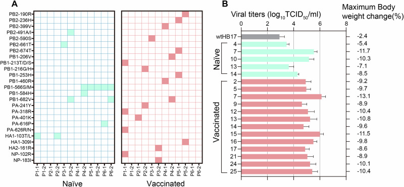
H9N2 avian influenza virus (AIV) is endemic in poultry worldwide and increasingly zoonotic. Despite the long-term widespread use of inactivated vaccines, H9N2 AIVs remain dominant in chicken flocks. We demonstrated that inactivated vaccines did not prevent the replication of H9N2 AIVs in the upper airway of vaccinated chickens. Viral transmission was enhanced during sequential passage in vaccinated chickens, which was attributed to the restricted production of defective interfering particles and the introduction of stable mutations (NP-N417D, M1-V219I, and NS1-R140W) which enhanced viral replication. Notably, the genetic diversity of H9N2 AIVs was greater and included more potential mammal/human-adapted mutations after passage through vaccinated chickens than through naïve chickens, which might facilitate the emergence of mammal-adapted strains. By contrast, vaccines inducing cellular/mucosal immunity in the upper respiratory tract effectively limit H9N2 AIV. These findings highlight the limitations of inactivated vaccines and the need for revised vaccination strategies to control H9N2 AIV.


Conflict of interest statement

Competing interests: The authors declare no competing interests.

Figures

Fig. 1
Fig. 1. Impact of inactivated vaccine on the replication and pathogenicity of AIVs.
A Naïve and inactivated-vaccinated chickens (HI = 7–8 log2) were intranasally inoculated with 106 EID50 of H5Ny, H7N9, or H9N2 AIV. The viral titers in the turbinate, larynx, trachea, bronchus, and lung were measured at 3 dpi. Statistical significance relative to H5Ny or H7N9 AIVs in turbinate and larynx of the naïve group was assessed with two-way ANOVA (*P < 0.05). Horizontal dashed black lines indicate the lower limits of detection. B Representative histopathological changes in H&E-stained turbinate, larynx, trachea, bronchus, and lung sections from HB17-infected groups at 5 dpi. Mucosal cells were swollen and vacuolated (▲); mucosal damage, loss of stratum corneum (△); congestion of capillaries and presence of inflammatory cells around blood vessels (black thick solid arrow); edema, desquamation, and lesion of tracheal epithelial cells (thick open arrow); bronchopneumonia and massive immune cell infiltrates around bronchi and blood vessels (black solid six-pointed star). Scale bar, 50 μm. C Electron microscopy (EM) analysis of larynges and bronchi in HB17-infected groups at 3 and 5 dpi. Scale bar, up, 50 μm; down, 10 μm. D Viral titers of recombinant viruses in the organs of naïve and vaccinated chickens. E IgG antibodies assessed in serum and saliva samples from chickens vaccinated with inactivated-virus vaccine.
Fig. 2
Fig. 2. Evolutionary dynamics of H9N2 AIV in naïve and vaccinated passage lines.
A Time course of H9N2 virus transmission through naïve and vaccinated passage chains. The schematic diagram was created using Adobe Illustrator and Canva (www.canva.com). B Serial intervals of viral transmission between chickens in the naïve and vaccinated passage chains, which refers to the number of days required for an uninfected animal to become infected after cohabitation with an infected animal. C Growth rate of viral replication in chickens at each passage in the naïve and vaccinated passage chains. D Nucleotide positions and absolute frequencies of mutations. E Numbers of nonsynonymous mutations at different passages. Statistical significance was assessed with two-way ANOVA (*P < 0.05; **P < 0.01).
Fig. 3
Fig. 3. Transmission bottleneck sizes within households.
A Frequency of intrahost-acquired single-nucleotide variants (iSNVs) in both donor and recipient samples. Frequencies <3% and >98% were set to 0% and 100%, respectively. B Estimated bottleneck sizes in 10 transmission events, calculated with the exact beta-binomial method. Statistical significance was assessed with two-way ANOVA (**P < 0.01).
Fig. 4
Fig. 4. Identification of defective interfering particle (DIP)-associated junctions in different influenza virus populations.
A Number of distinct junctions detected in each individual in three passage lineages at the indicated time points. B Number of distinct junctions detected in each viral segment in the naïve and vaccinated lineages. C Plots showing the viral DIP-associated junctions present in the overall viral populations from the naïve group. Each color indicates a particular DIP-associated junction, whose relative proportion corresponds to its abundance in the viral population in the indicated sample.
Fig. 5
Fig. 5. Viral variations in vaccinated chain and their impact on oropharyngeal shedding of H9N2 AIV.
A Linkage analysis of high-frequency (>10%) variants that emerged during homologous vaccinated passage. B Multistep growth curves of the indicated H9N2 AIV mutants in CEF infected at MOI of 0.001. All data are representative of or presented as means ± SD of three independent experiments. C Impact of NP-N417D, M1-V219I, and/or NS1-R140W on the replication and transmission of H9N2 AIV in chickens. Three chickens were intranasally inoculated with 106 EID50 of virus. After 24 h, the inoculated chickens were housed together with three contact chickens. The viral titers in oropharyngeal swabs from inoculated chickens (n = 3) were determined at 3, 5, and 7 dpi and at 3, 5, and 7 dpc for the contact chickens. Statistical significance relative to HB17 was assessed with two-way ANOVA (*P < 0.05; **P < 0.01).
Fig. 6
Fig. 6. Positive effects of NP-N417D, M1-V219I, and/or NS1-R140W mutations on replication pathway of H9N2 AIV.
A DF1 cells were infected with rgHB17 or rgHB17(NP-N417D) at MOI of 2. NP was localized with confocal microscopy at the indicated time points (scale bar: 50 μm). B Quantitative analysis of NP localization in virus-infected cells. C H9N2 viral morphology examined with transmission electron microscopy (scale bar: 500 nm). D Quantification of spherical and filamentous virions. E Nuclear-cytoplasmic localization of wild-type HB17 and mutant NS1 protein in DF1 cells during infection. Scale bar, 10 μm. F Quantitative localization of wild-type and mutant NS1 proteins in DF1 cells.
Fig. 7
Fig. 7. Evolution of H9N2 AIV in vaccinated chickens facilitates the emergence of novel viruses, posing increased threat to public health.
A Amino-acid substitutions related to mammal-adaptation detected in viral populations isolated from naïve and vaccinated animal chains. B Viral titers of monoclonal strains with significantly higher replication capacity than wild-type HB17 (P < 0.05) in naïve and vaccinated animal. Mice (n = 6) were inoculated with 106 TCID50 of different monoclonal strains. Three mice from each group were euthanized at 3 dpi, and viral titers in the lungs were determined by infection of MDCK cells. Statistical significance relative to wild-type HB17 was assessed with two-way ANOVA (*P < 0.05; **P < 0.01).
Fig. 8
Fig. 8. Vaccine protection afforded by rHVT-H9 and H9N2-LAIV against H9N2 influenza.
A Vaccinated chickens were intranasally inoculated with 106 EID50 of HB17 virus. At 24 h after challenge, in-contact vaccinated chickens were placed in physical contact with challenged birds. Viral titers on oropharyngeal swabs were determined at 3, 5, and 7 dpc. Horizontal dashed black line indicates the lower limits of detection. B HI antibody analysis. Sera collected were analyzed with HI assays to test the response to the corresponding vaccine strains. C Cellular immune response analysis. Chickens were vaccinated with different vaccines. Lungs (n = 6) were harvested at HI = 7–8 log2. IFN-γ+CD4+ and IFN-γ+CD8+ T cells were identified in lungs stimulated with corresponding vaccine strains using an intracellular cytokine staining assay. Percentages of IFN-γ+CD4+ or IFN-γ+CD8+ T cells among CD4+ or CD8+ T cells were analyzed. DE IgG and IgA antibodies measured in saliva samples from chickens vaccinated with either PBS (Mock) or different vaccines. F Numbers of spots of IFNγ-producing cells in turbinates of chickens vaccinated with either PBS or different vaccines. Statistical significance relative to inactivated vaccine group was assessed with two-way ANOVA (**P < 0.01).

References

    1. Zhang, J. et al. Mutational antigenic landscape of prevailing H9N2 influenza virus hemagglutinin spectrum. Cell Rep.42, 113409 (2023). - PubMed
    1. Peacock, T. H. P., James, J., Sealy, J. E. & Iqbal, M. A global perspective on H9N2 avian influenza virus. Viruses11, 10.3390/v11070620 (2019). - PMC - PubMed
    1. Gu, M., Xu, L., Wang, X. & Liu, X. Current situation of H9N2 subtype avian influenza in China. Vet. Res.48, 49 (2017). - PMC - PubMed
    1. Wang, Z. et al. Mixed selling of different poultry species facilitates emergence of public-health-threating avian influenza viruses. Emerg. Microbes Infect.12, 2214255 (2023). - PMC - PubMed
    1. Sun, Y. & Liu, J. H9N2 influenza virus in China: a cause of concern. Protein Cell6, 18–25 (2015). - PMC - PubMed

LinkOut - more resources