. 2025 Aug;30(8):3837-3845.
doi: 10.1038/s41380-025-03002-3. Epub 2025 Apr 4.

Impaired synaptosome phagocytosis in macrophages of individuals with autism spectrum disorder

Affiliations

Impaired synaptosome phagocytosis in macrophages of individuals with autism spectrum disorder

Yuki Nishi et al. Mol Psychiatry. 2025 Aug.

Abstract

Dendritic spine abnormalities are believed to be one of the critical etiologies of autism spectrum disorder (ASD). Over the past decade, the importance of microglia in brain development, particularly in synaptic elimination, has become evident. Thus, microglial abnormalities may lead to synaptic dysfunction, which may underlie the pathogenesis of ASD. Several human studies have demonstrated aberrant microglial activation in the brains of individuals with ASD, and studies in animal models of ASD have also shown a relationship between microglial dysfunction and synaptic abnormalities. However, there are very few methods available to directly assess whether phagocytosis by human microglia is abnormal. Microglia are tissue-resident macrophages with phenotypic similarities to monocyte-derived macrophages, both of which consistently exhibit pathological phenotypes in individuals with ASD. Therefore, in this study, we examined the phagocytosis capacity of human macrophages derived from peripheral blood monocytes. These macrophages were polarized into two types: those induced by granulocyte-macrophage colony-stimulating factor (GM-CSF MΦ, traditionally referred to as "M1 MΦ") and those induced by macrophage colony-stimulating factor (M-CSF MΦ, traditionally referred to as "M2 MΦ"). Synaptosomes purified from human induced pluripotent stem cell-derived neuron were used to assess phagocytosis capacity. Our results revealed that M-CSF MΦ exhibited higher phagocytosis capacity compared to GM-CSF MΦ, whereas ASD-M-CSF MΦ showed a marked impairment in phagocytosis. Additionally, we found a positive correlation between phagocytosis capacity and cluster of differentiation 209 expression. This research contributes to a deeper understanding of the pathobiology of ASD and offers new insights into potential therapeutic targets for the disorder.


Conflict of interest statement

Competing interests: Dr. Makinodan received research support from Sumitomo Pharma. Dr. Murayama, Dr. Ichikawa, and Dr. Nagata are employees of Sumitomo Pharma Co., Ltd. The remaining authors declare no conflict of interest.

Figures

Fig. 1
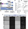
Fig. 1. Phagocytosis assay of synaptosomes purified from hiPSC-derived neurons by macrophages.
A Summary of the phagocytosis assay. Two hiPSC lines from healthy controls are differentiated into excitatory neurons and collected for synaptosome purification. B Representative images of phagocytosis of synaptosomes by GM-CSF MΦ and M-CSF MΦ of TD12 and ASD21. Images captured at the outset of the experiment and 90 min later in the phase contrast and Texas Red Filter, respectively, are presented. Scale bar: 30 μm. C Results of the phagocytosis assay. Phagocytosis capacity is significantly higher in M-CSF MΦ compared to GM-CSF MΦ. Furthermore, the phagocytosis capacity of ASD-M-CSF MΦ is significantly lower than that of TD-M-CSF MΦ. Two-way ANOVA test, F(1, 72) = 8.518, p = 0.0047 (interaction), F(1, 72) = 39.45, p < 0.0001 (MΦ polarity), F(1, 72) = 18.33, p < 0.0001 (group), with post hoc Tukey’s multiple comparison test. n (TD) = 18 and n (ASD) = 20. *p < 0.05, **p < 0.01, ***p < 0.001, ****p < 0.0001.
Fig. 2
Fig. 2. Gene expression analysis of phagocytosis-related receptors.
A–F Gene expression analysis using qRT-PCR. Only CD209 expression showed a pattern consistent with the phagocytosis capacity results. A Two-way ANOVA test, F(1, 72) = 4.841, p = 0.0310 (interaction), F(1, 72) = 15.57, p = 0.0002 (MΦ polarity), F(1, 72) = 9.536, p = 0.0029 (group), with post hoc Tukey’s multiple comparison test. B Two-way ANOVA test, F(1, 72) = 0.1652, p = 0.6856 (interaction), F(1, 72) = 68.51, p < 0.0001 (MΦ polarity), F(1, 72) = 0.04149, p = 0.8392 (group), with post hoc Tukey’s multiple comparison test. C Two-way ANOVA test, F(1, 72) = 0.4829, p = 0.4893 (interaction), F(1, 72) = 123.4, p < 0.0001 (MΦ polarity), F(1, 72) = 0.7115, p = 0.4017 (group), with post hoc Tukey’s multiple comparison test. D Two-way ANOVA test, F(1, 72) = 0.04004, p = 0.8420 (interaction), F(1, 72) = 32.16, p < 0.0001 (MΦ polarity), F(1, 72) = 3.595, p = 0.0620 (group), with post hoc Tukey’s multiple comparison test. E Two-way ANOVA test, F(1, 72) = 3.380, p = 0.0701 (interaction), F(1, 72) = 9.742, p = 0.0026 (MΦ polarity), F(1, 72) = 0.9949, p = 0.3219 (group), with post hoc Tukey’s multiple comparison test. F Two-way ANOVA test, F(1, 72) = 14.02, p = 0.0004 (interaction), F(1, 72) = 2.310, p = 0.1329 (MΦ polarity), F(1, 72) = 0.00005556, p = 0.9941 (group), with post hoc Tukey’s multiple comparison test. n (TD) = 18 and n (ASD) = 20. *p < 0.001, **p < 0.0001. G A simple linear regression analysis of phagocytosis capacity and CD209 expression of M-CSF MΦ indicated a constant correlation (Y = 0.1953X + 0.9080, R2 = 0.1126, p = 0.0395).
Fig. 3
Fig. 3. Knockdown experiment of CD209 in TD-M-CSF MΦ.
A Summary of the knockdown experiment by siRNA in M-CSF MΦ from three TD individuals. B Gene expression of CD209 is significantly reduced in CD209-siRNA-transfected M-CSF MΦ compared to non-targeting-siRNA-transfected M-CSF MΦ and sham-treated M-CSF MΦ. Repeated measures ANOVA test, F(2, 4) = 19.86, p = 0.0084, with post hoc Dunnett’s multiple comparison test. C Representative images of phagocytosis by CD209-siRNA-transfected, non-targeting-siRNA-transfected, and sham-treated M-CSF MΦ of TD10, respectively. Images captured at the outset of the experiment and 90 min later in the phase contrast and Texas Red Filter, respectively, are presented. Scale bar: 30 μm. D Results of the phagocytosis assay. Phagocytosis capacity is significantly lower in CD209-siRNA-transfected M-CSF MΦ compared to non-targeting-siRNA-transfected M-CSF MΦ and sham-treated M-CSF MΦ. Repeated measures ANOVA test, F(2, 4) = 32.92, p = 0.0033, with post hoc Dunnett’s multiple comparison test.

References

    1. Gyawali S, Patra BN. Autism spectrum disorder: trends in research exploring etiopathogenesis. Psychiatry Clin Neurosci. 2019;73:466–75. - PubMed
    1. Marchetto MC, Carromeu C, Acab A, Yu D, Yeo GW, Mu Y, et al. A model for neural development and treatment of Rett syndrome using human induced pluripotent stem cells. Cell. 2010;143:527–39. - PMC - PubMed
    1. Cheffer A, Flitsch LJ, Krutenko T, Roderer P, Sokhranyaeva L, Iefremova V, et al. Human stem cell-based models for studying autism spectrum disorder-related neuronal dysfunction. Mol Autism. 2020;11:99. - PMC - PubMed
    1. Penzes P, Cahill ME, Jones KA, VanLeeuwen JE, Woolfrey KM. Dendritic spine pathology in neuropsychiatric disorders. Nat Neurosci. 2011;14:285–93. - PMC - PubMed
    1. Forrest MP, Parnell E, Penzes P. Dendritic structural plasticity and neuropsychiatric disease. Nat Rev Neurosci. 2018;19:215–34. - PMC - PubMed

MeSH terms

Substances