

Effect of the location and severity of partial ureteral obstruction on urinary system stone disease formation
- PMID: 40185910
- PMCID: PMC11971389
- DOI: 10.1038/s41598-025-96879-7
Effect of the location and severity of partial ureteral obstruction on urinary system stone disease formation
Abstract
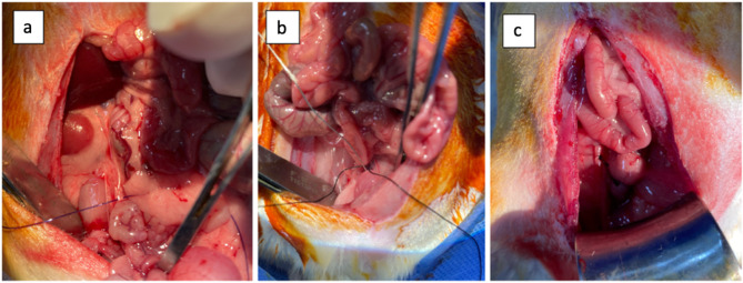
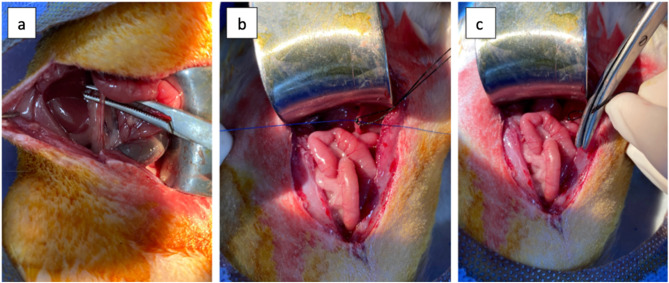
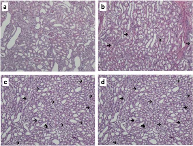
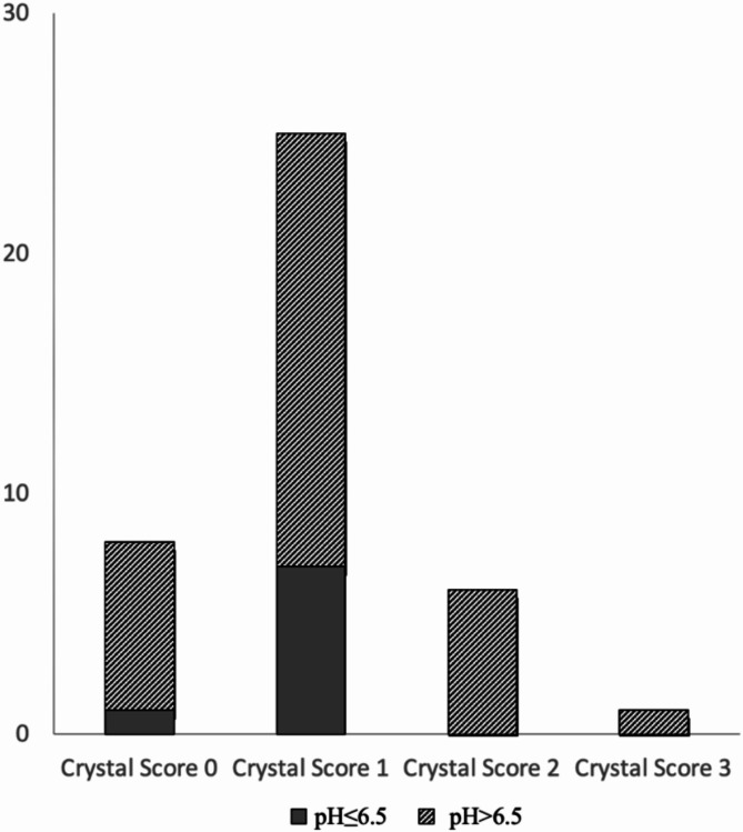
This study aimed to observe the effect of the location and severity of partial ureteral obstruction, based on ureteropelvic and ureterovesical junction strictures, on stone formation. Forty 8-week-old male rats were divided into five groups: control and mild/severe proximal or distal ureteral obstruction groups. A partial ureteral obstruction was created according to the type of obstruction by a surgical procedure. After 5 days of intraperitoneal glyoxylate injection, the urine, blood samples, and kidney tissues of the rats were examined. There were significant differences in urine citrate concentrations, pH values, and oxalate/creatinine, citrate/creatinine, and calcium/creatinine ratios between the groups (all p < 0.05). The mean citrate/creatinine ratio was 21.13 ± 0.44 in the control group, 17.31 ± 3.82 in the distal obstruction group, and 15.48 ± 1.87 in the proximal obstruction group. Regarding the degree of obstruction, urine citrate concentrations, pH values, and citrate/creatinine were lower, and the oxalate/creatinine ratio was higher in severe obstruction than in mild obstruction (p < 0.05). This study represents an initial attempt to evaluate a model of partial ureteral obstruction and urolithiasis. The findings indicate that obstruction alters urinary parameters, such as citrate and pH, indirectly increasing the risk of stone formation. Furthermore, stone formation in an obstructed urinary system appears to be a complex process. However, metabolic evaluation and treatment may help prevent stone formation in patients with ureteral obstruction.
Keywords: Kidney stone; Partial ureteral obstruction; Ureteropelvic junction obstruction; Ureterovesical junction obstruction; Urinary system stone disease.
© 2025. The Author(s).
Conflict of interest statement
Declarations. Competing interests: The authors declare no competing interests. Ethical standards: We confirm that this study is implemented in accordance with the Animal Research Reporting in Live Experiments (ARRIVE) guidelines. Ethics approval: The questionnaire and methodology for this study was approved by the Sakarya University Animal Experiments Local Ethics Committee. (Ethics approval number:22, 06/04/2022). A scanned copy of the ethics review approval document has been uploaded to the submission website.
Figures
Similar articles
-
Biochemical risk factors for stone formation in a Scottish paediatric hospital population.Ann Clin Biochem. 2010 Mar;47(Pt 2):125-30. doi: 10.1258/acb.2009.009146. Epub 2010 Feb 9. Ann Clin Biochem. 2010. PMID: 20144971
-
Is calcium oxalate nucleation in postprandial urine of males with idiopathic recurrent calcium urolithiasis related to calcium phosphate nucleation and the intensity of stone formation? Studies allowing insight into a possible role of urinary free citrate and protein.Clin Chem Lab Med. 2004 Mar;42(3):283-93. doi: 10.1515/CCLM.2004.052. Clin Chem Lab Med. 2004. PMID: 15080561
-
The role of overweight and obesity in calcium oxalate stone formation.Obes Res. 2004 Jan;12(1):106-13. doi: 10.1038/oby.2004.14. Obes Res. 2004. PMID: 14742848
-
[Calcium-dependent calcium oxalate lithiasis (type IIa). The importance of urinary citrate and the calcium/citrate ratio].Presse Med. 1999 Apr 17;28(15):784-6. Presse Med. 1999. PMID: 10325933 Review. French.
-
Dietary treatment of urinary risk factors for renal stone formation. A review of CLU Working Group.Arch Ital Urol Androl. 2015 Jul 7;87(2):105-20. doi: 10.4081/aiua.2015.2.105. Arch Ital Urol Androl. 2015. PMID: 26150027 Review.
References
-
- Singh, P., Harris, P. C., Sas, D. J. & Lieske, J. C. The genetics of kidney stone disease and nephrocalcinosis. Nat. Rev. Nephrol.18 (4), 224–240. 10.1038/s41581-021-00513-4 (2022). - PubMed
-
- Song, L., Maalouf, N. M. & Nephrolithiasis [Updated 2020 Mar 9]. In: Feingold KR, Anawalt B, Blackman MR, et al., editors. Endotext [Internet]. South Dartmouth (MA): MDText.com, Inc.; 2000-. https://www.ncbi.nlm.nih.gov/books/NBK279069/ Accessed 8 December 2024.
-
- Fontenelle, L. F. & Sarti, T. D. Kidney stones: treatment and prevention. Am. Family Phys.99 (8), 490–496 (2019). - PubMed
MeSH terms
Substances
LinkOut - more resources
Full Text Sources
