. 2025 Apr 4;23(1):270.
doi: 10.1186/s12951-025-03331-z.

Platycodon grandiflorum exosome-like nanoparticles: the material basis of fresh platycodon grandiflorum optimality and its mechanism in regulating acute lung injury

Affiliations

Platycodon grandiflorum exosome-like nanoparticles: the material basis of fresh platycodon grandiflorum optimality and its mechanism in regulating acute lung injury

Jingmin Fu et al. J Nanobiotechnology. .

Abstract

Background: Acute lung injury (ALI) is a severe respiratory disease accompanied by diffuse inflammatory responses induced by various clinical causes. Many fresh medicinal plants have shown better efficacy than their dried forms in preventing and treating diseases like inflammation. As a classical Chinese herb, platycodon grandiflorum (PG) has been demonstrated effective in treating pneumonia, but most of previous studies focused on the efficacy of processed or dried PG formats, while the specific benefits of its fresh form are still underexplored. Exosome-like nanoparticles derived from medicinal plants are expected to point out an important direction for exploring the material basis and mechanism of this fresh herbal medicine.

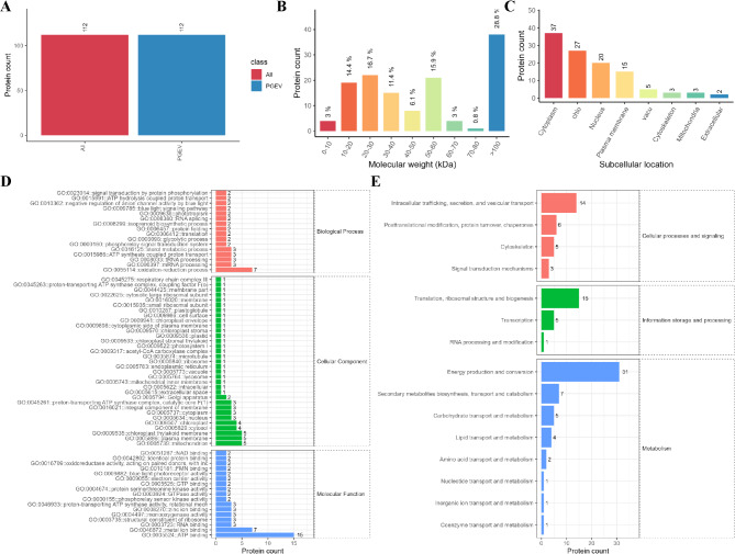
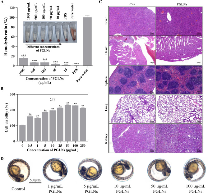
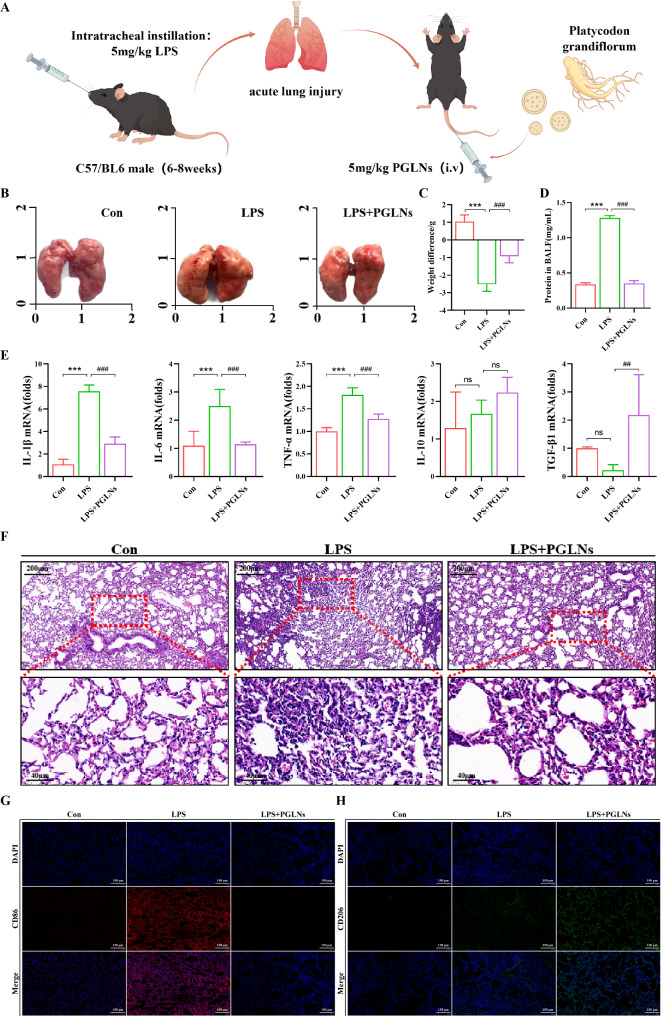
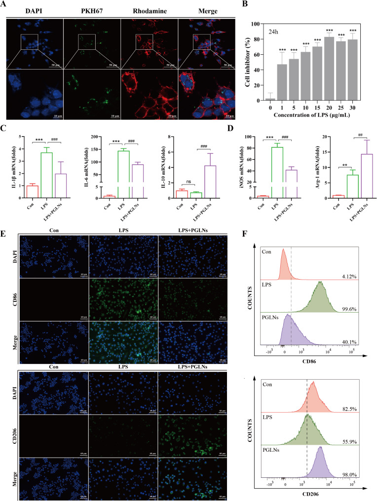
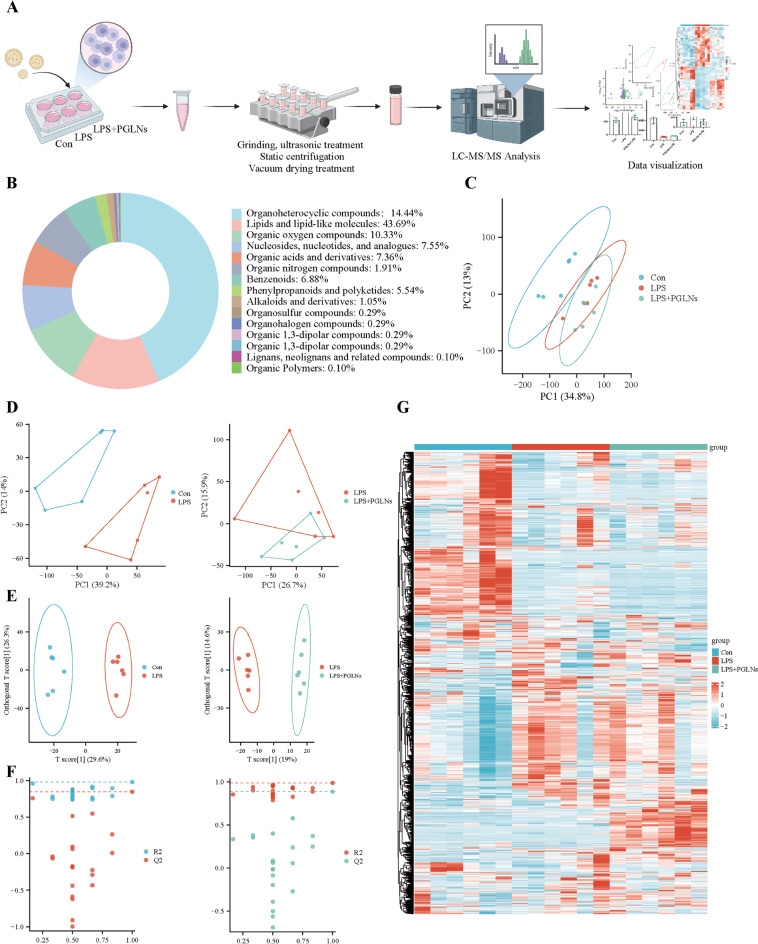
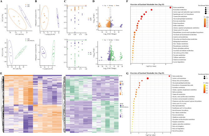
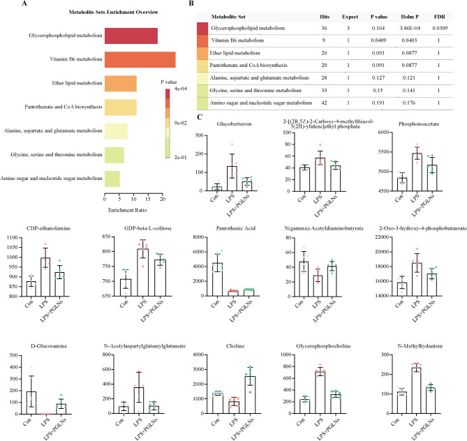
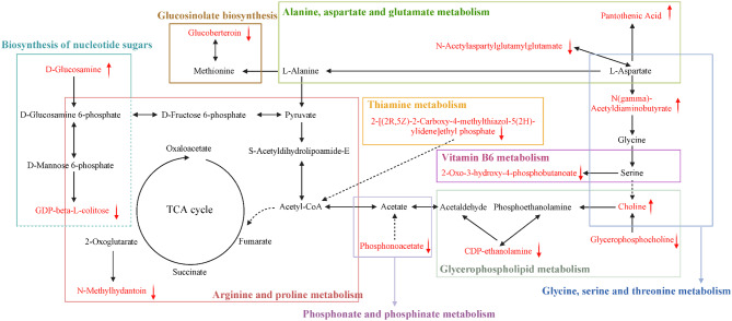
Results: The fresh form of PG could effectively improve ALI induced by lipopolysaccharide (LPS), relieve lung histopathological injury and weight loss, and reduce levels of inflammatory factors in mice, exhibiting better efficacy than dried PG in the treatment of ALI. Further extraction and purification of PG exosome-like nanoparticles (PGLNs) demonstrated that PGLNs had good biocompatibility, with characteristics consistent with general exosome-like nanoparticles. Besides, proteomic analysis indicated that PGLNs were rich in a variety of proteins. Animal experiments showed that PGLNs improved the pathological changes in LPS-induced lung tissues, inhibited the expression of inflammatory factors and promoted the expression of anti-inflammatory factors, and exerted a regulatory effect on the polarization of lung macrophages. Cell experiments further confirmed that PGLNs could be effectively taken up by RAW264.7 cells and repolarize M1 macrophages into M2 type, therefore reducing the secretion of harmful cytokines. Moreover, non-targeted metabolomics analysis reveals that PGLNs reduce inflammation and control macrophage polarization in a manner closely linked to pathways including glycolysis and lipid metabolism, highlighting a potential mechanism by which PGLNs protect the lungs from inflammatory damage like ALI.

Conclusion: Fresh PG has better anti-inflammatory and repair effects than its dried form. As one of the most effective active substances in fresh PG, PGLNs may regulate macrophage inflammation and polarization by regulating metabolic pathways including lipid metabolism and glycolysis, so as to reduce inflammation and repair lung injury.

Keywords: Acute lung injury; Fresh medicinal plant; Macrophage polarization; Non-targeted metabolomics; PGLNs; Platycodon grandiflorum.


Conflict of interest statement

Declarations. Ethics approval and consent to participate: All animal experiments were approved by the Ethics Committee of the Center for Animal Experiments of Hunan University of Chinese Medicine (Approval number: SLBH-202307190001). Consent for publication: All authors have approved the manuscript and agree with the submission. Competing interests: The authors declare no competing interests.

Figures

Fig. 1
Fig. 1
Fresh PG shows a better therapeutic effect than dried PG in alleviating LPS-induced ALI in mice. (A) Administration of LPS and PG(PF, PFW, PD, PDW) in ALI model mice. (B) Weight changes in each group within 7 days after treatment with PF, PFW, PD and PDW, respectively (n =4). (C) Protein concentration in the alveolar lavage fluid of mice in each group (n = 4). (D) mRNA levels of IL-1β, IL-6 and TNF-α in mouse lung tissues in each group (n = 4). (E) H&E staining results of mouse lung sections in each group. Scale: 200μm and 100μm
Fig. 2
Fig. 2
Isolation and characterization of PGLNs. (A) Differential centrifugation and sucrose density gradient centrifugation for PGLNs preparation. (B) Transmission electron microscope (TEM) images of PGLNs. Scale, 100 nm. (C) Nanoparticle tracking analysis (NTA) to detect the size and particle number of isolated PGLNs. (D) The surface charge of PGLNs. (E) The nucleic acid content of PGLNs detected by agarose gel electrophoresis. The standard DNA molecular weight is used as a size marker. (F) The protein contents of PGLNs detected by SDS-PAGE (10%) and Coomassie brilliant blue staining. (G) The protein concentration of isolated PGLNs. The standard protein molecular weight is used as a molecular weight (MW) marker
Fig. 3
Fig. 3
Characterization of PGLNs - proteomics. Histogram shows (A) the total protein quantity, (B) the total protein molecular weight distribution, and (C) the total protein subcellular location identified in PGLNs. (D) GO function analysis results based on PGLNs proteomics. (E) KEGG analysis results based on PGLNs proteomics
Fig. 4
Fig. 4
Biosafety assessment of PGLNs. (A) Hemolysis activity of PGLNs at different concentrations (n = 3). (B) Cell activity after RAW264.7 cells were incubated with different concentrations of PGLNs for 24 hours (n = 6). (C) H&E staining results of liver, heart, spleen, lung and kidney tissue slices (normal control vs. PGLNs group). Scale: 200μm. (D) Zebrafish fertilized eggs after intervention with different concentrations of PGLNs for 1 day. Scale: 500 μm
Fig. 5
Fig. 5
PGLNs alleviated LPS-induced ALI in mice, which may be related to the regulation of macrophage polarization. (A) Schematic diagram of LPS and PGLNs administration regimens in ALI mice. (B) Differences in body weight between the LPS group and LPS+PGLNs group (n = 5). (C) Macroscopic images of mouse lung tissues in each group. Unit: cm. (D) Protein concentration in the alveolar lavage fluid of mice detected by BCA (n = 4). (E) mRNA levels of IL-1β, IL-6, TNF-α, IL-10 and TGF-β1 in mouse lung tissues detected by RT-qPCR (n = 4). (F) H&E staining results of mouse lung sections. Scale: 200μm and 40μm. (G) Fluorescence expression of M1 pro-inflammatory macrophage marker CD86 and (H) M2 anti-inflammatory macrophage marker CD206 in mouse lung tissues detected by tissue immunofluorescence. Scale: 150 μm
Fig. 6
Fig. 6
PGLNs regulate macrophage polarization in vitro. (A) Laser confocal images. Uptake of PKH67-labeled PGLNs by RAW264.7 cells in vitro. Scale: 50 and 10 μm. (B) Activity of RAW264.7 cells induced by LPS at different concentrations for 24 hours (n = 4). (C) mRNA levels of IL-1β, IL-6 and IL-10 (n = 4). (D) mRNA levels of iNOS and Arg-1 (n = 4). (E) Fluorescence expression of M1 proinflammatory macrophage marker CD86 and M2 anti-inflammatory macrophage marker CD206. Scale: 60μm. Data analysis using ImageJ software. (F) Flow cytometry analysis of RAW264.7 cells
Fig. 7
Fig. 7
PGLNs influenced the metabolic disorder of LPS-induced RAW264.7 cells. (A) Illustration of metabolomics analysis. (B) Quantity and classification of metabolites. (C) PCA score plots for various groups (Con, LPS and LPS + PGLNs). (D) PCA score plots for Con vs. LPS and LPS vs. LPS + PGLNs. (E) OPLS-DA score plots for Con vs. LPS and LPS vs. LPS + PGLNs. (F) OPLS-DA permutation test for Con vs. LPS and LPS vs. LPS + PGLNs. (G) Heatmap of hierarchical cluster analysis of metabolite changes (“red” and “blue” represent increased and decreased metabolite contents, respectively)
Fig. 8
Fig. 8
Analysis of differential metabolites in RAW264.7 cells after treatment by LPS and LPS + PGLNs. (A) PCA score plots of different metabolites for Con vs. LPS groups, and LPS vs. LPS + PGLNs groups. (B) OPLS-DA score plots of different metabolites for Con vs. LPS groups and LPS vs. LPS + PGLNs groups. (C) OPLS-DA permutation test of different metabolites for Con vs. LPS groups and LPS vs. LPS + PGLNs groups. (D) Volcano plot analysis of Con vs. LPS groups and LPS vs. LPS + PGLNs groups. (E) Heatmap of differential metabolites for Con vs. LPS groups and LPS vs. LPS + PGLNs groups. (F) Bubble map of differential metabolite KEGG enrichment factor for Con vs. LPS groups. (G) Bubble map of differential metabolite KEGG enrichment factor for LPS vs. LPS + PGLNs groups
Fig. 9
Fig. 9
Clustering heatmap of differential metabolites for Con vs. LPS (A) and LPS vs. LPS + PGLNs (B)
Fig. 10
Fig. 10
Potential biomarkers of PGLNs for the treatment of LPS-induced RAW264.7 cell inflammation. (A) PCA score plots of potential biomarkers for differential metabolites among various groups (Con, LPS, and LPS + PGLNs). (B) Heatmap of hierarchical cluster analysis of potential biomarkers for Con vs. LPS, and LPS vs. LPS + PGLNs (“red” and “blue” represent increased and decreased metabolite contents, respectively). (C) Multiples of difference in potential biomarkers for Con vs. LPS. (D) Multiples of difference in potential biomarkers for LPS vs. LPS + PGLNs
Fig. 11
Fig. 11
Analysis of key metabolites of PGLNs in the treatment of LPS-induced RAW264.7 cell inflammation. (A) KEGG enrichment map of potential biomarkers. (B) Metabolic pathway enrichment information of potential biomarkers. (C) Quantitative analysis of key metabolites
Fig. 12
Fig. 12
Relationship diagram of metabolic pathways for the pharmacological mechanisms of PGLNs

References

    1. Zhang J, Guo Y, Mak M, Tao Z. Translational medicine for acute lung injury. J Transl Med. 2024;22(1):25. - PMC - PubMed
    1. Liu C, Xiao K, Xie L. Advances in the use of exosomes for the treatment of ALI/ARDS. Front Immunol. 2022;13:971189. - PMC - PubMed
    1. Meng J, Leung KS, Jiang Z, Dong X, Zhao Z. Establishment of GC-MS fingerprint of fresh houttuynia cordata. Chem Pharm Bull (Tokyo). 2005;53(11):1484–9. - PubMed
    1. Zheng Y, Lei L, Liang S, et al. Protective effect of fresh/dry dandelion extracts on APAP-Overdose-Induced acute liver injury. Chin J Integr Med. 2022;28(8):683–92. - PubMed
    1. Zhang L, Wang Y, Yang D, et al. Platycodon grandiflorus - an ethnopharmacological, phytochemical and Pharmacological review. J Ethnopharmacol. 2015;164:147–61. - PubMed

LinkOut - more resources