. 2025 May 29;188(11):2957-2973.e28.
doi: 10.1016/j.cell.2025.03.029. Epub 2025 Apr 4.

Molecular basis for shifted receptor recognition by an encephalitic arbovirus

Affiliations

Molecular basis for shifted receptor recognition by an encephalitic arbovirus

Xiaoyi Fan et al. Cell. .

Abstract

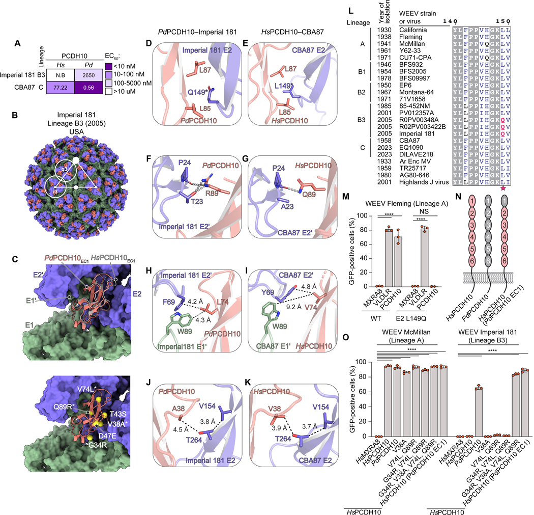
Western equine encephalitis virus (WEEV) is an arbovirus that historically caused large outbreaks of encephalitis throughout the Americas. WEEV binds protocadherin 10 (PCDH10) as a receptor, and highly virulent ancestral WEEV strains also bind low-density lipoprotein receptor (LDLR)-related proteins. As WEEV declined as a human pathogen in North America over the past century, isolates have lost the ability to bind mammalian receptors while still recognizing avian receptors. To explain shifts in receptor dependencies and assess the risk of WEEV re-emergence, we determined cryoelectron microscopy structures of WEEV bound to human PCDH10, avian PCDH10, and human very-low-density lipoprotein receptor (VLDLR). We show that one to three E2 glycoprotein substitutions are sufficient for a nonpathogenic strain to regain the ability to bind mammalian receptors. A soluble VLDLR fragment protects mice from lethal challenge by a virulent ancestral WEEV strain. Because WEEV recently re-emerged in South America after decades of inactivity, our findings have important implications for outbreak preparedness.

Keywords: alphavirus; arbovirus; emerging virus; encephalitis; receptor; viral glycoprotein.


Conflict of interest statement

Declaration of interests The authors declare no competing interests.

Figures

Figure 1.
Figure 1.. Structural basis for WEEV recognition of human PCDH10.
(A) Schematic diagrams of PCDH10 and Flag-tagged constructs. (B) Partial phylogenetic tree and summary of K562 infectivity assays with GFP-expressing RVPs. Strains newly tested in this study are marked with black triangles; others are from Li et al. See also Figure S1. (C) Cryo-EM maps of HsPCDH10EC1-Fc bound to WEEV CBA87 VLP with E2 in purple, E1 in green, and PCDH10 EC1 in pink. Icosahedral symmetry axes (i5, i3, i2) are indicated with a closed circle, triangle, and hexagon, respectively. The icosahedral i3 and q3 E2–E1 trimers are circled and indicated. (D) Ribbon diagram of a single WEEV E2–E1 heterodimer and PCDH10 EC1 fitted into its associated cryo-EM density map. The EC1 insertion angle relative to the E2–E1 trimer is shown. (E) CBA87 E2–E1 trimer bound by PCDH10 EC1 (surface rendered) with one EC1 as ribbons. WEEV E2–E1 residues that interact with EC1 are shown in yellow. Interaction details are in panels F to J. (F–J) PCDH10 EC1 interactions with WEEV E2–E1 (F and G) and E2’–E1’ protomers (H–J). Key polymorphic residue E2 L149 is marked with an asterisk. Hydrogen bonds and salt bridges are shown as dashed lines. (K) Interface between human PCDH10 EC1 and EC4 in a crystal structure of the PCDH10 EC1–4 homodimer (PDB: 6VFW). (L) Infection of K562 cells expressing WT (EC1-Flag) and mutant human PCDH10 EC1 constructs by GFP-expressing WEEV 71V RVPs at MOI=0.5, quantified by flow cytometry. Data are mean ± s.d. from three experiments performed in duplicates or triplicates (n = 3) (L). One-way ANOVA with Dunnett’s multiple comparisons test, ****P<0.0001 compared to stalk-Flag (L).
Figure 2.
Figure 2.. Structural basis for WEEV recognition of avian PCDH10.
(A) EC50 values from ELISAs with HsPCDH10EC1-Fc or PdPCDH10EC1-Fc on immobilized WEEV VLPs. (B) Cryo-EM map of PdPCDH10EC1-Fc bound to Imperial 181 VLP. E2 is in purple, E1 is in green, and PCDH10 EC1 is in pink. (C) Top panel: Top view of superposition of PdPCDH10 EC1–Imperial 181 E2–E1 trimer and HsPCDH10 EC1–CBA87 E2–E1 trimer. Bottom panel: Top view of PdPCDH10 EC1–Imperial 181 E2–E1 trimer, highlighting six polymorphisms between Hs and PdPCDH10 EC1. (D–K) Comparison of contact residues of PdPCDH10-bound Imperial 181 (D, F, H, J) and HsPCDH10-bound CBA87 (E, G, I, K), showing polar contacts (gray dashed lines) and the closest distances between PCDH10 residues and atoms on WEEV E2 (black dashed lines) (H–K). E2 residue L/Q149 is marked with an asterisk. (L) Partial WEEV E2 sequence alignment. E2 residue 149 is indicated with a star. Light gray background indicates completely conserved residues. Boxes indicate positions where a single majority residue or multiple chemically similar residues are present. E2 Q149 is colored pink. The panel was generated using ESPript 3.0. (M) K562 cells expressing HsMXRA8, HsVLDLR or HsPCDH10 were infected with GFP-expressing WT or mutant Fleming RVPs at MOI=1. (N) Schematic diagrams of WT HsPCDH10, PdPCDH10, and a HsPCDH10 chimeric construct with PdPCDH10 EC1. (O) K562 cells expressing HsMXRA8, HsPCDH10, PdPCDH10, HsPCDH10 (PdPCDH10 EC1), or HsPCDH10 mutants were infected with GFP-expressing McMillan or Imperial 181 RVPs at MOI=3. Data are mean ± s.d. from two experiments performed in duplicates or triplicates (n = 3) (M, O). Two-way ANOVA with Dunnett’s multiple comparisons test, ****P<0.0001; ***P<0.001 (M, O); NS: not significant. See also Figure S6E.
Figure 3.
Figure 3.. VLDLR recognition by ancestral WEEV strains.
(A) Infectivity of GFP-expressing RVPs for the indicated alphaviruses with K562 cells expressing VLDLR single LA repeat constructs (see Figures S6F–H). EEEV, SFV, and SINV data are from a prior study. “Entry” indicates that the construct mediates statistically significant (P < 0.05) RVP infection when compared to LBD-lacking control. (B) Cryo-EM map of VLDLRLBD-Fc bound to McMillan VLP. E2 is purple, E1 is in green, VLDLR LA1 in is light yellow, and LA2 is in dark yellow. See also Figure S10. (C) Ribbon diagram of a single WEEV E2–E1 protomer and VLDLR LA(1–2) fitted into the cryo-EM density map. E2–E1 domains are indicated. (D) McMillan E2–E1 trimer bound to VLDLR (surface rendered) with one VLDLR LA(1–2) protomer rendered as ribbons. (E and F) McMillan contact with VLDLR LA1 (E) or LA2 (F) showing polar contacts (dashed lines), Ca2+ ions (green spheres), and polymorphic interacting residues (asterisks). (G) K562 cells expressing human MXRA8, VLDLR, or PCDH10 were infected by GFP-expressing WT or mutant WEEV McMillan RVPs at MOI=0.5. (H) K562 cells expressing human MXRA8, VLDLR, or ApoER2 were infected with GFP-expressing E1 K227A (site 1) + E2 K190A (site 2) mutant Fleming RVPs at MOI=1. (I) Top view of the WEEV E2–E1 trimer showing three potential binding sites for VLDLR LA repeats. Key residues are: E1 K227 (site 1), E2 K181 (site 2), and E2 K81 (site 3). (J) K562 cells expressing human MXRA8, VLDLR, ApoER2, or PCDH10 were infected with GFP-expressing WT or E2 K81E (site 3) mutant Fleming RVPs at MOI=1. Data are mean ± s.d. from three experiments performed in triplicates (n = 3) (G, H, J). One-way ANOVA with Dunnett’s multiple comparison test (H). Two-way ANOVA with Dunnett’s multiple comparisons test (G and J). ****P<0.0001 (G, H, J). MXRA8 vs. ApoER2 ***P = 0.0001 (H); NS: not significant.
Figure 4.
Figure 4.. WEEV E2 protein polymorphisms affecting receptor recognition and neurotropism.
(A) Imperial 181 mutants generated and summary of K562 infectivity assay in (C). (B) Side view of the WEEV E2–E1 trimer highlighting mutated E2 residues. (C) K562 cells expressing MXRA8, VLDLR, ApoER2, or PCDH10 were infected with WT or mutant Imperial 181 RVPs at MOI=1. (D) K562 cells expressing HsPCDH10, PdPCDH10, or PdMXRA8 were infected with the indicated GFP-expressing RVPs at various MOIs. (E and F) Primary murine cortical neurons were infected with GFP-expressing WT or mutant Imperial 181 RVPs at MOI=2 in the absence of additional proteins (E and F) or in the presence of 316 μg ml−1 HsPCDH10EC1-Fc, 100 μg ml−1 RAP, or 316 μg ml−1 isotype control (F). Absolute infection levels in the absence of additional proteins are shown in (E) and relative infection levels in the presence of indicated proteins normalized to infection levels in the absence of additional proteins are shown in (F). Infection was monitored through a live cell imaging system. (G) Representative merged images of GFP and bright field from (E and F). Scale bars: 100 μm. Data are mean ± s.d. from three experiments performed in duplicates or triplicates (n = 3) (C, D, E, and F). One-way ANOVA with Dunnett’s multiple comparisons test (E). Two-way ANOVA with Dunnett’s multiple comparisons test (C, and F). ****P < 0.0001 (C, E and F). WT vs. Mut-1 **P = 0.0028; WT vs. Mut-2 *P < 0.0111; WT vs. Mut-3 *P = 0.038 (E). Mut-2 isotype control vs. RAP ***P = 0.0004; Mut-3 isotype control vs. PCDH10EC1-Fc ***P = 0.0005; Mut-5 isotype control vs. PCDH10EC1-Fc **P = 0.0045 (F).
Figure 5.
Figure 5.. Prediction of WEEV strain receptor usage based on E2 glycoprotein sequences.
(A) K562 cells expressing the indicated human receptors were infected with GFP-expressing RVPs for the indicated South American WEEV strains at MOI=0.5. (B) K562 cells expressing the indicated receptor orthologs were infected with GFP-expressing RVPs for the indicated South American WEEV strains at MOI=0.5. (C) K562 cells expressing the indicated human receptors were infected with the indicated GFP-expressing WEEV strain RVPs at MOI=0.5. (D) K562 cells expressing HsPCDH10, PdPCDH10, or PdMXRA8 were infected with GFP-expressing AG80–646 RVPs at indicated MOIs. Data are mean ± s.d. from three experiments performed in duplicates or triplicates (n = 3) (A, B, C and D). Two-way ANOVA with Dunnett’s multiple comparisons test (A, B, C). ****P < 0.0001 (A, B, and C).
Figure 6.
Figure 6.. A WEEV-related North American alphavirus uses PCDH10 as a receptor.
(A) Maximum likelihood phylogenetic tree of select alphaviruses based on the structural polyprotein coding sequences. WEEV and HJV strains are indicated in parentheses. IMP181: Imperial 181. McM: McMillan. See Table S1 for GenBank accession numbers. (B and C) K562 cells expressing indicated human or animal receptor orthologs were infected with GFP-expressing HJV 585–01 RVPs at MOI=1. (D) Viral replication for HJV (strain B 230) in transduced K562 cells at an MOI of 0.01. (E) Infection of K562 cells expressing PCDH10 orthologs with GFP-expressing WT and mutant HJV 585–01 RVPs at MOI=1. (F) AF3-predicted model of HJV 585–01 with an E2 A177K substitution bound by human PCDH10 EC1. See Figure S8C for predicted local distance difference test (pLDDT) scores. Data are mean ± s.d. from three experiments performed in duplicates or triplicates (n = 3) (B–E). One-way ANOVA with Dunnett’s multiple comparisons test (B and C). Two-way ANOVA with Šídák’s multiple comparisons test (D and E). ****P < 0.0001(B, D and E). HsMXRA8 vs. HsPCDH10 *P = 0.0299; HsMXRA8 vs. PdMXRA8 ***P = 0.0005; HsMXRA8 vs. PdPCDH10 ***P = 0.0003 (C). 24 h HsMXRA8 vs. HsPCDH10 *P = 0.0144 (D). EcPCDH10 WT vs. E2 A177K **P = 0.0015 (E).
Figure 7.
Figure 7.. Comparison of alphavirus interactions with receptors and protective effect of a VLDLR LA(1–2) decoy protein.
(A) K562 cells expressing indicated human or animal receptor orthologs were infected with GFP-expressing WT or mutant McMillan RVPs at MOI=1. Datasets are compared within groups (shown at the top) and with HsMXRA8 (above each bar graph). (B) McMillan E2–E1 trimer (surface rendered) bound by a chimeric Duck–D1–Mouse–D2 MXRA8 (ribbons) (PDB ID: 8SQN). MXRA8 contact residues on E2–E1 are in green. Shared contact residues between duck MXRA8 and PCDH10 (E2 L149, E2 V265, and E1 K227) are indicated. (C) CBA87 E2–E1 trimer (surface rendered) bound by one human PCDH10 EC1 (ribbons). PCDH10 contact residues on E2–E1 are in green. Shared contact residues between PCDH10 and VLDLR (E2 K177, E2 K224, and E1 K227) or PCDH10 and duck MXRA8 (E2 L149, E2 V265 and E1 K227) are indicated. (D–G) Comparison of alphavirus E2–E1 trimers (surface rendered) bound by LA repeats (ribbon). The key basic residues on WEEV McMillan (D), EEEV PE6 (E) (PDB ID: 8UFB), VEEV TC-83 (G) (PDB ID: 7FFF), and SFV SFV4 (F) (PDB ID: 8UA8), are in yellow. LA repeats are in magenta; E2–E1 glycoproteins are in different shades of gray; E3 is in light blue. Calcium ions are in green. D1: domain 1. (H and I) McMillan (H) or 71V (I) RVPs were pre-incubated with the indicated Fc fusion proteins and used to infect K562 cells expressing human PCDH10. (J and K) Six-week-old CD1 mice were administered VLDLR LA(1–2)-Fc fusion protein, an isotype control antibody, or a buffer diluent intraperitoneally 6 h before subcutaneous inoculation with 1000 PFU of WEEV McMillan rescued from a molecular clone. Survival (J) and weight change (K) of the mice were monitored. Data are mean ± s.d. from three experiments performed in duplicates or triplicates (n = 3) (A, H, and I). ****P < 0.0001; PdMXRA8 WT vs. K177A ***P = 0.0003; PdPCDH10 ET vs. R224A ***P = 0.0001; R224A HsMXRA8 vs. HsVLDLR *P = 0.0178 (A). For VLDLR LA(1–2)-Fc protection experiment (J): buffer only, n = 10; VLDLR LA(1–2)-Fc, n = 10; isotype control, n = 10 mice. Log-rank (Mantel–Cox) test comparing VLDLR LA(1–2)-Fc or isotype control to buffer. **P<0.01; NS, not significant.

Update of

References

    1. Arechiga-Ceballos N, and Aguilar-Setien A. (2015). Alphaviral equine encephalomyelitis (Eastern, Western and Venezuelan). Rev Sci Tech 34, 491–501. 10.20506/rst.34.2.2374. - DOI - PubMed
    1. Griffin DE (2013). Alphaviruses. In Virology Fields, Knipe DM, and Howley PM, eds. (Lippincott Williams & Wilkins; ), pp. 651–686.
    1. Ronca SE, Dineley KT, and Paessler S. (2016). Neurological sequelae resulting from encephalitic alphavirus infection. Front Microbiol 7, 959. 10.3389/fmicb.2016.00959. - DOI - PMC - PubMed
    1. Bergren NA, Haller S, Rossi SL, Seymour RL, Huang J, Miller AL, Bowen RA, Hartman DA, Brault AC, and Weaver SC (2020). “Submergence” of Western equine encephalitis virus: Evidence of positive selection argues against genetic drift and fitness reductions. PLoS Pathog 16, e1008102. 10.1371/journal.ppat.1008102. - DOI - PMC - PubMed
    1. Aviles G, Bianchi TI, Daffner JF, and Sabattini MS (1993). [Post-epizootic activity of Western equine encephalitis virus in Argentina]. Rev Argent Microbiol 25, 88–99. - PubMed

LinkOut - more resources