. 2025 Apr 5;16(1):3266.
doi: 10.1038/s41467-025-58671-z.

Molecular basis for the interaction between Saccharomyces cerevisiae Rtt103 and the Rat1-Rai1 complex

Affiliations

Molecular basis for the interaction between Saccharomyces cerevisiae Rtt103 and the Rat1-Rai1 complex

Hsu-Feng Chu et al. Nat Commun. .

Abstract

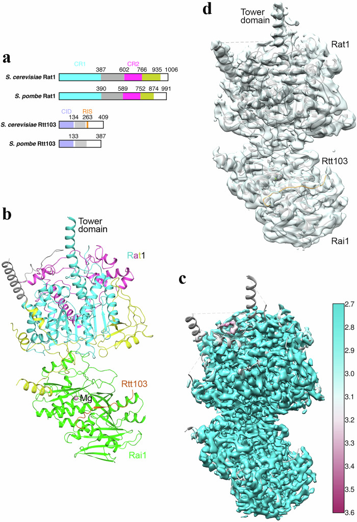
The Rat1 5'-3' exoribonuclease together with its partner Rai1 have important roles in Saccharomyces cerevisiae RNA polymerase II transcription termination. Rtt103 copurifies with Rat1-Rai1 in S. cerevisiae, but its mechanism of interaction with them is not known. We report here the cryo-EM structure of the S. cerevisiae Rat1-Rai1-Rtt103 ternary complex at 2.9 Å resolution. We found that a short segment of Rtt103 is in close contact with Rai1, while the rest of Rtt103, including its RNA polymerase II C-terminal domain interaction domain, shows no interactions with Rai1 or Rat1. This is in contrast to the observations on the Komagataella phaffii Rat1-Rai1-Rtt103 complex, where only the RNA polymerase II C-terminal domain interaction domain of Rtt103 has contacts with Rai1. Our structure reveals that S. cerevisiae Rtt103 Pro261 and Tyr263 have important contacts with Rai1, and we show that the P261G/Y263A mutation of Rtt103 blocks the interaction with Rat1-Rai1. Our structure suggests that, in yeast, this segment of Rtt103, which we have named the Rai1 interaction segment, likely helps the recruitment of Rat1-Rai1 to RNA polymerase II for termination.


Conflict of interest statement

Competing interests: The authors declare no competing interests.

Figures

Fig. 1
Fig. 1. Structure of the S. cerevisiae Rat1-Rai1-Rtt103 ternary complex.
a Domain organization of S. cerevisiae and S. pombe Rat1 and Rtt103. Conserved region 1 (CR1) of Rat1 is colored in cyan, CR2 in magenta, and the linker between them in gray. A poorly conserved segment following CR2 that is also observed in our structure is in yellow. The CTD interaction domain (CID) of Rtt103 is in light blue, and a helical region in gray. The Rai1 interaction segment (RIS) of S. cerevisiae Rtt103 is in orange. b Schematic drawing of the structure of S. cerevisiae Rat1-Rai1-Rtt103 ternary complex. Residues are colored according to panel (a). A putative metal ion in the active site of Rai1 is shown as a sphere (pink). c EM density of the Rat1-Rai1-Rtt103 complex colored by local resolution. The atomic model is also shown, and several segments on the surface have no EM density in this final map. d EM density of the Rat1-Rai1-Rtt103 complex after a heterogeneous refinement. Density is observed for several segments on the surface. All the structure figures were produced with PyMOL (www.pymol.org) unless otherwise noted. Panels c and d were produced with UCSF Chimera.
Fig. 2
Fig. 2. The Rai1-Rtt103 interface.
a EM density of the segment of Rtt103 observed at high resolution in the current structure. b Detailed interactions between Rtt103 (orange) and Rai1 (green). c Electrostatic surface of Rai1 in the interface with Rtt103. A highly positive surface patch may interact with a stretch of negatively charged residues in Rtt103. Red: negatively charged; Blue: positively charged. d Sequence conservation of the Rai1 interaction segment (RIS) of Rtt103 homologs. Sc: S. cerevisiae, Zr: Zygosaccharomyces rouxii; Kl: Kluyveromyces lactis; Eg: Eremothecium gossypii; Ca: Candida albicans; Kp: K. phaffii; Sp: S. pombe. Residue numbers of S. cerevisiae Rtt103 are shown. Pro261 and Tyr263 are highlighted in red. e Gel filtration profiles of wild-type Rtt103 co-expressed with Rat1-Rai1 (blue trace) and P261G/Y263A mutant of Rtt103 co-expressed with Rat1-Rai1 (red trace). Insets show SDS PAGE gels for peaks in the gel filtration profiles. Ni: eluates from the Ni column. Both Rat1 and Rtt103 carry a His tag. Peak A: Rat1-Rai1-Rtt103 complex, B and D: Rat1-Rai1 complex; C: Rtt103 alone. Rtt103 alone runs larger than the Rat1-Rai1 complex on gel filtration, possibly due to the fact that it is mostly disordered or it forms aggregates. This experiment was done one time. Source data are provided as a Source Data file.
Fig. 3
Fig. 3. Comparisons to related structure.
a Overlay of the S. cerevisiae Rat1-Rai1-Rtt103 ternary complex structure reported here (in color) and the S. cerevisiae Rat1-Rai1 binary complex structure together with Pol II (gray). The Pol II structure is not shown here for clarity, but shown in Supplementary Fig. 4. b Overlay of Rai1 in the Rtt103 binding region. Strands β1 and β2 of Rai1 in the Rat1-Rai1-Pol II complex are disordered, and the Rtt103 binding site does not exist. c Overlay of the S. cerevisiae Rat1-Rai1-Rtt103 ternary complex structure reported here (in color) and the S. pombe Rat1-Rai1 binary complex structure (gray). d Detailed interactions in the Rat1-Rai1 interface. S. cerevisiae Rat1 is shown as an electrostatic surface, and Rai1 residues in the interface are shown as stick residues (green for S. cerevisiae Rai1 and gray for S. pombe Rai1). In all panels, the current structure of S. cerevisiae Rat1-Rai1-Rtt103 complex is shown in color, while previously published structures are in gray.

References

    1. Kim, M. et al. The yeast Rat1 exonuclease promotes transcription termination by RNA polymerase II. Nature432, 517–522 (2004). - PubMed
    1. West, S., Gromak, N. & Proudfoot, N. J. Human 5’->3’ exonuclease Xrn2 promotes transcription termination at co-transcriptional cleavage sites. Nature432, 522–525 (2004). - PubMed
    1. Connelly, S. & Manley, J. L. A functional mRNA polyadenylation signal is required for transcription termination by RNA polymerase II. Genes Dev.2, 440–452 (1988). - PubMed
    1. Luo, W., Johnson, A. W. & Bentley, D. L. The role of Rat1 in coupling mRNA 3’-end processing to transcription termination: implications for a unified allosteric-torpedo model. Genes Dev.20, 954–965 (2006). - PMC - PubMed
    1. Proudfoot, N. J. Transcriptional termination in mammals: Stopping the RNA polymerase II juggernaut. Science352, aad9926 (2016). - PMC - PubMed

MeSH terms

LinkOut - more resources