. 2025 Jun:96:102140.
doi: 10.1016/j.molmet.2025.102140. Epub 2025 Apr 4.

Wolfram syndrome 2 gene (CISD2) deficiency disrupts Ca2+-mediated insulin secretion in β-cells

Affiliations

Wolfram syndrome 2 gene (CISD2) deficiency disrupts Ca2+-mediated insulin secretion in β-cells

Zhao-Qing Shen et al. Mol Metab. 2025 Jun.

Abstract

Objective: Diabetes, characterized by childhood-onset, autoantibody-negativity and insulin-deficiency, is a major manifestation of Wolfram syndrome 2 (WFS2), which is caused by recessive mutations of CISD2. Nevertheless, the mechanism underlying β-cell dysfunction in WFS2 remains elusive. Here we delineate the essential role of CISD2 in β-cells.

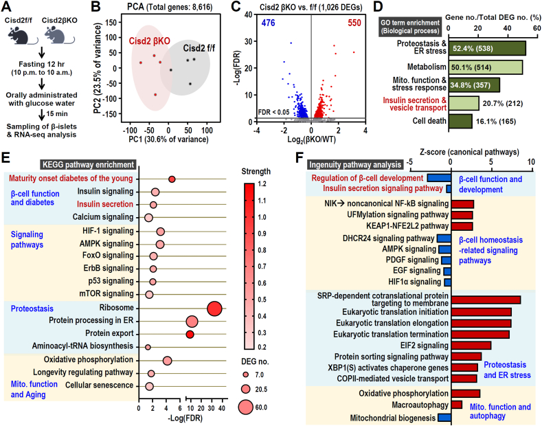
Methods: We use β-cell specific Cisd2 knockout (Cisd2KO) mice, a CRISPR-mediated Cisd2KO MIN6 β-cell line and transcriptomic analysis.

Results: Four findings are pinpointed. Firstly, β-cell specific Cisd2KO in mice disrupts systemic glucose homeostasis via impairing β-granules synthesis and insulin secretion; hypertrophy of the β-islets and the presence of a loss of identity that affects certain β-cells. Secondly, Cisd2 deficiency leads to impairment of glucose-induced extracellular Ca2+ influx, which compromises Ca2+-mediated insulin secretory signaling, causing mitochondrial dysfunction and, thereby impairing insulin secretion in the MIN6-Cisd2KO β-cells. Thirdly, transcriptomic analysis of β-islets reveals that Cisd2 modulates proteostasis and ER stress, mitochondrial function, insulin secretion and vesicle transport. Finally, the activated state of two potential upstream regulators, Glis3 and Hnf1a, is significantly suppressed under Cisd2 deficiency; notably, their downstream target genes are deeply involved in β-cell function and identity.

Conclusions: These findings provide mechanistic insights and form a basis for developing therapeutics for the effective treatment of diabetes in WFS2 patients.

Keywords: CISD2; Ca(2+) homeostasis; Diabetes; Mitochondrial function; Wolfram syndrome 2.


Conflict of interest statement

Declaration of competing interest The authors declare that they have no known competing financial interests or personal relationships that could have appeared to influence the work reported in this paper.

Figures

Figure 1
Figure 1
Cisd2 deficiency in β-cells disturbs systemic glucose homeostasis via an impairment of insulin secretion in mice. (A) Protocol used for the oral glucose tolerance tests (GTT) and insulin tolerance tests (ITT). For the oral GTT, mice were orally administrated with glucose water solution (1.5 mg/g body weight) by a feeding needle after 12 h fasting (10 pm–10 am). For the ITT, mice were intraperitoneally injected with insulin (0.75 U/kg body weight) after 2 h fasting (9 am–11 am). Blood samples were collected from tail vein before (0 min) and after the treatment at the indicated time points. (B) Basal blood glucose (fasting 2 h and 12 h) levels in the Cisd2 f/f and Cisd2βKO mice at 3-mo old (n = 4–5). (CE) The oral GTT assay for mice at 3-months (C) and 12-months (D) old. The quantification of GTT was carried out by calculating the area under curve (AUC) (n = 5–6) (E). (F–H) The ITT assay for mice at 3-months (F) and 12-months (G) old. The quantification of ITT was measured by calculating the area above curve (AAC) (n = 4–6) (H). (I) For the insulin secretion test, mice were orally administrated with glucose water solution (1.5 mg/g body weight) by a feeding needle after 12 h fasting (10 pm–10 am). The mouse blood samples were collected before (0 min) and after glucose administrated (2, 5, 15, and 30 min). Phase 1, Ph. 1; Phase 2, Ph. 2. (J) Serum insulin levels of Cisd2 βKO and Cisd2 f/f mice at 3-months old. (K) H&E staining of the pancreas from male mice at 6-months old. β-islet hypertrophy was found to be present in the pancreas of Cisd2 βKO mice. (L) Quantification of β-islet size from histological images. The β-islet size is classified into three subtypes, namely large (>12,000 μm2), medium (6000–12000 μm2) and small (<6000 μm2) and the distribution was then assessed. ∗p < 0.05 by chi-square test. (M) Immunofluorescence staining for insulin (red) and glucagon (green) present in pancreas from Cisd2f/f and Cisd2 βKO male mice at 6-months old. Data are presented as mean ± SD. ∗p < 0.05, ∗∗p < 0.005. The statistical analysis was performed using the Student's t test; not significant (n.s.). (For interpretation of the references to color in this figure legend, the reader is referred to the Web version of this article).
Figure 2
Figure 2
Cisd2 deficiency causes ER dilation and mitochondrial degeneration, as well as disturbing the maturation and secretion of β-granules. (A) Sampling schedule of the ultrastructural analysis of mouse β-islet by TEM. To analyze the glucose-induced insulin secretion, mice were orally administrated with glucose water solution (1.5 mg/g body weight) by a feeding needle after 12 h fasting (10 pm–10 am) and sacrificed before (−) or after 15 min of glucose treatment. (B) Schematic diagram of insulin biosynthesis, β-granule maturation and secretion. (CD) Cisd2 deficiency results in ultrastructural abnormalities including rough ER (RER) dilation and mitochondrial degeneration. (E) Quantification of β-granule density from the TEM images. Numbers of immature, mature and empty β-granule vesicles were quantified. (F) The number of mature β-granule vesicles within the readily releasable and reserve pools were quantified using TEM images. Data are presented as a mean ± SD. ∗p < 0.05, ∗∗p < 0.005. The statistical analysis was performed using one-way ANOVA with Bonferroni multiple comparison test; not significant (n.s.).
Figure 3
Figure 3
Cisd2 deficiency in mice (the MIN6 β-cell line) disrupts Ca2+-mediated insulin secretion, impairs mitochondrial function and disturbs intracellular Ca2+ homeostasis. (A) Schematic diagram of Ca2+-mediated insulin secretion in β-cell. Arginine treatment can stimulate membrane depolarization of β-cells. Gliclazide is a second-generation sulfonylurea that can inhibit KATP channels leading to membrane depolarization in β-cells. (B) For the Ca2+-mediated insulin secretion test, mice were intraperitoneally injected with arginine (0.3 g/kg body weight) or gliclazide (10 mg/kg body weight) after 2 h fasting (9 am–11 am). The mouse blood samples were collected before (0 min) and after arginine or gliclazide administrated (15 and 30 min). (C) Significant decreases in arginine-stimulated insulin secretion by the Cisd2 βKO mice at 3-months old. (D) Significant decreases in gliclazide-induced insulin secretion by the Cisd2 βKO mice at 3-months old. ∗p < 0.05, ∗∗p < 0.005. The statistical analysis was performed using Student's t test. (E) Decreased mitochondrial oxygen consumption rate (OCR) in MIN6-Cisd2KO cells. (F) Schematic diagram of intracellular Ca2+ regulation in β-cells. (G) Protocol for KCl-induced extracellular Ca2+ influx. (HJ) Significant increases in the basal cytosolic Ca2+ levels. Significant decreases in KCl-induced extracellular Ca2+ influx in the MIN6-Cisd2KO cells. (K) Protocol for assessing glucose-induced extracellular Ca2+ influx. (LM) Significant decreases in glucose-induced extracellular Ca2+ influx in MIN6-Cisd2KO cells. (N) Protocol for thapsigargin (TAG)-evoked store-operated calcium entry (SOCE). (OQ) Significant decreases in TAG-induced Ca2+ depletion and TAG-evoked SOCE in MIN6-Cisd2KO cells. Levels of cytosolic Ca2+ in single MIN6 β-cells were measured by fluorescence microscopy using Fura-2/AM staining. ∗p < 0.05, ∗∗p < 0.005. In C-E, data are presented as mean ± SD. In H–J, L, M, O–Q, data are presented as mean ± SEM.
Figure 4
Figure 4
Transcriptomic analysis of the β-islets reveals that Cisd2 modulates proteostasis and ER, mitochondrial function, insulin secretion and vesicle transport in order to maintain β-cell functioning. (A) Cisd2 f/f and Cisd2 βKO mice at 3-months old were orally administrated with glucose water solution (1.5 mg/g body weight) by a feeding needle after 12 h fasting (10 pm–10 am) and sacrificed after 15 min of glucose administration. Total RNAs were isolated from the β-islets of the mice. (B) Principal component analysis (PCA) of all transcriptomic data (total expressed 8,616 genes) in the β-islets of Cisd2 f/f and Cisd2 βKO mice (n = 4). (C) Volcano plot revealing transcriptome changes (Cisd2 βKO vs. f/f). Horizontal line shows the 5% false discovery rate (FDR) threshold. Red or blue plots identify genes above the indicated FDR threshold. A total of 1,026 DEGs are affected by Cisd2 deficiency (550 up-regulated and 476 down-regulated genes; Cisd2 βKO vs. f/f, FDR < 0.05). (D) The biological processes obtained from the Gene Ontology (GO) annotation of the transcriptome changes (1,026 DEGs) between β-islets of Cisd2βKO and f/f mice. The DEGs are grouped into proteostasis and ER stress, mitochondrial (Mito.) function and stress response, insulin secretion and vesicle transport and cell death associated pathways; the results are presented as a percentage of the total DEG number. (E) A bubble plot illustrating the enrichment analysis of KEGG pathway DEGs between the β-islets of Cisd2βKO and f/f mice. The grouping of the GO annotation and KEGG pathways were carried out by STRING v11.5 (https://string-db.org/). Pathway FDR < 0.05. (F) Canonical pathway analysis by Ingenuity Pathway Analysis (IPA) software using the transcriptome changes in the β-islets of Cisd2βKO mice (Cisd2βKO vs. f/f; pathway p-value < 0.05 and absolute Z-score > 0.5). (For interpretation of the references to color in this figure legend, the reader is referred to the Web version of this article).
Figure 5
Figure 5
Activation state of two potential upstream regulators, Glis3 and Hnf1a, was suppressed under Cisd2 deficiency. (AB) Significant inhibition of Glis3 transcriptional signaling based on the activation z-scores (z-score < −2.0 and p-value < 0.05) and the IPA upstream regulator analysis of the DEGs identified in the β-islets of Cisd2 βKO mice. Most of the mRNA levels of Glis3 downstream target genes are significantly downregulated under Cisd2 deficiency. The DEGs can be classified according to their functions as regulators of β cell function and identity and as regulators of insulin biosynthesis and insulin secretion signaling. (CD) Significant inhibition of Hnf1a transcriptional signaling based on activation z-score (z-score < −2.0 and p-value < 0.05) and the IPA upstream regulator analysis of the DEGs in the β-islets of Cisd2 βKO mice. Most of the mRNA levels of Hnf1a downstream target genes are significantly down-regulated under Cisd2 deficiency. The DEGs can be classified according to their functions as being involved in glucose metabolism DEGs, amino acid transport DRGs and metabolism DEGs, pancreas development (develop.) DEGs, vesicle transport DEGs, Na+ channel genes involved in insulin secretion, transcriptional regulators and more. (E) The mRNA levels of Glis3. (F) The mRNA levels of Hnf1a. (G) The mRNA levels of Mafa, Pdx1, Ins1 and Glut2. In (E)–(G). There are five independent biological replicates (n = 5) for each genotype of the cells. The mRNA levels were quantified by real-time RT-qPCR. Data are represented as mean ± SD. ∗p < 0.05, ∗∗p < 0.005. The statistical analysis was performed using one-way ANOVA with the Bonferroni multiple comparison test.
Figure 6
Figure 6
A graphic summary of the DEGs and pathways associated with the insulin secretion defect in the β-cells of Cisd2 βKO mice. (A) Cisd2 deficiency leads to suppression of Glis3 and Hnf1a, which are transcriptional regulators involved in β-cell functioning and identity. This, in turn, leads to downregulation of their target genes thereby impairing glucose uptake and the signaling associated with insulin biosynthesis and secretion. Moreover, Cisd2 deficiency causes ER stress, which then disturbs the maturation process of the β-granules. (B) Cisd2 deficiency results in downregulation of glucose transporter Glut2 expression leading to a decrease in glucose uptake and decreased ATP production. In addition, inhibition of AMPKα-Pgc-1α signaling, which is involved in mitochondrial biogenesis, is present in Cisd2KO-MIN6 β-cells and this is likely to contribute to mitochondrial dysfunction. (C) Cisd2 deficiency causes mitochondrial dysfunction and reduces ATP production thereby decreasing the ATP/ADP ratio; consequently, this disrupts KATP channel-mediated membrane depolarization and impairs Ca2+ influx in β-cells. (D) Downregulation of VGCC and various Ca2+-related signaling pathways, namely PLCγ, Camk2b and PKC, may further contribute to defects in exocytosis of insulin from β-granules. Together, all these defects, which are occurring in multiple processes and in various stages of insulin biogenesis/secretion, result in β-cell dysfunction within the Cisd2 βKO mice. Red boxes indicate upregulation of the mRNA levels; green and blue boxes indicate downregulation of the mRNA levels. (For interpretation of the references to color in this figure legend, the reader is referred to the Web version of this article).

References

    1. Fischer T.T., Ehrlich B.E. Wolfram syndrome: a monogenic model to study diabetes mellitus and neurodegeneration. Curr Opin Physiol. 2020;17:115–123. - PMC - PubMed
    1. Stone S.I., Balasubramanyam A., Posey J.E. Atypical diabetes: what have we learned and what does the future hold? Diabetes Care. 2024;47(5):770–781. - PMC - PubMed
    1. Tamaroff J., Kilberg M., Pinney S.E., McCormack S. Overview of atypical diabetes. Endocrinol Metabol Clin. 2020;49(4):695–723. - PMC - PubMed
    1. Loncke J., Vervliet T., Parys J.B., Kaasik A., Bultynck G. Uniting the divergent wolfram syndrome-linked proteins WFS1 and CISD2 as modulators. Sci Signal. 2021;14 - PubMed
    1. Tokarz V.L., MacDonald P.E., Klip A. The cell biology of systemic insulin function. J Cell Biol. 2018;217(7):2273–2289. - PMC - PubMed