. 2025 Jun 6;71(3):145-153.
doi: 10.1262/jrd.2024-077. Epub 2025 Apr 4.

CpG site methylation regulates mouse Rec8 gene promoter activity

Affiliations

CpG site methylation regulates mouse Rec8 gene promoter activity

Mei Rong et al. J Reprod Dev. .

Abstract

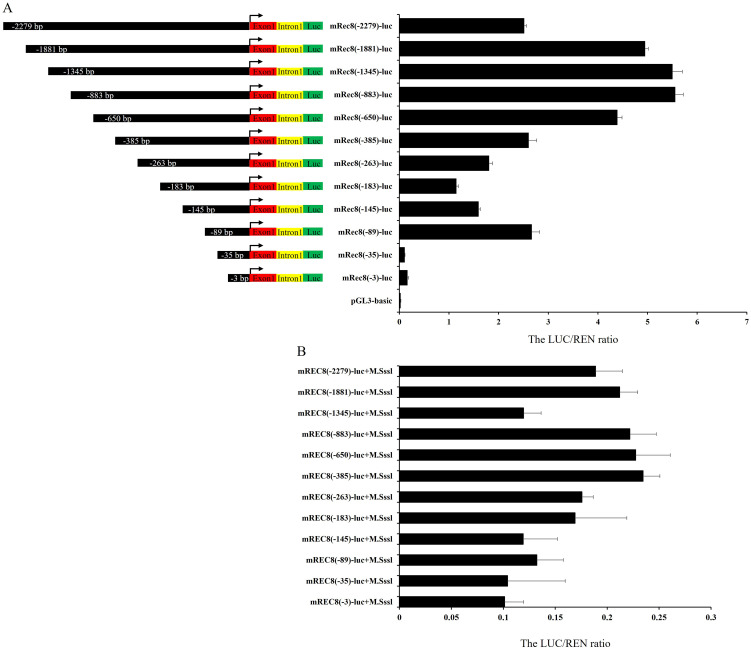
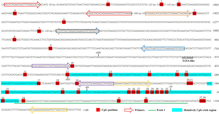
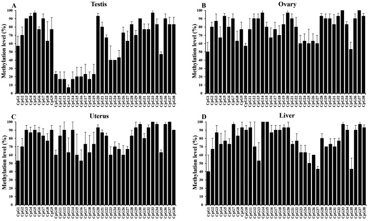
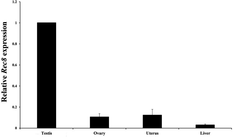
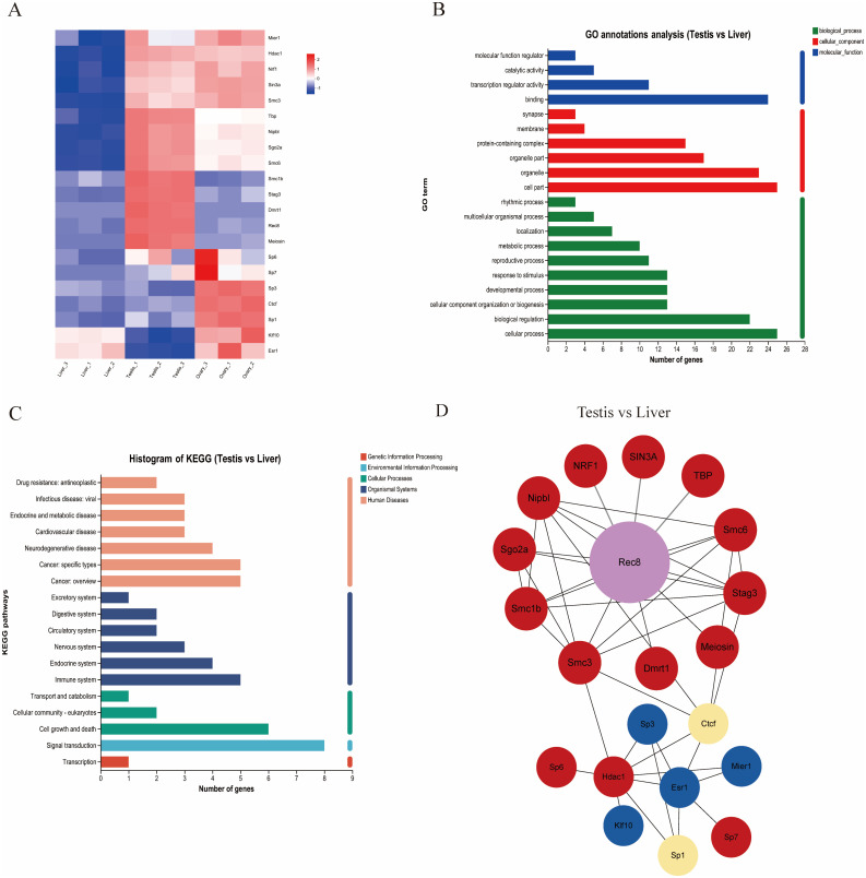
The Rec8 gene is specifically expressed in fetal and adult gonads. Although the importance of REC8 in gametogenesis is widely acknowledged, the mechanisms underlying its germ cell-specific expression remain unclear. In this study, we utilized the mouse Rec8 gene sequence to construct a 2577 bp sequence, which included intron 1 (180 bp), exon 1 (118 bp), and an upstream 2279 bp region. The dual-luciferase assay results showed significant differences in promoter activity between -650 bp and -385 bp and between -89 bp and -35 bp. This indicated that the core promoter region of the Rec8 gene may exist within these regions. Bisulfite sequencing PCR results showed that CpGs 10-19 were largely unmethylated in the testes but hypermethylated in other tissues. Interestingly, correlation analysis between CpG methylation status and Rec8 mRNA expression levels showed that methylation of CpGs 10 to 19 was negatively correlated with Rec8 mRNA expression levels (Pearson's r = -0.991, P = 0.009). Furthermore, RNA-Seq data and bioinformatic analyses suggested that the specific expression of Rec8 may be linked to the presence of TATA-like sequences within its core promoter region. Overall, these findings indicate that Rec8 expression is regulated by the low methylation of CpG sites and the presence of TATA-like sequences in its core promoter.

Keywords: Cohesin; DNA methylation; Gene expression; Promoter; Rec8.


Conflict of interest statement

The authors declare no competing interests

Figures

Fig. 1.
Fig. 1.
Analysis of Rec8 Promoter Activity. (A) Deletion analysis of the Rec8 promoter: Left panel shows a schematic representation of Rec8 promoter deletion constructs. Right panel displays the relative luciferase activities measured after the transfection of twelve Rec8 promoter deletion constructs. (B) Effect of in vitro methylation on Rec8 promoter activity: relative luciferase activity of the twelve Rec8 deletion constructs following in vitro methylation treatment. Each bar represents the mean ± SD (n = 3).
Fig. 2.
Fig. 2.
Locations of CpG dinucleotides on the mouse Rec8 target gene. The positions of the CpG motifs are marked as red boxes and are numbered sequentially from 1 to 38. Exon I is underlined in green, while the regions for bisulfite PCR primers are indicated by arrows. The aqua blue areas signify the relatively CpG-rich region, with numbers on the right showing nucleotide positions relative to the Rec8 gene transcription initiation site.
Fig. 3.
Fig. 3.
CpG methylation statuses in various mouse tissues. Bar charts depicting the percentage of methylation at each CpG position are presented. Tissues analyzed include the testis (A), ovary (B), uterus (C), and liver (D), with data obtained from three mice.
Fig. 4.
Fig. 4.
Mouse Rec8 gene expression in tissues. The data are presented as the means ± SDs and were obtained from three independent experiments in mice.
Fig. 5.
Fig. 5.
Transcriptome analysis results. (A) Heatmaps of 21 DEGs associated with Rec8 in the testes, ovaries, and liver. (B) GO analysis of the 21 DEGs enriched in the testes and liver. (C) KEGG pathway analysis of the 21 DEGs enriched in the testes and liver. (D) PPI network constructed using the DEGs identified in the testis and liver. Red represents upregulated proteins, blue represents downregulated proteins, and yellow indicates unaffected proteins.

References

    1. Ogushi S, Rattani A, Godwin J, Metson J, Schermelleh L, Nasmyth K. Loss of sister kinetochore co-orientation and peri-centromeric cohesin protection after meiosis I depends on cleavage of centromeric REC8. Dev Cell 2021; 56: 3100–3114.e4. - PMC - PubMed
    1. Sumara I, Vorlaufer E, Gieffers C, Peters BH, Peters JM. Characterization of vertebrate cohesin complexes and their regulation in prophase. J Cell Biol 2000; 151: 749–762. - PMC - PubMed
    1. Revenkova E, Eijpe M, Heyting C, Gross B, Jessberger R. Novel meiosis-specific isoform of mammalian SMC1. Mol Cell Biol 2001; 21: 6984–6998. - PMC - PubMed
    1. Eijpe M, Offenberg H, Jessberger R, Revenkova E, Heyting C. Meiotic cohesin REC8 marks the axial elements of rat synaptonemal complexes before cohesins SMC1beta and SMC3. J Cell Biol 2003; 160: 657–670. - PMC - PubMed
    1. Lee J, Iwai T, Yokota T, Yamashita M. Temporally and spatially selective loss of Rec8 protein from meiotic chromosomes during mammalian meiosis. J Cell Sci 2003; 116: 2781–2790. - PubMed