. 2025 Apr 7;13(1):71.
doi: 10.1186/s40478-025-01988-z.

Tet2 loss and enhanced ciliogenesis suppress α-synuclein pathology

Affiliations

Tet2 loss and enhanced ciliogenesis suppress α-synuclein pathology

Emmanuel Quansah et al. Acta Neuropathol Commun. .

Abstract

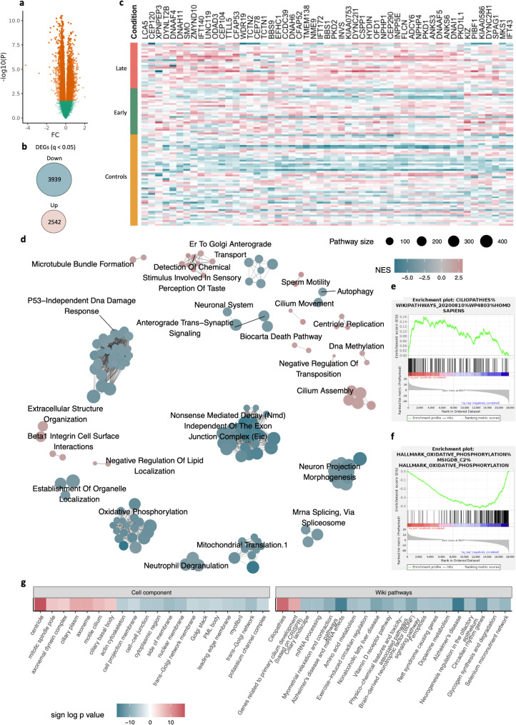
There are no approved treatments that slow Parkinson's disease (PD) progression and therefore it is important to identify novel pathogenic mechanisms that can be targeted. Loss of the epigenetic marker, Tet2 appears to have some beneficial effects in PD models, but the underlying mechanism of action is not well understood. We performed an unbiased transcriptomic analysis of cortical neurons isolated from patients with PD to identify dysregulated pathways and determine their potential contributions to the disease process. We discovered that genes associated with primary cilia, non-synaptic sensory and signaling organelles, are upregulated in both early and late stage PD patients. Enhancing ciliogenesis in primary cortical neurons via sonic hedgehog signaling suppressed the accumulation of α-synuclein pathology in vitro. Interestingly, deletion of Tet2 in mice also enhanced the expression of primary cilia and sonic hedgehog signaling genes and reduced the accumulation of α-synuclein pathology and dopamine neuron degeneration in vivo. Our findings demonstrate the crucial role of TET2 loss in regulating ciliogenesis and potentially affecting the progression of PD pathology.

Keywords: Parkinson’s disease; Primary cilia; Tet2; α-synuclein.


Conflict of interest statement

Declarations. Consent for publication: Not applicable. Competing interests: PB declares employment for F Hoffmann-La Roche and stock ownership for F Hoffmann-La Roche, Acousort AB, Axial Therapeutics, Enterin Inc and Kenai Therapeutics. Ethics approval: All experiments were done with approval from the ethics committee of the Van Andel Institute.

Figures

Fig. 1
Fig. 1
Dysregulation of primary cilia gene networks in cortical neurons from PD patients. Differentially expressed genes (DEGs) in the prefrontal cortex of PD neurons were identified in our RNAseq dataset using a robust linear regression model a Significantly altered genes in PD cortical neurons are highlighted in orange (q < 0.05, log(FC) ≥ 0.5, robust linear regression, multiple testing corrected). b Number of significantly dysregulated (up- and down-regulated) genes in all PD neurons relative to controls (q < 0.05). c Heatmap of dysregulated primary cilia-associated genes under the ‘ciliopathies’ hub. d Network and clustering of the significantly enriched pathways obtained via GSEA of the transcriptomic changes in PD neurons, with nodes representing altered pathways clustered into networks by EnrichmentMap and node colors constituting normalized enrichment score e, f Enrichment plots for ciliopathies (e) and oxidative phosphorylation (f), g Heatmap of dysregulated pathways in PD neurons as revealed by Cell component and Wiki pathways
Fig. 2
Fig. 2
Enhancing ciliogenesis via SHH signaling suppresses αSyn pathology in vitro. Altered primary cilia and αSyn pathology in cortical neurons exposed to αSyn PFF for 14 days and incubated with different doses of purmophamine (PM). a, b Representative images showing ACIII+ (a) and pS129 αSyn (b) puncta (scale bar 50 µM). Analysis of c NeuN+ cortical neuron count normalized to vehicle (DMSO) control, d Area covered by MAP2+ neurons, e Area covered by ACIII+ primary cilia normalized to NeuN+ cell count, f pS129 αSyn pathology area normalized to total MAP2 area. Data are mean ± s.e.m., for 9 independent wells from 3 separate experiments, *P < 0.05, **P < 0.01, ***P < 0.001, one way ANOVA with Tukey’s multiple comparisons test relative to respective controls
Fig. 3
Fig. 3
Tet2 inactivation reduces αSyn pathology. Representative images (scale bar 250 µM) and quantification of pS129 αSyn pathology in the frontal cortex, striatum, and substantia nigra at a, b 3 months (n = 5–7 mice per PFF group, n = 15 mice per PBS group) and c, d 6 months (n = 11–12 mice per PFF group, n = 13–14 mice per PBS group) after αSyn PFF injection. The percentage area with pS129 αSyn immunoreactive staining was quantified using the Aiforia platform. Data are mean ± s.e.m., analyzed using two-way ANOVA with Tukey’s multiple comparisons test relative to the respective controls, *P < 0.05, **P < 0.01, ***P < 0.001, ****P < 0.0001
Fig. 4
Fig. 4
Tet2 deletion enhances the expression of genes linked with ciliogenesis and SHH signaling. Six months after striatal αSyn PFF inoculation, we examined the cortical expression of multiple genes using qPCR, a mRNA expression of Tet1, bTet2, cTet3, dTalpid3, e, Gli1, fGli2, gGli3, and hPtch1 (n = 3–5 mice per group). Data are mean ± s.e.m., analyzed using two-sided t-test, *P < 0.05, **P < 0.01, ***P < 0.001, ****P < 0.0001
Fig. 5
Fig. 5
Tet2 inactivation protects against αSyn-induced dopamine neuron loss. a Representative image (scale bar 500 µM) and b quantification of TH-positive neurons in the substantia nigra 6 months after αSyn PFF injection (n = 11–13 mice per PFF group, n = 9 mice per PBS group). TH immunoreactive staining was quantified using the Aiforia platform. c, d Behavioral assessment i.e., wire hang test (c) and rotarod (d) were performed 6 months after αSyn PFF injection (n = 15–16 mice per PFF group, n = 17–20 mice per PBS group). Data are mean ± s.e.m., analyzed using two-way ANOVA with Tukey’s multiple comparisons test relative to the respective controls, **P < 0.01

References

    1. Rey NL, Steiner JA, Maroof N, Luk KC, Madaj Z, Trojanowski JQ et al (2016) Widespread transneuronal propagation of α-synucleinopathy triggered in olfactory bulb mimics prodromal Parkinson’s disease. J Exp Med 213:1759–1778 - PMC - PubMed
    1. Henderson MX, Cornblath EJ, Darwich A, Zhang B, Brown H, Gathagan RJ et al (2019) Spread of α-synuclein pathology through the brain connectome is modulated by selective vulnerability and predicted by network analysis. Nat Neurosci 22:1248–1257 - PMC - PubMed
    1. Braak H, Del Tredici K, Rüb U, de Vos RAI, Jansen Steur ENH, Braak E (2003) Staging of brain pathology related to sporadic Parkinson’s disease. Neurobiol Aging 24:197–211 - PubMed
    1. Steiner JA, Quansah E, Brundin P (2018) The concept of alpha-synuclein as a prion-like protein: ten years after. Cell Tissue Res 373:161–173 - PMC - PubMed
    1. Mastenbroek SE, Vogel JW, Collij LE, Serrano GE, Tremblay C, Young AL et al (2024) Disease progression modelling reveals heterogeneity in trajectories of Lewy-type α-synuclein pathology. Nat Commun 15:5133 - PMC - PubMed

Publication types

MeSH terms

LinkOut - more resources