. 2025 Sep;30(9):4274-4285.
doi: 10.1038/s41380-025-03008-x. Epub 2025 Apr 6.

Deconstructing a common pathway concept for Deep Brain Stimulation in the case of Obsessive-Compulsive Disorder

Affiliations

Deconstructing a common pathway concept for Deep Brain Stimulation in the case of Obsessive-Compulsive Disorder

Volker A Coenen et al. Mol Psychiatry. 2025 Sep.

Abstract

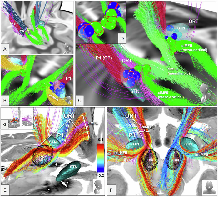
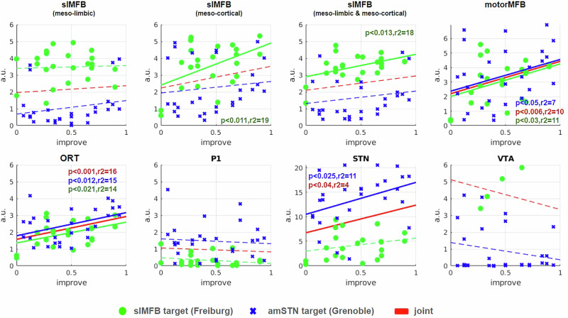
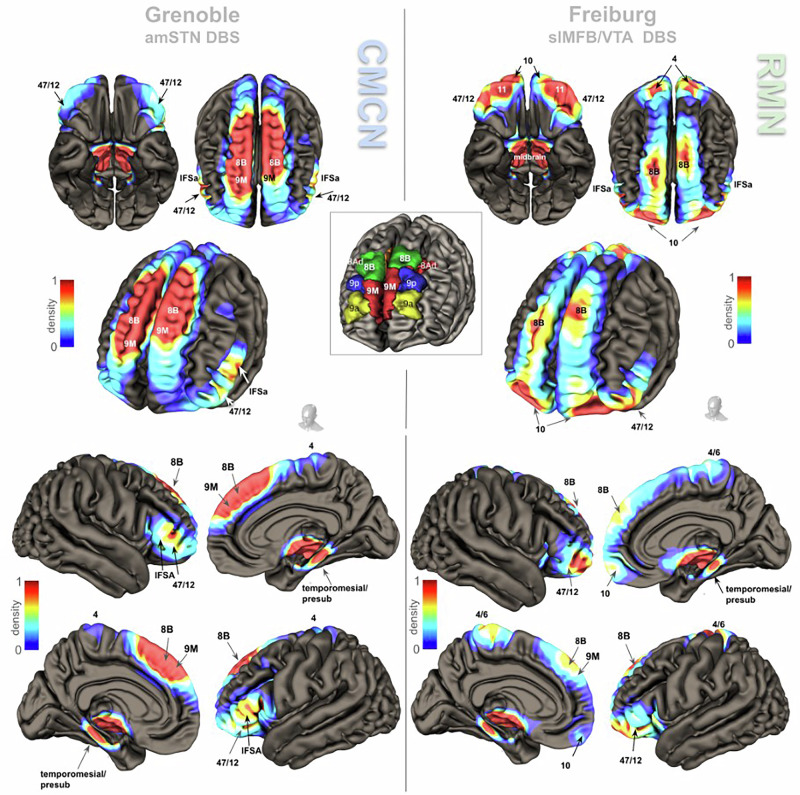
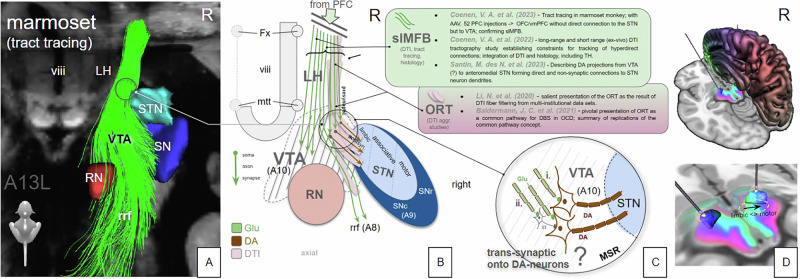
Deep Brain Stimulation (DBS) is a therapeutic option for treatment resistant (TR) obsessive-compulsive disorder (OCD). The OCD network comprises different sub-networks with homeostatic functions, altered under disease and modifiable with DBS. Connectomic analyses of DBS data sets have defined fiber selections explaining anti-OCD efficacy. This is a retrospective stimulation and outcome derived anatomical overlay analysis of 26 TR-OCD patients who received DBS at two academic centers. Grenoble, 14 anteromedial subthalamic nucleus (amSTN); Freiburg, 12 superolateral medial forebrain bundle (slMFB). Yale-Brown Obsessive Compulsive Scale improvement at 24 months served as outcome parameter. Structural proximity and outcomes were correlated using individual volumes of activated tissue for STN, slMFB, ORT (average OCD response tract) and further structures based on atlases or established connectomes. Connectomes (slMFB, ORT) were inspected for structural congruences. Normative connectomic data served to investigate cortical fiber penetration for the two target regions. Cortical sub-network conjugations were evaluated as peak levels. Our analyses revealed that ORT represents a fiber selection from the slMFB. DBS of amSTN and slMFB each address distinctive sub-networks while deep amSTN DBS can also address slMFB. Sub-network conjugations project amongst other regions onto the dorsomedial prefrontal cortex (dmPFC). The average ORT fiber selection is an integral part of the generic slMFB. Anti-OCD effects of amSTN DBS are not entirely explained by ORT overlay. The slMFB is dispersed and encompasses all OCD sub-networks and might qualify as a common DBS target when stimulated close to the ventral tegmental area. The dmPFC emerges as an interesting conjugation/hub between OCD sub-networks.


Conflict of interest statement

Competing interests: VAC and TES as employees of University of Freiburg, listed by the institution as inventors, have filed a U.S. provisional patent application generally related to highly focused DBS in the treatment of OCD (U.S. Patent Application Number 63/253740). Unrelated: VAC receives a collaborative grant from BrainLab (Munich, Germany). He is a consultant for Ceregate (Munich, Germany), Cortec (Freiburg, Germany) and Inbrain (Barcelona, Spain). He has an ongoing IIT with Boston Scientific (USA) and has received personal honoraria and travel support for lecture work from Boston Scientific (USA), ALEVA, UNEEG and PRECISIS. Unrelated: MP has received financial support for investigator-initiated trials from Boston Scientific and honoraria for lecturing or consultation from Lundbeck. Unrelated: SC has received honoraria for consultation from Medtronic, Boston Scientific. Unrelated: PCR has received research support from Else Kröner-Fresenius Foundation, Fraunhofer Foundation (ATTRACT), German Ministry for Economic Affairs and Energy, and Medical Faculty of the University of Freiburg. He has received personal honoraria for lectures or advice from Fraunhofer Foundation and is a consultant for Boston Scientific, Brainlab, and Inomed. DMD receives research support from Evangelisches Studienwerk e.V. and Wissenschaftliche Gesellschaft Freiburg im Breisgau. JCB is funded by the Else-Kröner-Fresenius-Stiftung (2022_EKES.23). BEAS received a research grant from Ceregate (Munich, Germany) and received honoraria as a consultant for Precisis, Heidelberg, both unrelated to this work. All other authors declare no conflicts of interest. Ethics approval and consent to participate: a) All methods were performed in accordance with relevant guidelines and regulations. b) Freiburg: Patients with TR-OCD who previously had received bilateral slMFB-DBS were selected for analysis, if they gave informed consent to our DBS registry that adheres to the principles of the Helsinki Declaration and received approval from institutional review board (Ethics Committee of Freiburg University; no.21-1274). Grenoble: The publication of results had previously been discussed [17] and waived on the basis of a national committee decision: Comité consultatif National d’ethique. La neurochirurgie fonctionnelle D affectionspsychiatriques severes 2002. c) Freiburg: Patients gave written informed consent. Grenoble: Written consent was obtained for surgery. For publication, based on national guidelines no written consent was necessary (see b).

Figures

Fig. 1
Fig. 1. Two OCD-DBS connectomes.
The ORT (A, purple) [23] and the slMFB (B,C, green) [, –50, 70] bear a clear amount of resemblance/overlap. A The group of yellow spheres indicate actual effective slMFB DBS VATs the Freiburg cohort [18]. Note how these spheres project in proximity to but not exactly onto the ORT [23] while perfectly hitting slMFB. B Superimposition of slMFB connectome (short-range) and C, long-range. slMFB connectome taken from [32]. Inset in C shows the distinction between ORT/slMFB as such that slMFB reaches further dmPFC regions. DF Details from upper corresponding panel but enriched with E, slMFB and F, motor MFB [39] as a third connectome. G, H View from posterior especially focussing on the connectome relations to STN, VTA and anterior limb of the internal capsule (ICa). Inset in lower panel taken from [21] showing the inferior and lateral position of the reward network in ICa. ORT and slMFB leave ICa at its deepest portion (superior to anterior commissure, ac) rather laterally. G Left ORT connectome touches STN superficially while on the right side fibers appear to enter the STN from out of the hypothalamus. Such fiber route is unphysiological according to [32, 33, 81] and presumably the result of an anatomically unsupervised fiber selection. H slMFB follows the fiber corridor and deconstructs into a medial (mesolimbic) and lateral (mesocortical) portion.
Fig. 2
Fig. 2. Volumes of activated tissue positions in the distinct target regions (amSTN VATs, blue spheres; slMFB VATs green spheres).
ORT purple, slMFB green. For both target regions, VATs present as disjunct groups of spheres. A View from posterior left. DBS electrode lead ensemble for Freiburg cohort only; B View from posterior right, including motorMFBs (yellow); C View from posterior left. VAT sizes (in A-C) are not individual but set to an arbitrary 2 mm diameter. DActual individual sizes of VTAs (halved for improved visualization) depending on stimulation settings. Legend: STN, subthalamic nucleus; RN, red nucleus; P1, fronto-pontine tract (red). EG Streamlines (slMFB) from the midbrain atlas proposed in [32] with the big-brain [44] as background. E View from lateral right, left streamlines shown only. F View from superior and back. G Inset showing overview. Subthalamic nuclei (STN) and red nuclei (RN) were taken from [40]. The streamlines are colored by their correlation values between VAT-overlap and response in both cohorts. The ORT (purple) is only displayed for orientation reasons and projects within most correlating “activated” streamlines of the slMFB.
Fig. 3
Fig. 3. Correlation of stimulated electrode contact proximity to respective anatomical structures and anti-OCD efficacy (details in methods).
Green regression line, MFB only; blue regression line amSTN only. Red regression line indicates joint analysis. Grenoble (blue crosses): Strong correlation for ORT AND for STN. Freiburg (green dots): Strong correlations for mesocortical (cort) slMFB, limbic & mesocortical(limb+cort) slMFB and motor MFB. Joint: Statistically significant findings for MFB (motor) and ORT. Joint correlation for mtrMFB appears almost equally strong as ORT (p < 0.006 vs. p < 0.001). Remember that Grenoble cohort was used to define ORT, introducing a certain bias for this analysis. Dots and crosses represent left and right hemispheres as such that each patient is represented by two dots or crosses, respectively. Legend: a.u., arbitrary unit (higher number indicates higher structure density moving toward center); slMFB, superolateral medial forebrain bundle; cort, mesocortical part; limb, mesolimbic part; mtrMFB, motor MFB; ORT OCD, unified connectomic tract for OCD; P1 cortico-pontine tract (Arnold’s Bundle), STN subthalamic nucleus, VTA ventral tegmental area.
Fig. 4
Fig. 4. Cortical fingerprints of factual amSTN (left) & slMFB DBS (right) in TR-OCD.
Upper panel gives views of both hemispheres, the lower panel shows individual hemispheres from medial and lateral. Group level connectomic analysis of cortical fiber penetration patterns. Inset identification of some selected prefrontal Brodman regions taken from [69]. In general, the patterns of cortical involvement address distinct but in part overlapping OCD sub-networks (amSTN target: cognitive / motor control sub-network; slMFB target: reward sub-network). Overlap/conjugation of cortical penetrations occur at BA8Bl (superior frontal gyrus), BA4/6, BA11 (lateral), BA47/12; and temporal mesial. See Table 1 and discussion for details.
Fig. 5
Fig. 5. Topographical relationship of OFC/vmPFC projection fiber terminals in the diencephalic-mesencephalic junction (summary).
The ORT (pink) presents itself as a DTI fiber selection belonging to the lateral slMFB. ORT additionally includes secondary neuronal connections to STN (and SN). A Three-dimensional reconstruction of a tract tracing from a single injection in a marmoset monkey (A13, orbitofrontal, example of OFC/vmPFC injections from [33, 81]. Limbic STN-direct (hyperdirect) connections were not replicated in this study. B, C Cartoonistic summary and integration into human anatomy. B Unconstrained DTI seeding approaches select lateral fibers of the slMFB which seem to enter the STN/SN region. After termination of descending glutamatergic fibers in the VTA, these fibers switch to neurons (interneurons, DA neurons) which in turn might connect to the STN (C). The fiber connection to the STN is therefore not a direct one but only picked up as such by DTI tractography which is agnostic for synapses or directionality (efferent or afferent) of the projection. D Demonstration of the medial/lateral limbic/motor gradient (arrow) with the SPECTRE [82] technology. Electrodes are located in the sensorimotor STN. Green coloring denotes more prefrontal (limbic) connectivity. Blue coloring denotes motor connectivity. Legend: viii, third ventricle; LH lateral hypothalamus, STN subthalamic nucleus, SN substantia nigra, SNc SN pars compacta, SNr SN pars reticulata, VTA ventral tegmental area, RN red nucleus, rrf retro-rubral field, mtt mammillothalamic tract, Fx fornix, MSR medial subthalamic region, DA Dopamine, Glu Glutamate, slMFB superolateral branch of the medial forebrain bundle, ORT unified connectomic pathway, DTI diffusion-tensor magnetic resonance imaging.

References

    1. Hirschtritt ME, Bloch MH, Mathews CA. Obsessive-compulsive disorder: advances in diagnosis and treatment. JAMA. 2017;317:1358–67. - PubMed
    1. Atmaca M. Treatment-refractory obsessive compulsive disorder. Prog Neuro-Psychopharmacol Biol Psychiatry. 2016;70:127–33. - PubMed
    1. Wu H, Hariz M, Visser-Vandewalle V, Zrinzo L, Coenen VA, Sheth SA, et al. Deep Brain Stimulation for refractory obsessive-compulsive disorder (OCD): emerging or established therapy? Mol Psychiatr. 2021;26:60–65. - PMC - PubMed
    1. Luyck K, Bervoets C, Deblieck C, Nuttin B, Luyten L Deep Brain Stimulation in the bed nucleus of the stria terminalis: a symptom provocation study in patients with obsessive-compulsive disorder. Medrxiv:2021.03.12.21253450 [Preprint]. 2021. - PubMed
    1. Greenberg BD, Gabriels LA, Malone DA, Rezai AR, Friehs GM, Okun MS, et al. Deep Brain Stimulation of the ventral internal capsule&ventral striatum for obsessive-compulsive disorder: worldwide experience. Mol Psychiatry. 2010;15:64–79. - PMC - PubMed

LinkOut - more resources