

The Kappa Free Light Chains Index and Central Vein Sign: Two New Biomarkers for Multiple Sclerosis Diagnosis
- PMID: 40189723
- PMCID: PMC12089642
- DOI: 10.1007/s40120-025-00737-7
The Kappa Free Light Chains Index and Central Vein Sign: Two New Biomarkers for Multiple Sclerosis Diagnosis
Abstract
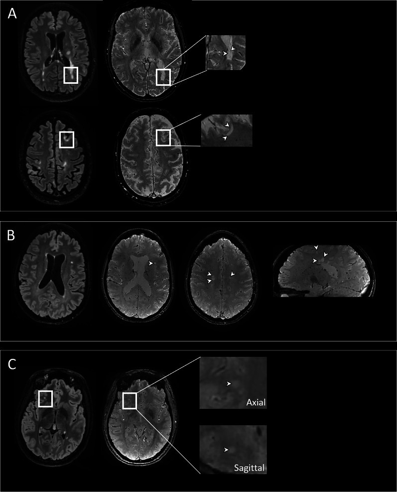
At the last European Committee for Treatment and Research in Multiple Sclerosis (ECTRIMS) meeting, Montalban and the experts' panel announced the 2024 revision of the McDonald criteria. The optic nerve has been added as a fifth dissemination in space (DIS) criteria topography among the main changes. Multiple sclerosis (MS) can be diagnosed in patients with radiologically isolated syndrome (RIS) if specific biomarkers are added to at least two of the five DIS criteria. Among the particular MS biomarkers, the kappa free light chain index (kFLC index) positivity will be added to oligoclonal band (OCB) detection to fulfill intrathecal immunoglobulin synthesis, and the central vein sign (CVS) will be able to ensure the specificity of the lesions detected on MRI scans. This review summarises the knowledge on the kFLC index and the CVS in people with MS and RIS, allowing them to be incorporated into the proposed revision of the McDonald criteria.
Keywords: Central vein sign; Kappa free light chains index; Multiple sclerosis; Radiologically isolated syndrome.
© 2025. The Author(s).
Conflict of interest statement
Declarations. Conflict of Interest: Michael Levraut, Cassandre Landes-Chateau, Mikael Cohen, and Lydiane Mondot have nothing to disclose related to this study. Christine Lebrun-Frenay was part of the expert panel that reviewed and proposed the 2024 update of the McDonald criteria. Ethical Approval: This article is based on previously conducted studies and does not contain any new studies with human participants or animals performed by any of the authors. Patient Consent: Patients' consent was obtained for their MR images to be published anonymously.
Figures
Similar articles
-
Kappa free light chain index as a diagnostic biomarker in multiple sclerosis: A real-world investigation.J Neurochem. 2021 Nov;159(3):618-628. doi: 10.1111/jnc.15500. Epub 2021 Sep 15. J Neurochem. 2021. PMID: 34478561
-
The Persisting Significance of Oligoclonal Bands in the Dawning Era of Kappa Free Light Chains for the Diagnosis of Multiple Sclerosis.Int J Mol Sci. 2018 Nov 29;19(12):3796. doi: 10.3390/ijms19123796. Int J Mol Sci. 2018. PMID: 30501024 Free PMC article.
-
The kappa free light chain index and oligoclonal bands have a similar role in the McDonald criteria.Brain. 2022 Nov 21;145(11):3931-3942. doi: 10.1093/brain/awac220. Brain. 2022. PMID: 35727945
-
Biological Markers in Early Multiple Sclerosis: the Paved Way for Radiologically Isolated Syndrome.Front Immunol. 2022 Apr 27;13:866092. doi: 10.3389/fimmu.2022.866092. eCollection 2022. Front Immunol. 2022. PMID: 35572543 Free PMC article. Review.
-
Oligoclonal bands and kappa free light chains: Competing parameters or complementary biomarkers?Autoimmun Rev. 2025 Apr 30;24(5):103765. doi: 10.1016/j.autrev.2025.103765. Epub 2025 Feb 11. Autoimmun Rev. 2025. PMID: 39947571 Review.
Cited by
-
Kappa Free Light Chain Index Correlates With Prognostic Biomarkers in Multiple Sclerosis and Decreases Slowly Following Treatment.Eur J Neurol. 2025 Jul;32(7):e70291. doi: 10.1111/ene.70291. Eur J Neurol. 2025. PMID: 40643211 Free PMC article.
-
Multiple sclerosis: 2024 update.Free Neuropathol. 2025 Jul 8;6:14. doi: 10.17879/freeneuropathology-2025-6762. eCollection 2025. Free Neuropathol. 2025. PMID: 40636815 Free PMC article. Review.
References
-
- Thompson AJ, Banwell BL, Barkhof F, Carroll WM, Coetzee T, Comi G, et al. Diagnosis of multiple sclerosis: 2017 revisions of the McDonald criteria. Lancet Neurol. 2018;17(2):162–73. - PubMed
-
- Filippi M, Preziosa P, Meani A, Dalla Costa G, Mesaros S, Drulovic J, et al. Performance of the 2017 and 2010 revised McDonald criteria in predicting MS diagnosis after a clinically isolated syndrome: a MAGNIMS study. Neurology [Internet]. 2022. 10.1212/WNL.0000000000013016. - PubMed
Publication types
LinkOut - more resources
Full Text Sources
