. 2020 Nov 13;21(1):298-312.
doi: 10.5114/aoms.2020.100833. eCollection 2025.

Adipose tissue-derived mesenchymal stem cells have better restorative capacity than bone marrow-derived cells in a cerebellar ataxic rat model

Affiliations

Adipose tissue-derived mesenchymal stem cells have better restorative capacity than bone marrow-derived cells in a cerebellar ataxic rat model

Rasha M Atta et al. Arch Med Sci. .

Abstract

Introduction: Regenerative treatment using stem cells represents a potentially effective therapy for cerebellar ataxia (CA). We compared the therapeutic potential of adipose tissue stem cells (ASCs) and bone marrow mesenchymal stem cells (BM-MSCs) in a rodent monosodium glutamate (MSG)-induced CA cell (BM-MSC) model.

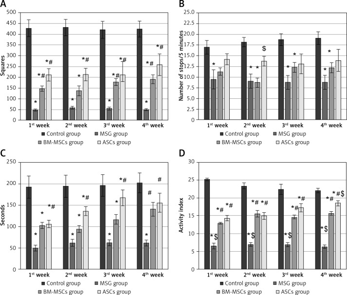
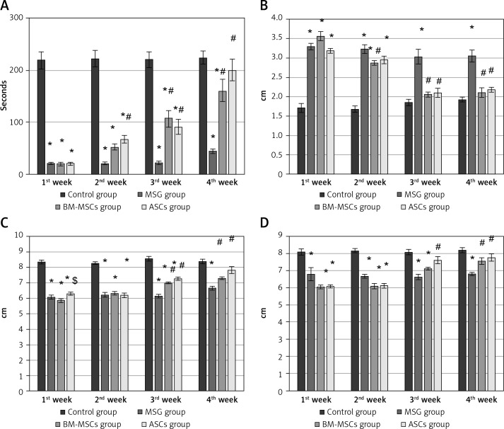
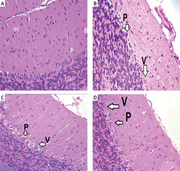
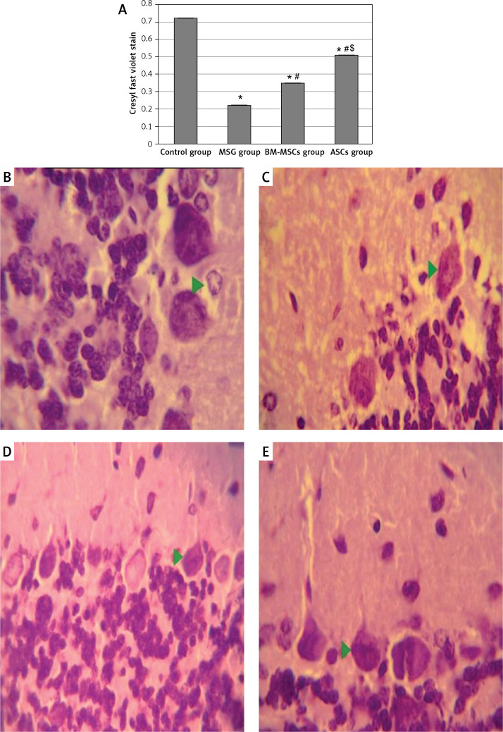
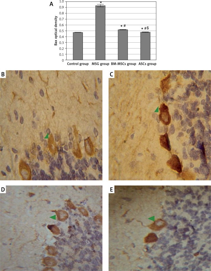
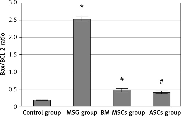
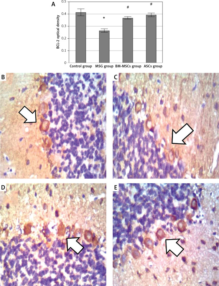
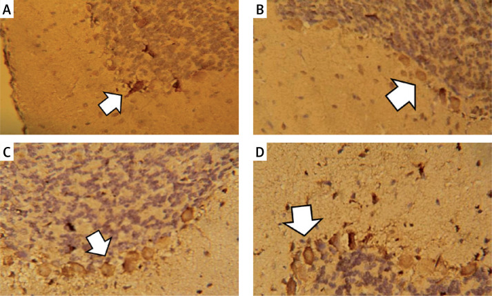
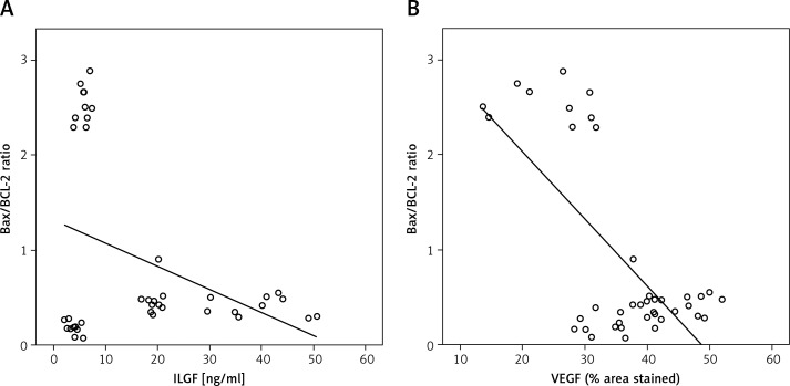
Material and methods: Female Wistar rats (n = 40) were equally divided into a saline-treated control group and 3 MSG-induced CA groups randomly treated with either saline, or 1 × 10⁶ ASCs or BM-MSCs. We assessed the following: 1) cerebellar motor functions in vivo (by Rotarod test, open-field test, and Quantitative gait analysis); 2) cerebellar histological architecture; and 3) cerebellar immunohistochemical examination of the Bax/Bcl-2 ratio as in indicator of apoptosis, and the levels of vascular endothelial growth factor (VEGF) and insulin-like growth factor-1 (IGF-1) as neuroprotective factors.

Results: Treatment with either of the MSCs improved MSG-induced poor motor performance, restored the disrupted Purkinje cell layer, decreased neuronal apoptosis and enhanced cerebellar VEGF and IGF-1 levels observed in CA rats. Adipose tissue stem cells showed superiority over BM-MSCs in the improvement of some motor performance parameters and cerebellar VEGF and IGF-1 levels.

Conclusions: In conclusion, both stem cell types induced structural, physiological, and biochemical improvement, with ASCs being best for treatment of CA.

Keywords: Purkinje cells; adipose tissue stem cells; bone marrow mesenchymal stem cells; cerebellar ataxia; monosodium glutamate.


Conflict of interest statement

The authors declare no conflict of interest.

Figures

Figure 1
Figure 1
The motor performance of rats in open field tests. Rats were observed for number of squares crossed (A), number of stops (B), mobility duration (C), and locomotor activity (D) in a 5-minute session. The monosodium glutamate (MSG) group showed a decrease motor performance compared to the control group started in the first week. Either type of stem cells improved all these parameters except the number of stops, as compared to the MSG group. Mean ± SEM were analysed using one-way analysis of variance and Tukey post-hoc test Data were expressed as mean ± SEM, *significant p-value in comparison to control group, #significant p-value in comparison to MSG group and $significant p-value in comparison to bone marrow mesenchymal stem cells group.
Figure 2
Figure 2
The motor performance in rotarod test and quantitative gait analysis. Rats were observed for time spent on a rotating rode (A), stance width (B), fore-limb stride length (FSL) (C), and hind-limb stride length (D). The monosodium glutamate group showed deterioration compared to the control group started at the first week. Adipose tissue stem cells group showed significant improvement in time spent in rotarod test started at the second week and all gait parameters started at the third week. Bone marrow mesenchymal stem cells group showed significant increase in time spent in rotarod test and FSL started at the third week, stance width in the second week, and hind limb stride length started at the fourth week. Mean ± SEM were analysed using one-way analysis of variance and Tukey post-hoc test Data were expressed as mean ± SEM, *significant p-value in comparison to control group, #significant p-value in comparison to monosodium glutamate group and $significant p-value in comparison to bone marrow mesenchymal stem cells group.
Figure 3
Figure 3
Haematoxylin and eosin (H&E) stained cerebellar sections of study groups A–D. A – Control group. B – Monosodium glutamate group. C – Bone marrow mesenchymal stem cells group. D – Adipose tissue stem cells group (H&E 400×). Control group showed normal appearance with scattered small stellate (SC) and basket cells (B) in the molecular layer. Crowded small cells with deeply stained nuclei (G) in the granular cell layer and large flask shaped cells, uniformly arranged Purkinje cell layer. Monosodium glutamate group showed widely displaced, distorted, and shrunken Purkinje cells (P), leaving vacuoles (V) in intercellular spaces. Both stem cell groups showed restoration of the normal architecture of the cortex with decreased number of distorted Purkinje cells
Figure 4
Figure 4
Optical density of Cresyl fast violet stain in study groups (A) photomicrographs of sections in the cerebellar cortex of rats (B–E). Control group showed purple Nissl granules (Δ) in the perikarya of Purkinje cells (B). The monosodium glutamate (MSG) group showed decreased purple Nissl granules (C). Bone marrow mesenchymal stem cell (BM-MSCs) group (D) and adipose tissue stem cell group (E) showed increased purple Nissl’s granules. Mean ± SEM were analysed using one-way analysis of variance and Tukey post-hoc test *Significant p-value compared to control group, #significant p-value compared to MSG group and $significant p-value compared to BM-MSC group.
Figure 5
Figure 5
Quantitative analysis of Bax optical density in the study groups (A). Bax immunostaining in the cerebellum of study groups (B–E). Control rat (B) showed positive brownish immunoreaction of Bax stain in the perikarya of Purkinje cells. The monosodium glutamate (MSG) group (C) showed an increase in positive brownish immunoreaction in the perikarya of Purkinje cells. Bone marrow mesenchymal stem cell (BM-MSC) group (D) and adipose tissue stem cell group (E) showed a decrease in positive brownish immunoreaction. Results are mean ± SEM and were analysed using one-way analysis of variance and Tukey post-hoc test *Significant p-value in comparison to control group, #significant p-value in comparison to MSG group and $significant p-value in comparison to BM-MSCs group.
Figure 6
Figure 6
Bax/Bcl-2 ratio in study groups. The monosodium glutamate (MSG) group was significantly increased compared to control group. Both stem cell groups showed significantly low Bax/Bcl-2 ratio when compared to the MSG group. Results are mean ± SEM and were analysed using one-way analysis of variance and Tukey post-hoc test Data were expressed as mean ± SEM, *significant p-value in comparison to control group, #significant p-value in comparison to MSG group.
Figure 7
Figure 7
Quantitative analysis of BCL-2 optical density. A – BCL-2 immunostaining in the cerebellum (B–E). Control rat (B) showed positive brownish immunoreaction of BCL-2 stain in the perikarya of Purkinje cells. The monosodium glutamate (MSG) group (C) showed decrease in positive brownish immunoreaction in the perikarya of Purkinje cells. Bone marrow mesenchymal stem cells group (D) and adipose tissue stem cells group (E) showed increase in positive brownish immunoreaction. Mean ± SEM was analysed using one-way analysis of variance and Tukey post-hoc test *Significant p-value compared to control group, #significant p-value compared to MSG group.
Figure 8
Figure 8
Vascular endothelial growth factor immunostaining in the cerebellar cortex. A – Control group. B – Monosodium glutamate group. C – Bone marrow mesenchymal stem cell group. D – Adipose tissue stem cell group
Figure 9
Figure 9
Linear regression between cerebellar IGF-I and Bax/Bcl-2 ratio in Purkinje cells (A) r2 = –0.374, p = 0.017* and between cerebellar vascular endothelial growth factor and Bax/Bcl-2 ratio (B) r2 = –0.683, p < 0.0018*

Similar articles

References

    1. Farghaly WM, El-Tallawy HN, Shehata GA, Rageh TA, Hakeem NA, Abo-Elfetoh NM. Population-based study of acquired cerebellar ataxia in Al-Kharga district, New Valley, Egypt. Neuropsychiatr Dis Treat 2011; 7: 183–7. - PMC - PubMed
    1. Braga-Neto P, Pedroso J, Kuo S, et al. . Current concepts in the treatment of hereditary ataxias. Arq Neuropsiquiatr 2016; 74: 244–52. - PMC - PubMed
    1. Reimers D, Osuna C, Gonzalo-Gobernado R, et al. . Liver growth factor promotes the survival of grafted neural stem cells in a rat model of Parkinson’s disease. Curr Stem Cell Res Ther 2012; 7: 15–25. - PubMed
    1. Sullivan MO, Gordon-Evans WJ, Fredericks LP, et al. . Comparison of mesenchymal stem cell surface markers from bone marrow aspirates and adipose stromal vascular fraction sites. Front Vet Sci 2015; 2: 82. - PMC - PubMed
    1. Pittenger MF, Mackay AM, Beck SC, et al. . Multilineage potential of adult human mesenchymal stem cells. Science 1999; 284: 143–7. - PubMed