. 2025 Apr 1:20:3977-3997.
doi: 10.2147/IJN.S512509. eCollection 2025.

Adjuvanticity of Tannic Acid-Modified Nanoparticles Improves Effectiveness of the Antiviral Response

Affiliations

Adjuvanticity of Tannic Acid-Modified Nanoparticles Improves Effectiveness of the Antiviral Response

Martyna Janicka et al. Int J Nanomedicine. .

Abstract

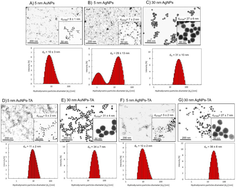
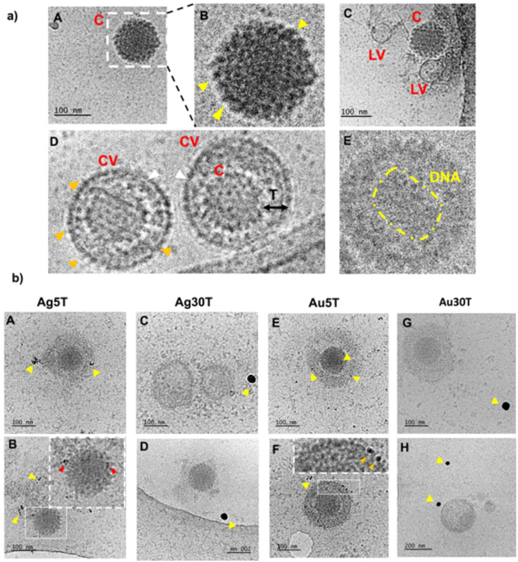
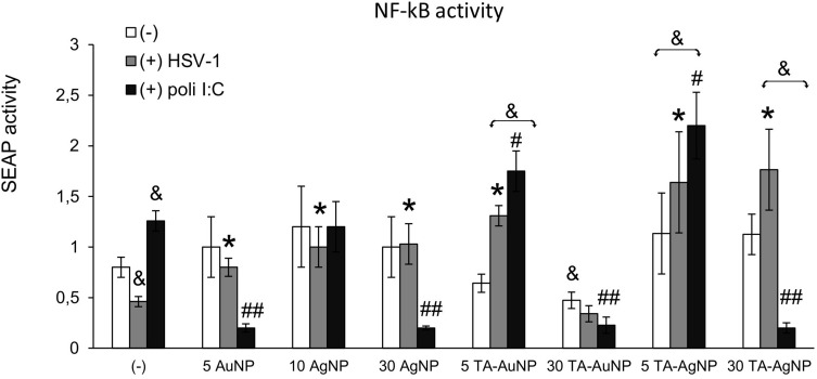
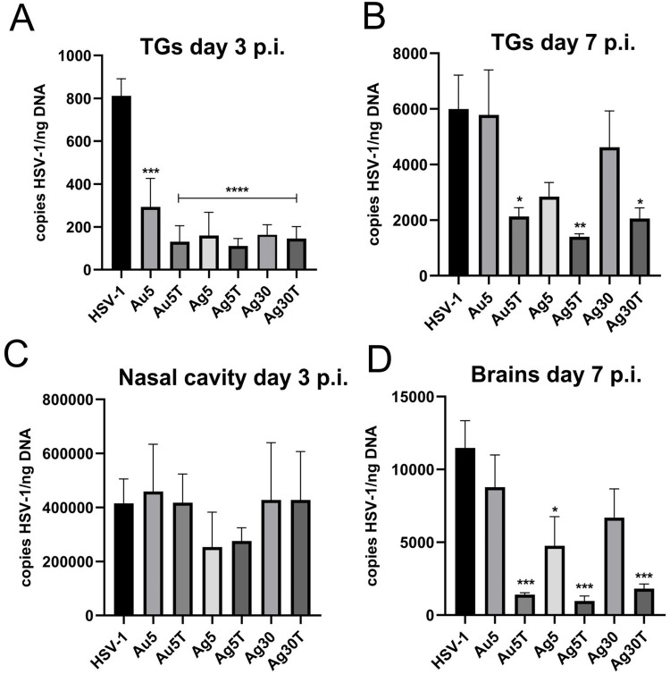
Introduction: Herpes simplex virus type 1 (HSV-1) causes recurrent infections of skin and mucosal tissues with high global prevalence. HSV-1 also invades the nervous system where it establishes a lifelong latency-making infection poorly treatable We previously showed that both tannic acid-modified silver and gold nanoparticles (TA-Ag/AuNPs) inhibit HSV-1 infection in vitro.

Methods: We used an in vitro and in vivo model of HSV-1 infection to study how metal type, size and tannic acid modification of nanoparticles can influence development of the early innate response and the mounting of specific anti-HSV-1 response upon treatment of the nasal mucosa.

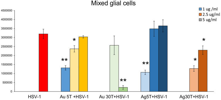
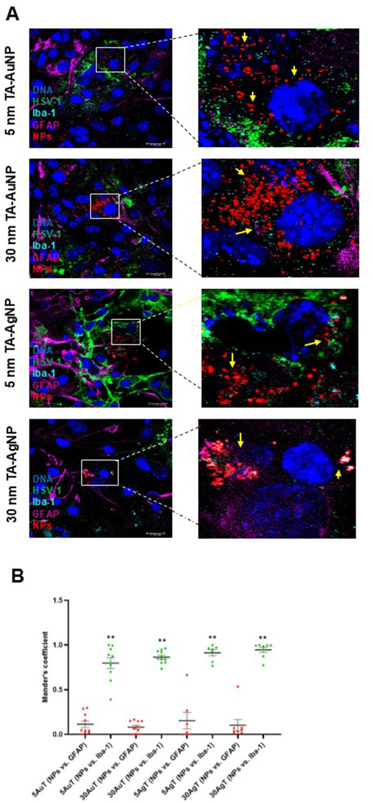
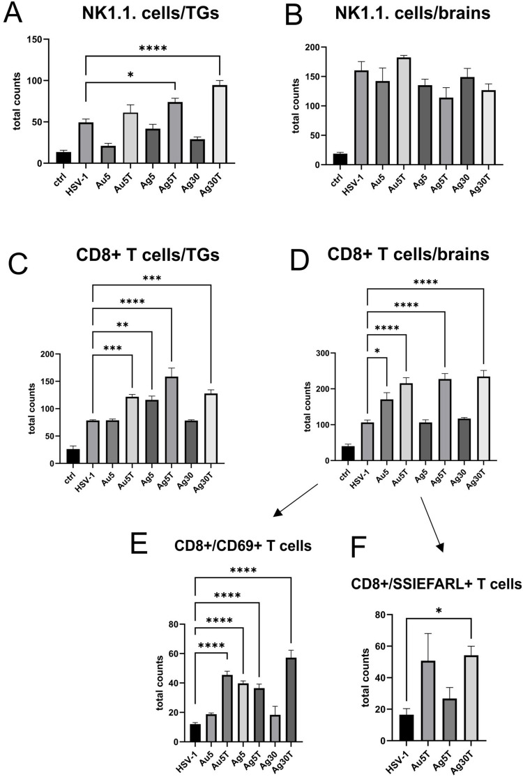
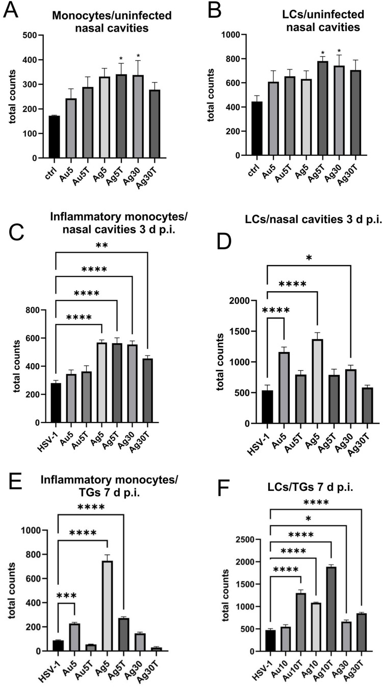
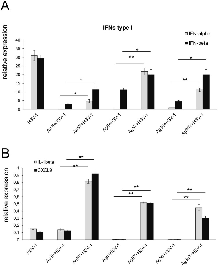
Results: We found that tannic acid is necessary for binding with HSV-1, with smaller sizes independent of the NPs composition, whereas for larger NPs, only TA-AgNPs can inhibit HSV-1 infection. Intranasal treatment of HSV-1 infection with TA-Ag/AuNPs results in lower viral titers and a better antiviral response, followed by increased IFN-α, CXCL9, and CXCL10 levels as well as infiltration of T cells and NK cells in the infected sites. We also found that the application of TA-NPs to the nasal cavities of infected mice induced infiltration of both monocytes and Langerhans cells (LCs), which lasted longer compared to the application of unmodified NPs. Furthermore, TA-NPs activated monocytes and microglia to produce antiviral cytokines and chemokines better than unmodified NPs, except for the large TA-AuNPs.

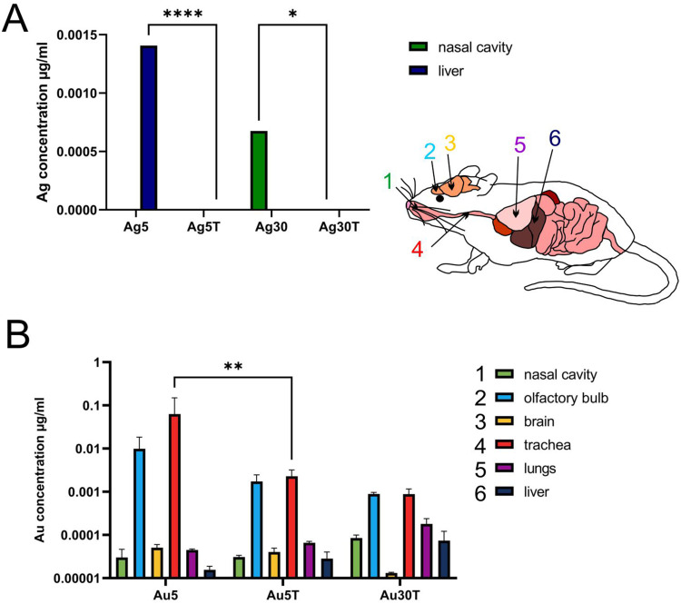
Discussion: Treatment of the mucosal tissues at the early stage of HSV-1 infection helps to modulate specific and effective antiviral immune response by attracting cytotoxic lymphocytes and inducing the production of antiviral cytokines and chemokines. Furthermore, tannic acid modification is helpful for the removal of nanoparticles from the respiratory tract, which increases the safety of nanoparticle applications to treat infections.

Keywords: AgNPs; AuNPs; HSV-1; microglia; tannic acid.


Conflict of interest statement

The authors report no conflicts of interest in this work.

Figures

Figure 1
Figure 1
HR-STEM images and DLS size distribution histograms of the mNPs: citrate-(A-C) and tannic acid-modified (D-G).
Figure 2
Figure 2
Cryo-TEM imaging of HSV-1 treated with nanoparticles. The HSV-1 virus was treated with nanoparticles for 5 minutes at room temperature (RT), followed by preparation for Cryo-TEM imaging as described in the Materials and Methods section. Panel (a) illustrates the structure of HSV-1 in the absence of nanoparticle treatment. Various morphological forms of HSV-1 were observed - icosahedral capsids (A–C) were identified, with yellow arrows indicating the presence of spike proteins on the capsid surface. Additionally, empty virions (C) were noted, along with complete virions containing an envelope, capsid, and tegument (D). White arrows highlight the viral envelope, while Orange arrows indicate envelope-associated proteins. In some instances, the genetic material of the virus was visible (E). Panel (b) shows HSV-1 cells treated with nanoparticles (A and B) Ag5T; (C and D) Ag30T; (E and F) Au5T; (G and H) Au30T). Yellow arrows indicate the nanoparticles under investigation, whereas red and orange arrows highlight their interactions with the structural elements of the virus. Red arrows indicate interactions with the capsid and orange arrows indicate interactions with the viral envelope.
Figure 3
Figure 3
The dose of tannic-acid modified Ag/AuNPs (TA- Ag/AuNPs) necessary to inhibit HSV-1 infection of glial cells is size and metal-dependent. The mixed glial cells were infected for 6 h, then washed and treated with a range of tannic acid modified AgNPs or AuNPs sized 5 and 30 nm concentrations (1–5 µg/mL). HSV-1 copies titration by qPCR was performed at 24 h p.i. Data from three independent experiments are presented as mean ± SEM. *Represents significant differences with p ≤ 0.05, **p ≤ 0.01 in comparison to untreated infected control (two-way ANOVA test).
Figure 4
Figure 4
TA-Au/AgNPs are internalized by microglia. The mixed glial culture were plated on slides and infected for 6 h with HSV-1, then washed and treated with a range of tannic acid modified AgNPs, or AuNPs sized 5 and 30 nm. (A) Twenty-four hours post infection, the fixed cells were stained for microglia (Iba-1+, turquoise), astrocytes (GFAP+, purple), HSV-1 (green), counterstained with DAPI for DNA (blue) and analyzed in confocal microscope. Nanoparticles were visualized in a reflection mode (red). Yellow arrows point to nanoparticles. (B) The overlap of nanoparticle signal with GFAP- or Iba-1-positive cells calculated as Mander’s Colocalization Coefficients. Data from three independent experiments are presented as mean ± SEM. Two-way ANOVA test p ≤ 0.01 **.
Figure 5
Figure 5
Tannic acid modified NPs significantly activate NF-κB activity in HSV-1- infected RAW-Dual™ cells. The RAW cells were uninfected, infected with HSV-1 (1 PFU/cell) or treated with poly (I:C) at 1 μg/mL for 2 h, then tannic acid-modified or unmodified 5 nm AgNPs, 30 nm AgNPs, 5 nm AuNPs and 30 nm AuNPs were added. After 6 h, the activation of NF-κB pathway was measured as SEAP activity. Data from three independent experiments are presented as mean ± SEM. Two-way ANOVA test. *Represents p ≤ 0.05 in comparison to HSV-1-infected control, &p ≤ 0.05 in comparison to uninfected control, and p #≤ 0.05, ##p ≤ 0.01 in comparison to poly (I:C) treated control.
Figure 6
Figure 6
Treatment with tannic acid-modified Ag/AuNPs of both 5 and 30 nm reduces HSV-1 infection in vivo much better than with unmodified NPs. C57BL/6 mice infected intranasally with HSV-1 were treated two times every 24 h with tannic acid-modified or unmodified 5 nm AgNPs, 30 nm AgNPs, 5 nm AuNPs or 0.9% NaCl (control). Trigeminal ganglia (TG), nasal cavities and brains were collected at 3 (A and C) and 7 days p.i. (B and D), and subjected to measurement of HSV-1 gB titers (copies/ng DNA) by qPCR (N = 10). The bars represent means ± SEM. * represents significant differences with p ≤ 0.05, **p ≤ 0.01, ***p ≤ 0.001 and extremely significant at p ≤ 0.0001 **** in comparison to untreated infected tissues.
Figure 7
Figure 7
Tannic acid-modified Ag/AuNPs of both 5 and 30 nm activate early antiviral response in HSV-1 infection much better than with unmodified NPs. Total counts of (A and B) NK cells, (C and D) CD8+ T cells, (E) CD8+CD69+ T cells and (F) CD8+SSIEFARL+ T cells isolated from TGs and brains of HSV-1-infected mice at 7 days p.i., and treated with tannic acid-modified or unmodified 5 nm AgNPs, 5 nm AuNPs or 0.9% NaCl. Results are expressed as mean ± SEM for N = 7. *Represents significant differences with p ≤ 0.05, **p ≤ 0.01, ***p ≤ 0.001 and extremely significant at p ≤ 0.0001 **** in comparison to untreated infected tissues.
Figure 8
Figure 8
Tannic acid-modified Ag/AuNPs of both 5 and 30 nm modulate local inflammatory and early antiviral response in HSV-1 infection. Total counts of (A) monocytes, (C and E) inflammatory monocytes and (B, D, F) Langerhans cells (LCs) isolated from nasal cavity, trigeminal ganglia (TGs) and brains of uninfected and HSV-1-infected mice at 3 or 7 days p.i., and treated with tannic acid-modified or unmodified 5 nm AgNPs, 30 nm AgNPs, 5 nm AuNPs or 0.9% NaCl. Results are expressed as mean ± SEM for N = 7. *Represents significant differences with p ≤ 0.05, **p ≤ 0.01, ***p ≤ 0.001 and extremely significant at p ≤ 0.0001 **** in comparison to HSV-1 infected tissues.
Figure 9
Figure 9
Tannic acid-modified Ag/AuNPs of both 5 and 30 nm modulate mucosal inflammatory response in HSV-1 infection better than unmodified NPs. Cytokine and chemokine expression in the nasal cavities at 3 days p.i. after treatment with tannic acid-modified or unmodified 5 nm AgNPs, 30 nm AgNPs, 5 nm AuNPs or 0.9% NaCl. Levels of IFN-α, IFN-β (A) as well as CXCL1 and CXCL9 (B) mRNAs are shown as expression relative to control based on the 2-∆∆Ct method. N = 7. *Represents significant differences with p ≤ 0.05, **p ≤ 0.01 in comparison to uninfected tissues.
Figure 10
Figure 10
Tannic acid-modified and unmodified Ag/AuNPs of both sizes - 5 and 30 nm accumulate in tissues differently. C57BL/6 mice were treated intranasally two times every 24 h with tannic acid-modified or unmodified 5 nm AgNPs, 30 nm AgNPs, 5 nm AuNPs or 30 nm TA-AuNPs. Nasal cavities, brains with olfactory bulbs, trachea, lungs and livers were collected at 2 days post-treatment and subjected to inductively coupled plasma mass spectrometry (ICP-MS) for Au (A) and Ag (B) content in extracts prepared from collected organs. Data from three independent experiments are presented as mean ± SEM. Two-way ANOVA test with p ≤ 0.05 *, p ≤ 0.01 **, and extremely significant at p ≤ 0.0001 **** in comparison to unmodified NPs (TA-AuNPs vs AuNPs, etc.).

Similar articles

Cited by

References

    1. Rodrigues I, Campo KN, Arns CW, Gabriel LP, Webster TJ, Lopes ÉSN. From bulk to nanoparticles: an overview of antiviral materials, its mechanisms, and applications. Part Part Syst Charact. 2021;38(8):2100044. doi:10.1002/ppsc.202100044 - DOI
    1. Lin N, Verma D, Saini N, et al. Antiviral nanoparticles for sanitizing surfaces: a roadmap to self-sterilizing against COVID-19. Nano Today. 2021;40:101267. doi:10.1016/j.nantod.2021.101267 - DOI - PMC - PubMed
    1. Krzyzowska M, Janicka M, Tomaszewska E, et al. Lactoferrin-conjugated nanoparticles as new antivirals. Pharmaceutics. 2022;14(9):1862. doi:10.3390/pharmaceutics14091862 - DOI - PMC - PubMed
    1. Luceri A, Francese R, Lembo D, Ferraris M, Balagna C. Silver nanoparticles: review of antiviral properties, mechanism of action and applications. Microorganisms. 2023;11(3):629. doi:10.3390/microorganisms11030629 - DOI - PMC - PubMed
    1. Almatroudi A. Unlocking the potential of silver nanoparticles: from synthesis to versatile bio-applications. Pharmaceutics. 2024;16(9):1232. doi:10.3390/pharmaceutics16091232 - DOI - PMC - PubMed

MeSH terms