. 2025 Mar 21:16:1493329.
doi: 10.3389/fimmu.2025.1493329. eCollection 2025.

Multi-targeted, NOT gated CAR-T cells as a strategy to protect normal lineages for blood cancer therapy

Affiliations

Multi-targeted, NOT gated CAR-T cells as a strategy to protect normal lineages for blood cancer therapy

Breanna DiAndreth et al. Front Immunol. .

Abstract

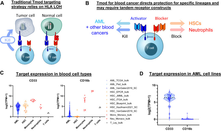
Introduction: Despite advances in treatment of blood cancers, several-including acute myeloid leukemia (AML)-continue to be recalcitrant. Cell therapies based on chimeric antigen receptors (CARs) have emerged as promising approaches for blood cancers. However, current CAR-T treatments suffer from on-target, off-tumor toxicity, because most familiar blood cancer targets are also expressed in normal lineages. In addition, they face the common problem of relapse due to target-antigen loss. Cell therapeutics engineered to integrate more than one signal, often called logic-gated cells, can in principle achieve greater selectivity for tumors.

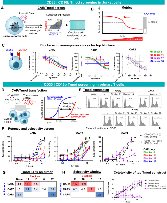
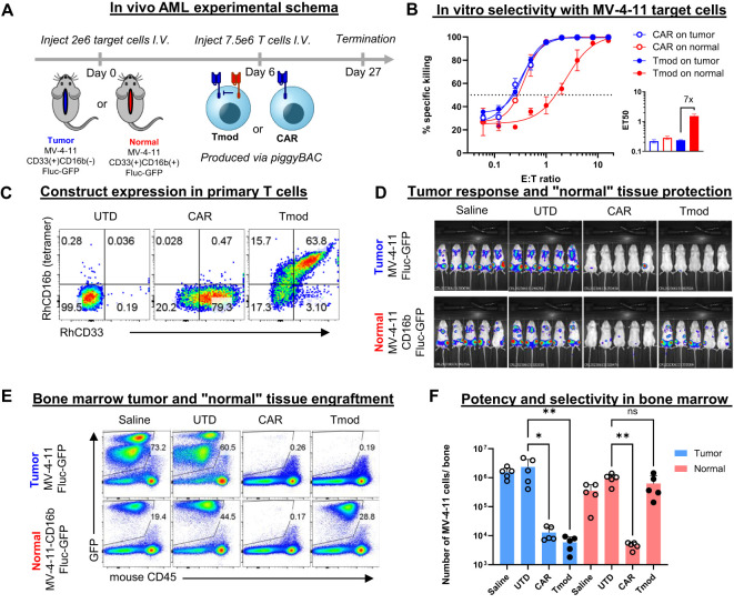
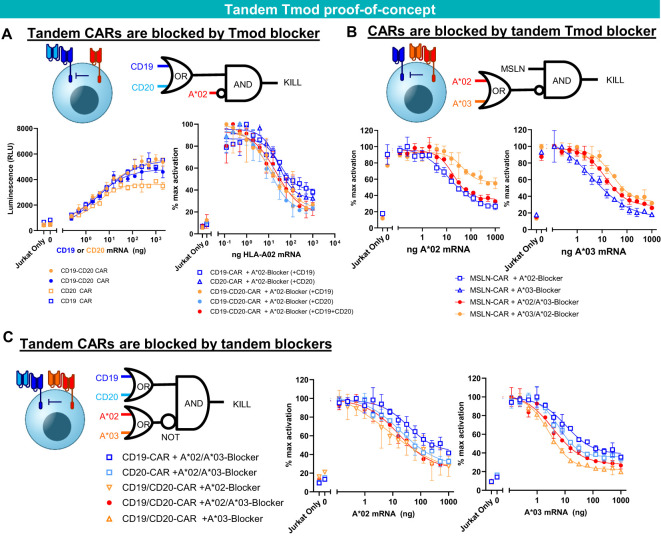
Methods: We applied such a technology, a NOT gated system called Tmod™ that is being developed to treat solid-tumor patients, to the problem of therapeutic selectivity for blood cancer cells.

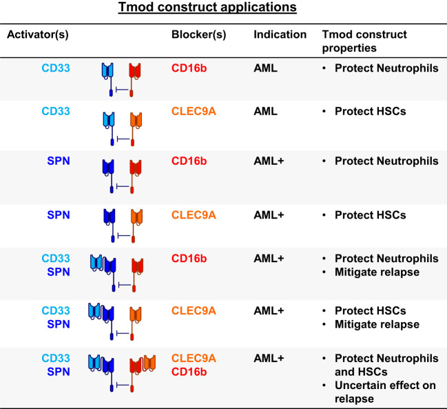
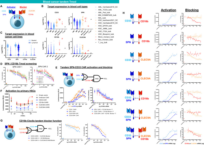
Results: Here we show that Tmod cells can be designed to target 2-4 antigens to provide different practical and conceptual options for a blood cancer therapy: (i) mono- and bispecific activating receptors that target CD33, a well-known AML antigen expressed on the majority of AML tumors (as well as healthy myeloid cells) and CD43 (SPN), an antigen expressed on many hematopoietic cancers (and normal blood lineages); and (ii) mono- and bispecific inhibitory receptors that target CD16b (FCGR3B) and CLEC9A, antigens expressed on key normal blood cells but not on most blood cancers.

Discussion: These results further demonstrate the robust modularity of the Tmod system and generalize the Tmod approach beyond solid tumors.

Keywords: AML; CAR-T cells; CD16; CD33; LIR-1 (LILRB1); SPN; blood cancer; logic gate.


Conflict of interest statement

All authors are current or former employees and shareholders of A2 Biotherapeutics, Inc.

Figures

Figure 1
Figure 1
Tmod can be adapted for blood cancer. (A) Diagram for Tmod system showing the two receptors that comprise the NOT gate targeting HLA loss of heterozygosity (LOH) in solid tumors. (B) Diagram for Tmod utilizing tandem receptors for blood cancer. (C) CD33 and CD16b mRNA expression in primary AML and healthy blood cells including T cells, neutrophils, monocytes, and hematopoietic stem cells (HSC) (data from sources shown; see Supplementary Table 1 ). (D) mRNA expression of targets in AML cell lines (n=43; DepMap).
Figure 2
Figure 2
CD33 | CD16b Tmod functions robustly in Jurkat and primary T cells. (A) Diagram of functional screen in Jurkat reporter cell line cocultured with K562 target cells transfected with different amounts of CD16b mRNA. Tmod transgene expression in Jurkat cells was detected by staining with recombinant human (rh) CD16b and CD33. (B) Diagram of functional parameters estimated from the Jurkat cell assay data. (C) Functional readout from 8-point mRNA titration curves. Three CARs combined with 4 blockers, that were selected for further analysis, are shown in color. Data are shown as mean ± standard deviation of technical replicates (n=2), normalized to each sample’s maximum activation. (D) Left, diagram of non-viral construct-screening in primary T cells using PiggyBac transposase and single vectors (BA vectors). Right, metrics used to quantify the potency and selectivity of the Tmod pair. (E) Flow cytometry analysis of stable integrants via staining with labeled recombinant human CD33 (see Methods). (E, F) T cell cytotoxicity curves generated from GFP signal at 48 hour time point with each well normalized to the zero time point. Tumor (CD33(+)CD16(-)) target-cell curves are shown with dashed lines and “normal” (CD33(+)CD16(+)) target-cell curves with solid lines. Black lines are CAR constructs and colored lines are Tmod constructs. Tumor cells are K562 cells engineered with CD33 and normal cells are K562 cells engineered to overexpress CD33 and CD16b. Data are shown as mean ± standard deviation of technical replicates (n=3). (G) Potency calculated as ET50 of Tmod cells cocultured with tumor cells. (H) Selectivity ratios are calculated as ET50 on normal cells divided by ET50 on tumor cells. (I) Kinetic cytotoxicity analysis of the most selective and potent construct compared to the CAR-T. GFP(+) area was used as proxy for target cell viability. Data are shown as mean ± standard deviation of technical replicates (n=3).
Figure 3
Figure 3
CD33 | CD16b Tmod cells selectively kill tumor but not “normal” cells in vivo. (A) Schema for in vivo experiment. 2 million MV-4-11 AML cells or MV-4-11 cells that overexpress CD16b were injected into NSG-SGM3 mice and 6 days later 7.5 million T cells were injected. (B) Selectivity in vitro using MV-4-11 cells. Surrogate normal cells were generated by overexpression of CD16b in the AML cells. E:T cytotoxicity curves were generated from firefly luciferase bioluminescence at 48 hours. Data are shown as mean ± standard deviation of technical replicates (n=3). Inset: ET50 values of depicted curves. Data shown are interpolated values with 95% CI. (C) Flow cytometry analysis of construct expression by staining with labeled recombinant human CD16b and CD33. (D) Bioluminescence imaging (BLI) at 20 days post target-cell injection. (E) Flow cytometry analysis of MV-4-11 cells in the bone marrow 27 days post target-cell injection. (F) Quantification of data shown in panel. (E) Statistics were calculated using a non-parametric Kruskal-Wallis H test; *: 0.01 < adjusted p < 0.05; **: adjusted p value < 0.01; ns: not significant (adjusted p > 0.05).
Figure 4
Figure 4
Tandem construct proof of concept. (A) A*02 blocker inhibits CD19-CD20 bispecific CAR. Jurkat functional readout from activator titration curves and blocker titration curves. (B) A*02-A*03 tandem blocker inhibits monospecific MSLN CAR. Jurkat (B2M KO) functional readout from blocker titration curves (either A*02 or A*03). (C) Tandem A*02-A*03 blocker inhibits tandem CD19-CD20 CAR. Jurkat (B2M KO) functional readout from blocker titration curves (either A*02 or A*03) with constant amounts of CD19 and CD20 mRNA. Data are shown as mean ± standard deviation of technical replicates (n=2).
Figure 5
Figure 5
Blood cancer applications for Tmod constructs. SPN (CD43 protein); FCGR3B (CD16b protein); HSCs, hematopoietic stem cells; AML+ refers to blood cancers beyond AML.
Figure 6
Figure 6
Tandem Tmod constructs for blood cancer. (A) Diagram of a Tmod cell with bispecific activator to target AML (CD33) and other blood cancers (SPN) and bispecific blocker to protect HSCs (CLEC9A) and neutrophils (CD16b). (B) Target expression in primary blood cancers and healthy blood cells (data from sources shown; see Supplementary Table 1 ). (C) Target expression in blood cancer cell lines (DepMap). (D) Jurkat cell (SPN KO) functional readout of SPN | CD16b Tmod with blocker titration curves. (E) Jurkat cell (SPN KO) functional readout of SPN-CD33 tandem CAR activation and blocking by CD16b blocker in the presence of SPN and CD33 antigens. (F) Jurkat functional readout of binders cloned as CARs with titration of primary HSCs. (G) Jurkat functional readout of CD33 CAR4 blocked by tandem CLEC9A-CD16b blocker. (H) Jurkat functional readout of CD33 and/or SPN monospecific or bispecific activators paired with CD16b and/or CLEC9A monospecific or bispecific blockers. Data are shown as mean ± standard deviation of technical replicates (n=2).

Similar articles

Cited by

References

    1. Kuykendall A, Duployez N, Boissel N, Lancet JE, Welch JS. Acute myeloid leukemia: the good, the bad, and the ugly. Am Soc Clin Oncol Educ Book. (2018) 38:555–73. doi: 10.1200/EDBK_199519 - DOI - PubMed
    1. Dohner H, Estey E, Grimwade D, Amadori S, Appelbaum FR, Buchner T, et al. . Diagnosis and management of AML in adults: 2017 ELN recommendations from an international expert panel. Blood. (2017) 129:424–47. doi: 10.1182/blood-2016-08-733196 - DOI - PMC - PubMed
    1. Mardiros A, Dos Santos C, McDonald T, Brown CE, Wang X, Budde LE, et al. . T cells expressing CD123-specific chimeric antigen receptors exhibit. specific cytolytic effector functions antitumor effects against Hum acute myeloid leukemia. Blood. (2013) 122:3138–48. doi: 10.1182/blood-2012-12-474056 - DOI - PMC - PubMed
    1. Gill S, Tasian SK, Ruella M, Shestova O, Li Y, Porter DL, et al. . Preclinical targeting of human acute myeloid leukemia and myeloablation using chimeric antigen receptor-modified T cells. Blood. (2014) 123:2343–54. doi: 10.1182/blood-2013-09-529537 - DOI - PMC - PubMed
    1. Brinkman-Van-der-Linden EC, Angata T, Reynolds SA, Powell LD, Hedrick SM, Varki A. CD33/Siglec-3 binding specificity, expression pattern, and consequences of gene deletion in mice . Mol Cell Biol. (2003) 23:4199–206. doi: 10.1128/MCB.23.12.4199-4206.2003 - DOI - PMC - PubMed

MeSH terms

Substances

LinkOut - more resources