. 2025;17(1):2486962.
doi: 10.1080/17590914.2025.2486962. Epub 2025 Apr 7.

G-Ratio Commentary-Why You've Been Doing It Wrong

Affiliations

G-Ratio Commentary-Why You've Been Doing It Wrong

R Douglas Fields et al. ASN Neuro. 2025.
No abstract available


Figures

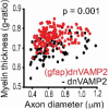
Figure 1.
Figure 1.
G-ratio of axons in adult mouse optic nerve expressing a transgene to reduce vesicle release from astrocytes (gfap dnVAMP2, in red), or not (-dnVAMP2, in black) showing the relation between g-ratio and axon diameter. Note the positive slope in both experimental and control groups. The myelin sheath thickness is reduced when exocytosis from astrocytes is decreased (higher g-ratio, in red). Reprinted from Dutta et al. (2018).
Figure 2.
Figure 2.
In contrast to g-ratios, there is a linear relation between the number of myelin wraps and axon caliber with a positive slope when plotted against either fiber diameter (above) or axon diameter (below) in both experimental (Dox off) and control (Dox on) conditions in adult mouse optic nerve. Data are from experiments by Dutta et al. (2018), which were not published previously, and can be compared with g-ratio plots from the same study in Figure 1.

References

    1. Assaf, Y., & Basser, P. J. (2005). Composite hindered and restricted model of diffusion (CHARMED) MR imaging of the human brain. NeuroImage, 27(1), 48–58. 10.1016/j.neuroimage.2005.03.042 - DOI - PubMed
    1. Assaf, Y., Blumenfeld-Katzir, T., Yovel, Y., & Basser, P. J. (2008). AxCaliber: A method for measuring axon diameter distribution from diffusion MRI. Magnetic Resonance in Medicine, 59(6), 1347–1354. 10.1002/mrm.21577 - DOI - PMC - PubMed
    1. Skoven, M., Andersson, M., Pizzolato, Hartwig, R., Siebner, & Dyrby, T. B. (2023). Mapping axon diameters and conduction velocity in the rat brain – Different methods tell different stories of the structure-function relationship. bioRxiv 2023. 10.1101/2023.10.20.558833 - DOI
    1. Caminiti, R., Ghaziri, H., Galuske, R., Hof, P. R., & Innocenti, G. M. (2009). Evolution amplified processing with temporally dispersed slow neuronal connectivity in primates. Proceedings of the National Academy of Sciences of the United States of America, 106(46), 19551–19556. 10.1073/pnas.0907655106 - DOI - PMC - PubMed
    1. Campbell, J. S. W., Leppert, I. R., Narayanan, S., Boudreau, M., Duval, T., Cohen-Adad, J., Pike, G. B., & Stikov, N. (2018). Promise and pitfalls of g-ratio estimation with MRI. NeuroImage, 182, 80–96. 10.1016/j.neuroimage.2017.08.038 - DOI - PubMed

LinkOut - more resources