. 2025 Apr 7;23(4):e3003063.
doi: 10.1371/journal.pbio.3003063. eCollection 2025 Apr.

Completing the BASEL phage collection to unlock hidden diversity for systematic exploration of phage-host interactions

Affiliations

Completing the BASEL phage collection to unlock hidden diversity for systematic exploration of phage-host interactions

Dorentina Humolli et al. PLoS Biol. .

Abstract

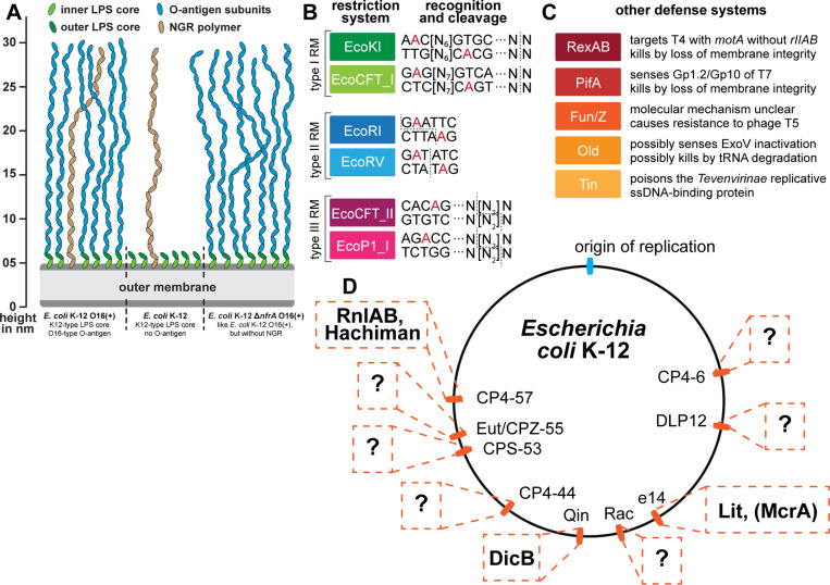
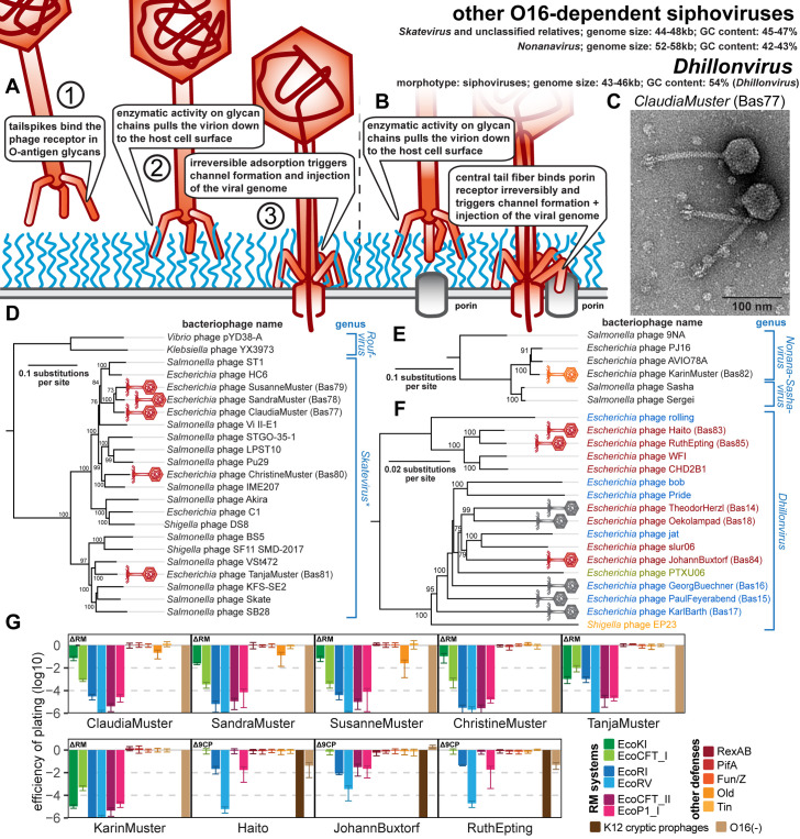
Research on bacteriophages, the viruses infecting bacteria, has fueled the development of modern molecular biology and inspired their therapeutic application to combat bacterial multidrug resistance. However, most work has so far focused on a few model phages which impedes direct applications of these findings in clinics and suggests that a vast potential of powerful molecular biology has remained untapped. We have therefore recently composed the BASEL collection of Escherichia coli phages (BActeriophage SElection for your Laboratory), which made a relevant diversity of phages infecting the E. coli K-12 laboratory strain accessible to the community. These phages are widely used, but their assorted diversity has remained limited by the E. coli K-12 host. We have therefore now genetically overcome the two major limitations of E. coli K-12, its lack of O-antigen glycans and the presence of resident bacterial immunity. Restoring O-antigen expression resulted in the isolation of diverse additional viral groups like Kagunavirus, Nonanavirus, Gordonclarkvirinae, and Gamaleyavirus, while eliminating all known antiviral defenses of E. coli K-12 additionally enabled us to isolate phages of Wifcevirus genus. Even though some of these viral groups appear to be common in nature, no phages from any of them had previously been isolated using E. coli laboratory strains, and they had thus remained largely understudied. Overall, 37 new phage isolates have been added to complete the BASEL collection. These phages were deeply characterized genomically and phenotypically with regard to host receptors, sensitivity to antiviral defense systems, and host range. Our results highlighted dominant roles of the O-antigen barrier for viral host recognition and of restriction-modification systems in bacterial immunity. We anticipate that the completed BASEL collection will propel research on phage-host interactions and their molecular mechanisms, deepening our understanding of viral ecology and fostering innovations in biotechnology and antimicrobial therapy.


Conflict of interest statement

The authors have declared that no competing interests exist.

Figures

Fig 1
Fig 1. O-antigen glycans as a barrier or host receptor for phage infections.
(A) The illustration shows cell surface glycans of E. coli K-12 laboratory strains, including rough lipopolysaccharide (LPS) without O-antigen and smooth LPS (with genetically restored O16-type O-antigen; see details in Materials and methods). The O-antigen can be a barrier to phage infection (green), used as a host receptor (blue), or be bypassed by phages that target the N4 glycan receptor (NGR) chains (brown). (B) Serial dilutions of different phages characterized previously [10] were spotted on the E. coli K-12 ΔRM strain without (left) or with (right) genetically restored O-antigen expression to illustrate the three different modes of interaction with these surface glycans (same color code as in panel A).
Fig 2
Fig 2. Expanding the BASEL collection with phages dependent on O-antigen or inhibited by K-12 cryptic prophages.
(A) Phages dependent on O16-type O-antigen were isolated and characterized as shown in the illustration (analogous to our previous work [10]). (B) O-antigen dependence of diverse newly isolated phages characterized in this work compared to JakobBernoulli and IrisVonRoten from our previous study that can infect E. coli K-12 without (left) and with (right) O-antigen [10]. (C) Phages inhibited by the cryptic prophages in the K-12 genome were isolated analogously to those dependent on O-antigen (see panel A) with help of the E. coli K-12 Δ9CP strain (details outlined in Materials and methods).
Fig 3
Fig 3. Overview of new phages included in the BASEL collection.
The illustration shows new phage isolates included in the BASEL collection with their Bas## identifiers. Isolates are sorted by morphotype and their current ICTV classification [68]. The color code distinguishes phages based on morphotype and genome size. For siphoviruses, small phages (ca. 30–50 kb) are shown in red and medium-sized phages (ca. 50–100 kb) are shown in orange. Small myoviruses (ca. 30–100 kb) are shown in green. For podoviruses, small phages (ca. 30–50 kb) are shown in violet and medium-sized phages (ca. 50–100kb) are shown in pink.
Fig 4
Fig 4. Overview of E. coli surface glycan variants and the immunity systems used in this study.
(A) Surface glycans of different E. coli K-12 variants are shown schematically (compare Fig 1A and see further details in Materials and methods). None of the newly isolated phages in this work showed any dependence on the NGR glycan (see S1A Fig) which has thus been omitted from the other illustrations. (B, C) Restriction-modification (B) and other defense systems (C) used to characterize phage isolates are summarized schematically (same as used in our previous work [10]). (D) The genome of E. coli K-12 contains nine cryptic prophages that encode all remaining antiviral defense systems predicted by DefenseFinder or PADLOC after deletion of all restriction systems in our previous work (including McrA; see details on these defense systems in Materials and methods under Bacterial strains) [10,46,47]. Due to the known accumulation of antiviral defenses in prophages [48,49], it seems likely that also many of the possibly still unknown defense systems of E. coli K-12 would be encoded in these loci (indicated by question marks). The E. coli K-12 Δ9CP variant of Wang and colleagues [51], which we use in the current study, has been cured of all nine cryptic prophages.
Fig 5
Fig 5. Overview of Guernseyvirinae genus Kagunavirus phages.
(A) Schematic model of host recognition by Kagunavirus phages (O-antigen chains in blue). In analogy to other siphoviruses infecting E. coli like lambda or T5, we anticipate that these phages also use a phage-derived conduit for DNA injection across the outer membrane after the tail fibers recognizing the terminal host receptor have bent off [110,141]. (B) Representative TEM micrograph of phage SonjaBuckley (Bas74). (C) Maximum-likelihood phylogeny of Kagunavirus phages and relatives based on a curated whole-genome alignment with bootstrap support of branches shown if > 70/100. Newly isolated phages of the BASEL collection are highlighted by red phage icons. The phylogeny was rooted between the genera Kagunavirus and Jerseyvirus. (D) The results of quantitative phenotyping experiments with Kagunavirus phages regarding sensitivity to altered surface glycans and bacterial immunity systems are presented as efficiency of plating (EOP). Small notes of “ΔRM” or “Δ9CP” indicate on which host strain the respective phage has been characterized (see Materials and methods). Data points and error bars represent the average and standard deviation of at least three independent experiments. Raw data and calculations are available in S1 Data.
Fig 6
Fig 6. Overview of other O16-dependent siphoviruses of genera Skatevirus and Nonanavirus as well as new isolates of siphoviral genus Dhillonvirus.
(A) Schematic model of host recognition by O16-dependent small siphoviruses just as shown for Kagunavirus phages in Fig 5A. (B) Schematic model of host recognition by small siphoviruses of the Dhillonvirus genus that use first a lateral tail fiber to overcome the O-antigen barrier before binding a porin receptor with the central tail fiber (instead of only glycan-targeting tailspikes for the LPS-dependent small siphoviruses; see also our previous work [10]). (C) Representative TEM micrograph of Skatevirus ClaudiaMuster (Bas77). (D) Maximum-likelihood phylogeny of the Skatevirus genus and relatives based on a curated whole-genome alignment with bootstrap support of branches shown if > 70/100. The phylogeny was rooted between genus Roufvirus and the others. Note that most reference phages presented here as Skatevirus have no formal ICTV classification while others are scattered over several seemingly paraphyletic genera, prompting us to show all phages as Skatevirus [68]. (E) Maximum-likelihood phylogeny of the Nonanavirus genus and relatives based on a curated whole-genome alignment with bootstrap support of branches shown if >70/100. The phylogeny was rooted between phage 9NA and the other relatives based on their much closer relationship. Note that Sasha and Sergei are formally classified as Sashavirus while 9NA and AVIO78A are classified as Nonanavirus [68], which—according to this phylogeny—would be paraphyletic. (F) Maximum-likelihood phylogeny of the Dhillonvirus genus based on a curated whole-genome alignment with bootstrap support of branches shown if > 70/100. The phylogeny was rooted as previously [10] between the clade containing phage WFI and the others. In (D-F), newly isolated phages of the BASEL collection are highlighted by red or orange phage icons. The color code of phage names in (F) refers to porin host receptors with blue indicating FhuA, red LptD, green BtuB, and orange LamB [10] (see also S2B Fig). (G) The results of quantitative phenotyping experiments with Skatevirus, Nonanavirus, and new Dhillonvirus isolates regarding sensitivity to altered surface glycans and bacterial immunity systems are presented as efficiency of plating (EOP). Small notes of “ΔRM” or “Δ9CP” indicate on which host strain the respective phage has been characterized (see Materials and METHODS). Data points and error bars represent average and standard deviation of at least three independent experiments. Raw data and calculations are available in S1 Data.
Fig 7
Fig 7. Overview of Gordonclarkvirinae phages.
(A) Schematic model of host recognition by Gordonclarkvirinae phages as an interpretation of different molecular studies [57,117]. (B) Representative TEM micrograph of phage LorandFenyves (Bas91). (C) Maximum-likelihood phylogeny of the Gordonclarkvirinae subfamily based on a curated whole-genome alignment with bootstrap support of branches shown if > 70/100. Phages of the BASEL collection are highlighted by little violet phage icons. The phylogeny was rooted between the genus Suseptimavirus and the genera Nieuwekanaalvirus + Kuravirus based on closer relationship of the latter two. (D) The results of quantitative phenotyping experiments with Gordonclarkvirinae phages regarding sensitivity to altered surface glycans and bacterial immunity systems are presented as efficiency of plating (EOP). Small notes of “ΔRM” or “Δ9CP” indicate on which host strain the respective phage has been characterized (see Materials and methods). Data points and error bars represent average and standard deviation of at least three independent experiments. Raw data and calculations are available in S1 Data.
Fig 8
Fig 8. Overview of rare O-antigen-dependent phage groups (Xuquatrovirus relatives, Gamaleyavirus, Jilinvirus).
(A) Schematic model of host recognition by podoviruses with enzymatic tailspikes targeting O-antigen (e.g., Gamaleyavirus) based on previous work on phage T7 and P22 [71,101,203]. (B) Schematic model of host recognition by myoviruses targeting O-antigen with a tail fiber (e.g., Jilinvirus). Note that Jilinvirus HeidiAbel encodes two sets of tail fibers (see below) whose relationship is unknown, so only a simple schematic with one set is shown. (C) Representative TEM micrograph of Jilinvirus phage HeidiAbel (Bas97). This phage forms myovirus virions with similar appearance to other Jilinvirus phages and relatives that have been studied previously [79,80,82]. (D) Maximum-likelihood phylogeny of the genus Xuquatrovirus and relatives based on a curated concatenated alignment of major capsid protein, portal protein, and terminase large subunit protein sequences with bootstrap support of branches shown if >70/100. The phylogeny was rooted between two marine Vibrio phages and the others based on a large phylogenetic distance between these groups. (E) Maximum-likelihood phylogeny of the Enquatrovirinae subfamily of Schitoviridae and relatives based on a curated whole-genome alignment with bootstrap support of branches shown if >70/100. The phylogeny was rooted between subfamilies Rothmandenesvirinae and subfamily Enquatrovirinae. (F) Maximum-likelihood phylogeny of the Iiscvirinae subfamily based on a curated whole-genome alignment with bootstrap support of branches shown if > 70/100. The phylogeny was rooted between the genus Jilinvirus and the other genera. The exemplary prophages of E. coli and Aeromonas media were identified in strains USECESBL382 (NCBI GenBank AATBGZ010000031) and T0.1-19 (NCBI GenBank CP038441), respectively, using PHASTEST [204]. In (D-F), phages of the BASEL collection are highlighted by pink and green (new) or gray (old) phage icons. (G) The results of quantitative phenotyping experiments with the three newly described phage isolates regarding sensitivity to altered surface glycans and bacterial immunity systems are presented as efficiency of plating (EOP). Small notes of “ΔRM” or “Δ9CP” indicate on which host strain the respective phage has been characterized (see Materials and methods). Data points and error bars represent average and standard deviation of at least three independent experiments. Raw data and calculations are available in S1 Data.
Fig 9
Fig 9. Overview of Wifcevirus phages.
(A) Schematic model of host recognition by Wifcevirus myoviruses targeting host O-antigen (blue) using tail fibers. Note that these phages carry two sets of tail fibers (see below) whose relationship is unclear, so only a simple schematic with one set is shown. (B) Maximum-likelihood phylogeny of the Wifcevirus genus and relatives based on a curated whole-genome alignment with bootstrap support of branches shown if > 70/100. The phylogeny was midpoint-rooted between the distantly related Wifcevirus and Pbunavirus genera. Phages of the BASEL collection are highlighted by little green phage icons. (C) Representative TEM micrograph of phage HermannAdler (Bas98) with a virion morphology very similar to other representatives that have been visualized previously [58]. (D) Wifcevirus isolate ManiWeber (Bas99) was streaked for single plaques on E. coli K-12 strains with and without cryptic prophages. Kuravirus SkaterBoy (Bas90; see Fig 7) is shown as a control without sensitivity to the presence of cryptic prophages. (E) The results of quantitative phenotyping experiments with Wifcevirus phages regarding sensitivity to altered surface glycans and bacterial immunity systems are presented as efficiency of plating (EOP). Small notes of “ΔRM” or “Δ9CP” indicate on which host strain the respective phage has been characterized (see Materials and methods). Data points and error bars represent average and standard deviation of at least three independent experiments. Raw data and calculations are available in S1 Data.
Fig 10
Fig 10. Overview of Autographiviridae: Studiervirinae and Lederbergvirus phages.
(A) Schematic model of host recognition by podoviruses with enzymatic tailspikes targeting O-antigen (e.g., Kayfunavirus and Lederbergvirus) based on previous work on phage T7 and P22 [71,101,203]. (B) Maximum-likelihood phylogeny of the Studiervirinae subfamily of Autographiviridae based on a curated whole-genome alignment with bootstrap support of branches shown if > 70/100. The phylogeny was midpoint-rooted between distantly related genus Kayfunavirus and the other genera. Phages of the BASEL collection are highlighted by violet (new) or gray (old) phage icons. (C) Representative TEM micrograph of Kayfunavirus ElsieAttenhofer (Bas105). (D) Maximum-Likelihood phylogeny of the Lederbergvirus genus based on an alignment of their structural genes with bootstrap support of branches shown if >70/100. This phylogeny was midpoint-rooted between two major clades of this genus. Phage CurroJimenez (Bas106) is highlighted by a violet phage icon while phages Huey, Dewey, and Louie are highlighted by gray phage icons. (E) The results of quantitative phenotyping experiments with Autographiviridae phages regarding sensitivity to altered surface glycans and bacterial immunity systems are presented as efficiency of plating (EOP). Small notes of “ΔRM” or “Δ9CP” indicate on which host strain the respective phage has been characterized (see Materials and methods). Data points and error bars represent average and standard deviation of at least three independent experiments. Raw data and calculations are available in S1 Data.
Fig 11
Fig 11. Phylogenetic analyses of tailspikes and tail fibers.
(A) Maximum-likelihood phylogeny of pectin lyase-like receptor-binding domains (InterPro accession IPR011050 [114]) of tailspikes encoded by our new BASEL phages and relevant controls. The phylogeny was midpoint-rooted between previously characterized tailspikes targeting the O:4 O-antigen of Salmonella Typhimurium (blue) and those of our new isolates targeting E. coli O16-type O-antigen (orange; including a few additional previously published phages with very similar tailspikes). Bootstrap support of branches is shown if >70/100. Phages of the BASEL collection are highlighted by colorful phage icons. (B) Maximum-likelihood phylogeny of Gp12-like receptor-binding domains (InterPro accession SSF88874 [114]) of tail fibers encoded by our new BASEL phages and relevant controls. The phylogeny was midpoint-rooted between a clade of distant Gordonclarkvirinae tail fibers at the top (blue) and those of Wifcevirus, Tequintavirus, and the Gordonclarkvirinae presented in this study (orange). Note that a clade with Wifcevirus receptor-binding domains (bottom) includes diverse phages from the database as well as Tequintavirus IrisVonRoten (Bas32 [10]) scattered between the Wifcevirus isolates characterized in the current study.
Fig 12
Fig 12. Predicted structures of representative tailspikes and tail fibers.
(A) AlphaFold-Multimer [198] predicted structures of homotrimeric tailspikes from selected BASEL phages (see also Fig 11A). (B) Predicted structures of the representative T-shaped tail fiber complexes of IrisVonRoten (Bas32, Tequintavirus), AnnaHegner (Bas87, Gordonclarkvirinae), and ManiWeber (Bas99, Wifcevirus) which all feature the dual Gp12-like RBD1 and T5-like RBD2 (predicted with the C-terminal intramolecular chaperone domain (IMC) attached; see also Figs 11B and S10). While ManiWeber features an N-terminal anchor domain for baseplate attachment, the T-shaped tail fibers of IrisVonRoten and AnnaHegner attach to proximal fibers (Gp22 and Gp9, respectively), which connect the complete fiber structure to the distal baseplate complex. The proximal fiber protein Gp22 of IrisVonRoten contains an additional galectin-like domain (inset box; colored N- (blue) to C- (red) terminus) that protrudes from the C-terminus of the fiber and could contribute to host receptor interaction as an additional RBD. While the overall setup of the T-shaped tail fibers is highly conserved, the RBDs are highly variable and seem to be frequently transferred horizontally (Figs 11B, S9C, and S10D). (C) Predicted structures of other tail fibers with single RBDs (more shown in S7 Fig). The short-tail fiber (Gp13) of AnnaHegner (Bas87, Gordonclarkvirinae) and the second tail fiber (Gp32) of ManiWeber (Bas99, Wifcevirus) have an elongated tip similar to the long-tail fiber of phage T4 including conserved HxH motifs for ion coordination [118]. Tail fibers of JakobBernoulli (Bas07, Drexlerviridae; Gp36) and RuthEpting (Bas85, Dhillonvirus; Gp29) both contain elongated N-terminal coiled coils connected to receptor-binding T5-like fiber tips with C-terminal IMC domains. In (A)-(C), N-terminal anchor domains and fiber shafts are colored blue, while the RBDs are colored red. A ~5-nm vertical scale bar is provided for reference. In all panels, viral morphotypes are annotated by phage icons analogous to Fig 11A and 11B.
Fig 13
Fig 13. Host range of new phage isolates in the BASEL collection.
(A) Lysis host range of all new BASEL phages and controls (same as in S1B Fig) as determined by qualitative spot test assays with high-titer phage stocks (>109 pfu/ml if possible; see Materials and methods). The O-antigen type of different E. coli and Salmonella Typhimurium strains and possibly rough LPS (as O(-)) are highlighted at the top, while phages with O16-targeting tail spikes or tail fibers are distinguished on the left side. Lysis of a host strain is shown by a red cell lysis icon while the inability to lyse a strain is shown as a green icon in the shape of a cell with full integrity. (B) Plating host range of all new BASEL phages and controls (like in (A)) as determined by quantitative efficiency-of-plating (EOP) assays using serial dilutions of phage stocks. Data points and error bars represent average and standard deviation of at least three independent experiments. Raw data and calculations are available in S1 Data. In (A) and (B), K-12 strains have been selected differently for phages inhibited by its cryptic prophages (Δ9CP) and for those who are not (ΔRM).
Fig 14
Fig 14. GtrABS-mediated O-antigen glycosylation inhibits phage infection.
(A) O16-type O-antigen of E. coli K-12 with (left) or without (right) the GtrABS system of CPS-53 differs in the presence or absence of the glucose side chain, respectively [129,130]. (B) Serial dilutions of different phages were spotted on variants of E. coli K-12 BW25113 with restored O-antigen expression as well as deletion of the GtrS glucosyltransferase (ΔgtrS) or the nine cryptic prophages including CPS-53 (Δ9CP). Three Dhillonvirus phages (Bas83−85), two Gordonclarkvirinae (Bas87 + Bas91), and the Wifcevirus phages (Bas98−101) are inhibited by the K-12 cryptic prophages if the GtrABS system in CPS-53 is intact (see S11 Fig for the same experiment without O-antigen expression). Gordonclarkvirinae phage SkaterBoy (Bas90) and Tequintavirus IrisVonRoten (Bas32) were used as GtrABS-insensitive controls that do or do not depend on O16-type O-antigen, respectively. A small growth defect of the three Kagunavirus phages in the ΔgtrS mutant compared to the Δ9CP strain (top right) is indicative of a slight inhibition of these phages by other factor(s) encoded in the cryptic prophages (C) Serial dilutions of lambda PaPa and Ur-lambda were spotted on variants of E. coli K-12 BW25113 either in its wildtype form (left) or with restored O-antigen expression and either active (middle) or inactivated (right) GtrABS system. Phages SkaterBoy (Bas90, Gordonclarkvirinae) and IrisVonRoten (Bas32, Tequintavirus) were added as controls that do or do not depend on the O16-type O-antigen and are both insensitive to GtrABS.
Fig 15
Fig 15. Overview of the complete BASEL collection.
The illustration shows all isolates of the complete BASEL collection with their Bas## identifiers. Isolates are sorted by morphotype and their current ICTV classification [68]. The color code distinguishes phages based on morphotype and genome size. For siphoviruses, small phages (ca. 30–50 kb) are shown in red, medium-sized phages (ca. 50–100 kb) are shown in orange, and large phages (>100 kb) are shown in yellow. For myoviruses, small phages (ca. 30–100 kb) are shown in green, medium-sized phages (ca. 100–150kb) are shown in cyan, and large phages (>100kb) are shown in blue. For podoviruses, small phages (ca. 30–50 kb) are shown in violet, and medium-sized phages (ca. 50–100kb) are shown in pink.

References

    1. Dion MB, Oechslin F, Moineau S. Phage diversity, genomics and phylogeny. Nat Rev Microbiol. 2020;18(3):125–38. doi: 10.1038/s41579-019-0311-5 - DOI - PubMed
    1. Cobián Güemes AG, Youle M, Cantú VA, Felts B, Nulton J, Rohwer F. Viruses as winners in the game of life. Annu Rev Virol. 2016;3(1):197–214. doi: 10.1146/annurev-virology-100114-054952 - DOI - PubMed
    1. Ofir G, Sorek R. Contemporary phage biology: from classic models to new insights. Cell. 2018;172(6):1260–70. doi: 10.1016/j.cell.2017.10.045 - DOI - PubMed
    1. Gordillo Altamirano FL, Barr JJ. Phage therapy in the postantibiotic era. Clin Microbiol Rev. 2019;32(2):e00066-18. doi: 10.1128/CMR.00066-18 - DOI - PMC - PubMed
    1. Schaechter M, Kolter R, editors. Small Things Considered [Internet]. 2022. https://schaechter.asmblog.org/schaechter/2022/03/the-phage-treaty.html2022. [cited 2022]. Available from: https://schaechter.asmblog.org/schaechter/2022/03/the-phage-treaty.html

LinkOut - more resources