. 2025 Apr 7;16(1):259.
doi: 10.1038/s41419-025-07595-z.

The SIRT1 activator SRT2104 exerts exercise mimetic effects and promotes Duchenne muscular dystrophy recovery

Affiliations

The SIRT1 activator SRT2104 exerts exercise mimetic effects and promotes Duchenne muscular dystrophy recovery

Matteo Giovarelli et al. Cell Death Dis. .

Abstract

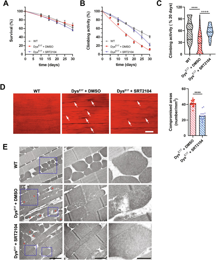
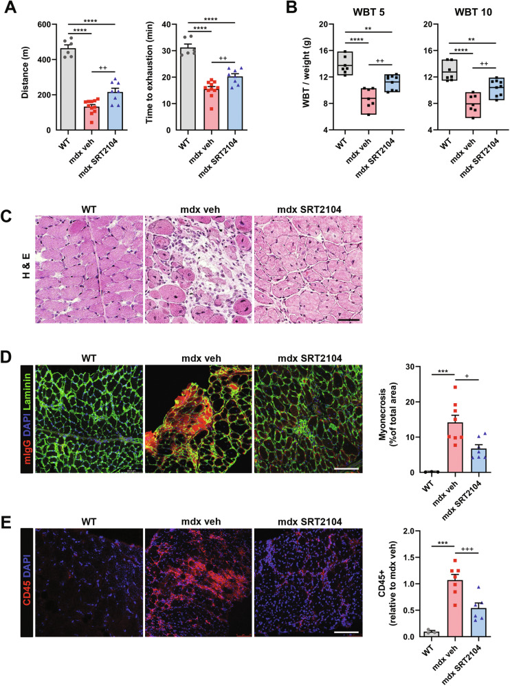
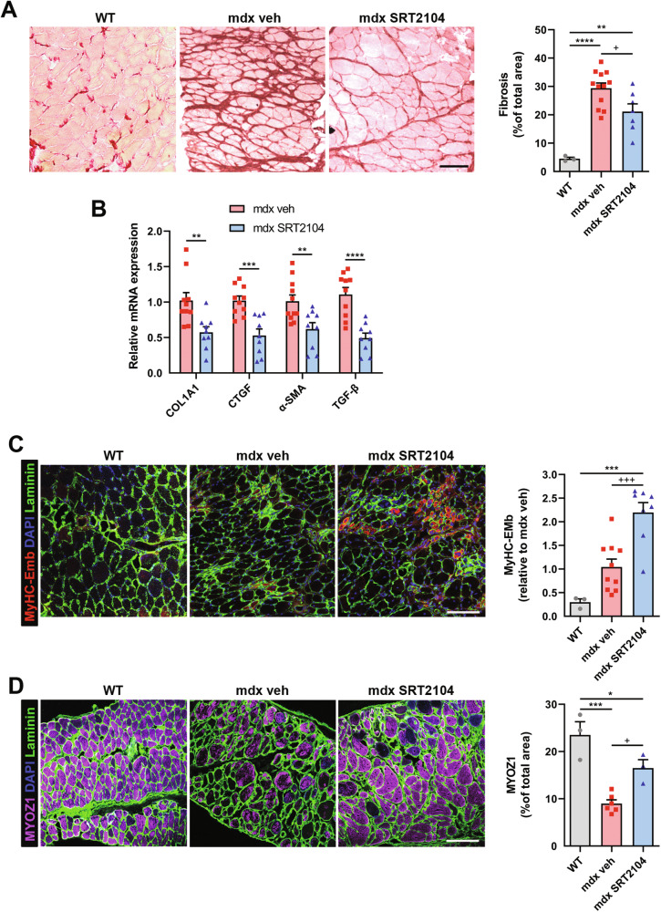
Duchenne muscular dystrophy (DMD) is a devastating genetic disorder, whose management is still a major challenge, despite progress in genetic and pharmacological disease-modifying treatments have been made. Mitochondrial dysfunctions contribute to DMD, however, there are no effective mitochondrial therapies for DMD. SIRT1 is a NAD+-dependent deacetylase that controls several key processes and whose impairment is involved in determining mitochondrial dysfunction in DMD. In addition to well-known resveratrol, other potent selective activators of SIRT1 exist, with better pharmacokinetics properties and a safer profile. Among these, SRT2104 is the most promising and advanced in clinical studies. Here we unveil the beneficial effects of SRT2104 in flies, mice, and patient-derived myoblasts as different models of DMD, demonstrating an anti-inflammatory, anti-fibrotic, and pro-regenerative action of the drug. We elucidate, by molecular dynamics simulations, that a conformational selection mechanism is responsible for the activation of SIRT1. Further, the impact of SRT2104 in reshaping muscle proteome and acetylome profiles has been investigated, highlighting effects that mimic those induced by exercise. Overall, our data suggest SRT2104 as a possible therapeutic candidate to successfully counteract DMD progression.


Conflict of interest statement

Competing interests: The authors declare no conflicts of interest. Ethical approval: All procedures of animal experiments comply with ethical standards and were approved by the Italian Ministry of Health (approval no. 761/2022-PR). The two pediatric patient-derived immortalized cell lines were granted by biobank MyoLine and used according to the MTA with Prof. Mouly, director of MyoLine biobank in the frame of the agreement with Professor Ornella Cappellari and Professor Matteo Giovarelli respectively at the University of Bari and Milan.

Figures

Fig. 1
Fig. 1. SRT2104 administration to dystrophic flies improves muscle functionality and mitochondrial morphology.
A SRT2104 administration to dystrophic (DysE17) D. melanogaster does not affect the survival rate compared to both WT and DMSO-treated flies. B 30 days of SRT2104 administration improves muscular performances in dystrophic flies measured by climbing activity. C Climbing activity measurement after 20 days of treatment is provided (right panel). * versus WT flies (**** P < 0.0001), + versus DysE17 DMSO flies (++++ P < 0.0001) (WT n = 84; DysE17 + DMSO n = 78; DysE17 + SRT2104 n = 18). D Representative phalloidin immunostaining of WT and DysE17 flies treated with either DMSO or SRT2104 (scale bar = 20 μm). Damaged fiber areas are indicated (arrows). Muscle-compromised areas for DysE17 flies SRT2104 treated or not are provided (right panel) (DysE17 + DMSO n = 26; DysE17 + SRT2104 n = 17). * versus DysE17 DMSO flies (**** P < 0.0001). E Electron microscopy analysis of WT, DysE17, and DysE17 + SRT2104 D. melanogaster thorax muscles. Representative insets showing mitochondrial morphologies (red asterisks) are provided (scale bar = 2 μm). Values are expressed as mean ± SEM.
Fig. 2
Fig. 2. SRT2104 administration to mdx mice enhances muscle performance and reduces muscle damage.
SRT2104 administration in mdx mice improves muscular performances, assessed by exhaustion treadmill running test, measuring distance ran (A left panel) and time to exhaustion (A right panel), as well as force, assessed by WBT test, with WBT 5 (B left panel) and 10 (B right panel), representing respectively the best 5 or the best 10 forward pulling tension normalized on body weight (WT n ≥ 6; mdx veh n ≥ 7; mdx SRT2104 n ≥ 8). Histological analyses in diaphragm (DP) muscle of WT, mdx vehicle, and mdx upon SRT2104 treatment: hematoxylin and eosin (H&E) staining (scale bar = 50 μm) (C); mouse IgG staining labeling myonecrotic fibers (scale bar = 100 μm) (D); CD45 staining to identify immune infiltrate (scale bar = 100 μm) (E). Percentage of myonecrosis area (WT n = 3; mdx veh n = 8; mdx SRT2104 n = 7) and CD45+ fibers relative to mdx vehicle (WT n = 3; mdx veh n = 7; mdx SRT2104 n = 6) are provided. * versus WT mice, (** P < 0.01, *** P < 0.001, **** P < 0.0001). + versus vehicle-treated mdx mice (+ P < 0.05, ++ P < 0.01, +++ P < 0.001). Values are expressed as mean ± SEM.
Fig. 3
Fig. 3. SRT2104 treatment reduces fibrosis and promotes regeneration in mdx mice.
A Sirius red staining labeling fibrotic tissue in DP muscle of WT, mdx vehicle, and mdx upon SRT2104 treatment (scale bar = 100 μm). Quantification of fibrosis is provided (right panel) (WT n = 3; mdx veh n = 12; mdx SRT2104 n = 7). B RT-qPCR analysis of fibrotic genes COL1A1, CTGF, α-SMA, and TGF-β in DP of vehicle- and SRT2104-treated mdx mice (mdx veh n ≥ 10; mdx SRT2104 n = 9). C, D Representative immunostaining of embryonic myosin heavy chain (MyHC3-Emb) to identify regenerating fibers and MYOZ1 to assess myofibers maturity (scale bars = 100 μm). Quantification of MyHC-Emb regeneration foci relative to mdx vehicle (WT n = 3; mdx veh n = 10; mdx SRT2104 n = 8) and MYOZ1 staining on total sections area (WT n = 3; mdx veh n = 6; mdx SRT2104 n = 3) are provided. AD * versus WT mice (* P < 0.05, ** P < 0.01, *** P < 0.001, **** P < 0.0001); + versus vehicle-treated mdx mice (+ P < 0.05, +++ P < 0.001.) B * versus vehicle-treated mdx mice (** P < 0.01, *** P < 0.001, **** P < 0.0001). Values are expressed as mean ± SEM.
Fig. 4
Fig. 4. SRT2104 treatment activates SIRT1 leading to deacetylated protein pattern reshape.
A Fluorescence-based SIRT1 activity assay using crude nuclear extracts of DP muscles, normalized to protein content (WT n = 4; mdx veh n = 9; mdx SRT2104 n = 7). + versus vehicle-treated mdx mice (+++ P < 0.001). Values are expressed as mean ± SEM. B Maps of the populations of the three-dimensional configurations explored by the open apo form of SIRT1 (left, oS1) and by the open holo form of SIRT1 (right, oS1-SRT2104) through molecular dynamics (MD) simulations. The geometric states are reported in the form of correlation maps between the intermolecular number of contacts and the overall radius of gyration. The populations have been normalized to 1 corresponding to the highest population value for each distribution. The family of structures (cS1*) characterized by more compactness and a high number of contacts have been highlighted by a gray rectangle. C Distribution of the backbone heavy atoms (Ca, C’, N) root mean square deviations (RMSDs) of the holo form of two closed configurations of SIRT1, i.e., the one obtained docking with SRT2014 the active structure reported in the literature (black line: cS1-SRT2104, adapted from the PDB ID 5zzh) and the one obtained docking the compact configuration obtained simulating the dynamics of the apo form of the inactive/open conformation (red line: cS1-SRT2104*). D Scheme of all the possible interconversion mechanisms between the open/inactive form and the closed/active form of SIRT1 upon SRT2104 binding (NTD: N terminal domain; CD: catalytic domain): the apo forms of the open and closed conformations of SIRT1 (oS1, cS1); the holo forms of the open and closed conformations of SIRT1 complexed with SRT2104 (oS1-SRT2104, cS1-SRT2104); the apo and holo forms of the final configurations after MD simulations of the open conformation of SIRT1 (cS1*, cS1-SRT2104*). The configurations that do not directly enter the activation mechanisms are shaded. E Representative immunoblot using Acetyl-lysine antibody to detect lysine acetylation pattern in GC muscle homogenates of mdx mice treated or not with SRT2104. Quantification of protein acetylation pattern is provided (right panel) (mdx veh n = 9; mdx SRT2104 n = 8). * versus vehicle-treated mdx mice (** P < 0.01). Values are expressed as mean ± SEM. FH Gene Ontology (GO) terms enrichment analyses of deacetylated proteins after SRT2104 treatment.
Fig. 5
Fig. 5. SRT2104 boosted dystrophic muscle metabolism improving mitochondrial bioenergetics.
AC GO terms enrichment analysis of differentially expressed proteins (upregulated for Biological Processes and Cellular Components; downregulated for Biological Processes) after SRT2104 treatment in GC muscle. D Luminescence-based ATP quantification measured in GC homogenates from mdx mice treated or not with SRT2104 (mdx veh n = 7; mdx SRT2104 n = 5). E Mitochondrial respiratory rates of permeabilized fibers’ bundles of DP muscles from mdx mice, treated or not with SRT2104, measured by high-resolution respirometry (HRR) (L: leak state; CI: complex I respiration; ETS: electron transfer system maximal capacity; CII: complex II respiration; CIV: complex IV activity) (mdx veh n ≥ 9; mdx SRT2104 n = 6). Representative immunostainings (red: myosin heavy chain; blue: DAPI nuclei. Scale bar = 50 μm) (F) and TMRM fluorescence levels (indicating basal mitochondrial membrane potential) (G) of human CTR and DMD myotubes in differentiation medium for 72 h (hCTR n = 3; hDMD n = 3). * versus hCTR (* P < 0.05). Representative immunostainings (red: myosin heavy chain; blue: DAPI nuclei. Scale bar = 50 μm) (H) and TMRM fluorescence levels (I) of human DMD myotubes treated with either DMSO (vehicle) or SRT2104 (3 μM) in differentiation medium for 72 h (hDMD veh n = 4; hDMD SRT2104 n = 4). * versus vehicle-treated hDMD (* P < 0.05). L RT-qPCR analysis of MyoD and MyoG genes in DMD patient-derived myoblasts treated or not with SRT2104 (individual values are the average of the 2 myoblast lines and the experiment has been repeated three times, n = 3). M Representative immunoblot for MyoD and MyoG proteins extracted from hDMD myoblasts treated or not with SRT2104. Molecular weights (kDa) are indicated on the left. Values are expressed as mean ± SEM.

References

    1. Emery AE. Population frequencies of inherited neuromuscular diseases-a world survey. Neuromuscul Disord. 1991;1:19–29. - PubMed
    1. Mah JK, Korngut L, Dykeman J, Day L, Pringsheim T, Jette N. A systematic review and meta-analysis on the epidemiology of Duchenne and Becker muscular dystrophy. Neuromuscular Disord. 2014;24:482–91. - PubMed
    1. Bez Batti Angulski A, Hosny N, Cohen H, Martin AA, Hahn D, Bauer J, et al. Duchenne muscular dystrophy: disease mechanism and therapeutic strategies. Front Physiol. 2023;14:1183101. - PMC - PubMed
    1. Kourakis S, Timpani CA, Campelj DG, Hafner P, Gueven N, Fischer D, et al. Standard of care versus new-wave corticosteroids in the treatment of Duchenne muscular dystrophy: can we do better? Orphanet J Rare Dis. 2021;16:117. - PMC - PubMed
    1. Heydemann A, Siemionow M. A brief review of Duchenne muscular dystrophy treatment options, with an emphasis on two novel strategies. Biomedicines. 2023;11:830. - PMC - PubMed

Grants and funding

LinkOut - more resources