. 2025 Apr 7;15(1):11832.
doi: 10.1038/s41598-025-96050-2.

Dexamethasone: a double-edged sword in the treatment of osteoarthritis

Affiliations

Dexamethasone: a double-edged sword in the treatment of osteoarthritis

Karyna Tarasova et al. Sci Rep. .

Abstract

Glucocorticoids are widely used to manage osteoarthritis (OA) symptoms, but long-term safety concerns exist. This study investigates the therapeutic potential of dexamethasone (DEX) and triamcinolone acetonide (TA) in chondrocytes, evaluating their anti-inflammatory effects and potential detrimental actions. This study evaluated the effects of DEX and TA on the expression of pro-inflammatory genes in inflamed chondrocytes. In addition, the effects of DEX treatment on chondrocytes were analyzed using next-generation sequencing, high-resolution mass spectrometry, proliferation and metabolic rate, wound healing capacity and senescence-associated B-galactosidase assays. A single therapeutic dose of DEX (40nM) effectively reduced the expression of inflammatory genes in chondrocytes, while TA showed no such effect. DEX significantly reduced inflammation but also ECM production in inflamed chondrocytes. At 24 h, DEX treatment led to 168 differentially expressed genes (DEGs) compared to untreated inflamed cells, decreasing to 5 DEGs by 48 h, indicating a rapidly diminishing anti-inflammatory effect. Conversely, the difference between DEX-treated and healthy cells increased over time, from 666 DEGs at 24 h to 1317 DEGs at 48 h. Pathway analysis revealed potential disruptions in cell cycle, mitosis, and ECM homeostasis in DEX-treated cells compared to both healthy and inflamed controls. Interestingly, repeated DEX administration at both a therapeutic (40nM) and a high dose (1µM) induced senescence in healthy cells but not in inflamed cells. In contrast, repeated high-dose DEX reduced apoptosis marker Caspase 3/7 in inflamed but not healthy cells. Despite the transient suppression of inflammation achieved with DEX treatment, the observed decrease in ECM production and induction of senescence in healthy chondrocytes at therapeutic doses, along with apoptosis in inflamed cells at higher doses, underscore the need for caution in its intra-articular administration.

Keywords: Apoptosis; Dexamethasone; Glucocorticoids; Osteoarthritis; Senescence; Triamcinolone.


Conflict of interest statement

Declarations. Competing interests: The authors declare no competing interests. Ethics approval: No human or animal participants were involved in this study; ovine chondrocytes had been obtained and biobanked from sheep euthanised for reasons unrelated to this study. Based on the “Good Scientific Practice. Ethics in Science und Research” regulation implemented at the University of Veterinary Medicine Vienna, the Institutional Ethics Committee (“Ethics and Animal Welfare Committee”) of the University of Veterinary Medicine Vienna does not require approval of in vitro cell culture studies, if the cells were isolated from tissue, which was obtained either solely for diagnostic or therapeutic purposes or in the course of institutionally and nationally approved experiments. The sheep from which the cells were obtained had been euthanised in the course of a study for which approval of the national (“Commission for Animal Research” of the Austrian Federal Ministry of Science, Research and Economy) and institutional. (“Ethics and Animal Welfare Committee” of the University of Veterinary Medicine Vienna”) animal welfare committees (ethical approval number: 68.205/0100-V/3b/2018) had been granted and which had been reported according to ARRIVE guidelines. All methods were carried out in accordance with the relevant guidelines and regulations.

Figures

Fig. 1
Fig. 1
Treatment effect of Dexamethasone (DEX 40nM) on inflamed chondrocytes (mRNA, miRNA). (A) DEX 40nM treatment significantly reduced inflammation related genes like matrix metalloproteinase-1 (MMP-1: logFC=-1.91, FDR = 5.50E−01), matrix metalloproteinase-12 (MMP-12: logFC=−1.15, FDR = 8.80E−05), chitinase-3 like-protein-1 (CHI3L1: logFC=−0.50, FDR = 1.68E−03) and complement factor B (CFB: logFC=−1.15, FDR = 2.97E−04)). (B) Extracellular matrix (ECM)-related genes COL2A1 and ACAN showed similar significantly decreased expression in both DEX-treated and untreated inflamed chondrocytes compared to healthy controls at T48 (COL2A1: DEX treated: logFC=−5.28, FDR = 9.78E−08, untreated: logFC=−4.76, FDR = 3.56E−03; ACAN: DEX treated: logFC=−2.10 FDR = 1.53E−05, untreated: logFC=−2.28, FDR = 6.10E−03). (C) DEX treatment downregulated inflammation- related miRNAs miR-147-3p (T24: logFC=−0.13, FDR = 9.99E−01; T48: logFC=−0.29,FDR = 9.52E−01), miR-146a-5p (T24:logFC = 0.35,FDR = 9.99E−01; T48: logFC=−0.36, FDR = 9.52E−01), miR-146b-5p (T24: logFC=−0.35, FDR = 9.99E−01; T48: logFC=−0.36 ,FDR = 9.52E−01) and miR-34a-5p (T24: logFC = 0.08, FDR = 9.99E−01; T48: logFC=−0.08, FDR = 9.52E−015) compared to inflamed untreated controls. (D) DEX treatment significantly downregulated pro-regenerative miRNAs miR-140-3p (logFC=−0.73, FDR = 2.30E−02) and miR-1290 (logFC=−1, FDR = 2.40E−02) in DEX-treated cells compared to healthy chondrocytes at T48.
Fig. 2
Fig. 2
Ingenuity Pathway Analysis of differentially expressed genes at 24 h. (A) Graphical summary of of the Ingenuity Pathway Analysis (IPA) core analysis for genes differentially expressed between inflamed and healthy chondrocytes at T24. The graphical summary selects and connects a subset of the most significant entities predicted in the analysis, including canonical pathways, upstream regulators, diseases and biological functions, to visualize related biological activities. (B) Graphical summary of the genes differentially regulated between DEX treated and untreated inflamed chondrocytes at T24. (C) Graphical summary of the genes differentially regulated between DEX treated and healthy chondrocytes at T24. (D) Comparative analysis of the canonical pathways enriched in the DEGs of all 3 groups (healthy, inflamed untreated, DEX treated inflamed) at both time points (T24 and T48). Pathways are ranked accordingly to their z-score. orange … predicted activation (based on the z-score), blue … predicted inhibition, solid line: direct interaction, dashed line: indirect interaction;
Fig. 3
Fig. 3
Treatment effect of Dexamethasone (DEX 40nM) on inflamed chondrocytes (proteomics data analysis). (A) DEX treatment led to a decreased abundance of inflammatory mediators such as matrix metalloproteinase − 1 (MMP1, T24: q = 0.746, T48: q = 0.000), matrix metalloproteinase − 3 (MMP3, T24: q = 0.000, T48: q = 0.000) and superoxide dismutase 3 (SOD3, T24: q = 0.072, T48: q = 1.00) compared to inflamed chondrocytes, but DEX treatment significantly increased chitinase-3 like-protein-1 (CH3L1, q = 0.000) at T48 compared to healthy cells. (B) DEX treatment non-significantly improved the secretion of extracellular matrix (ECM) proteins such as aggrecan (ACAN), procollagen C-proteinase enhancer (PCOLCE), hyaluronan and proteoglycan link protein 1 (HAPLIN1) and collagen type VI alpha 1 (COL6a1) compared to inflamed untreated chondrocytes, but their abundance remained significantly lower than in healthy controls at T24 (ACAN, T24: q = 0.048, T48: q = 0.283; PCOLCE, T24: q = 0.050, T48: q = 0.202) or T48 (HAPLIN1, T24: q = 0.349, T48: q = 0.000; COL6A1, T24: q = 0.276, T48: q = 0.048). ns…not significant (p ≥ 0.05), *…p < 0.05, **…p < 0.01, ***…p < 0.001.
Fig. 4
Fig. 4
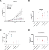
Long-term implications of Dexamethasone (DEX) (40nM and 1µM) treatments. (A) All conditions proliferated significantly slower (p < 0.0001) compared to healthy untreated over a period of 9 days. (B) Intracellular reactive oxygen species (ROS) levels significantly increased in inflamed (p = 0.0169) compared to healthy chondrocytes. DEX treatment did not reverse ROS accumulation in inflamed cells (40nM p = 0.8795, 1µM p = 0.4403), leaving ROS levels of DEX treated inflamed cells significantly higher than in healthy controls (40nM p = 0.0162, 1µM p = 0.0412). DEX treatment had no significant effect on ROS levels of healthy chondrocytes (40nM p = 0.1741, 1µM p = 0.0818). (C) SA-β Gal activity significantly increased in healthy chondrocytes treated with DEX (40nM:p = 0.0411 and 1µM:p = 0.0078) compared to healthy untreated chondrocytes. Neither inflammation (p = 0.3310) nor DEX treatment of inflamed chondrocytes (40nM:p = 0.8390, 1µM:p = 0.7526) increased SA-β Gal activity compared to healthy chondrocytes. (D) Caspase 3/7 activity significantly decreased in inflamed (p = 0.0401) but not in healthy (p = 0.5299) chondrocytes treated with DEX 1µM compared to healthy controls. Neither inflammation itself (p = 0.6766) nor DEX treatment 40nM significantly altered caspase 3/7 activity in either inflamed (p = 0.1282) or healthy (p = 0.9497) chondrocytes. ns…not significant (p ≥ 0.05), *…p < 0.05, **…p < 0.01, ***…p < 0.001.

References

    1. Kolasinski, S. L. et al. American College of Rheumatology/Arthritis Foundation Guideline for the Management of Osteoarthritis of the Hand, Hip, and Knee. Arthr. Rheumatol.72, 220–233 (2020). (2019). - PMC - PubMed
    1. Bannuru, R. R. et al. OARSI guidelines for the non-surgical management of knee, hip, and polyarticular osteoarthritis. Osteoarthr. Cartil.27, 1578–1589 (2019). - PubMed
    1. Mautner, K. et al. Cell-based versus corticosteroid injections for knee pain in osteoarthritis: A randomized phase 3 trial. Nat. Med.29, 3120–3126 (2023). - PMC - PubMed
    1. Lu, Y. C., Evans, C. H. & Grodzinsky, A. J. Effects of short-term glucocorticoid treatment on changes in cartilage matrix degradation and chondrocyte gene expression induced by mechanical injury and inflammatory cytokines. Arthr. Res. Ther.13, R142 (2011). - PMC - PubMed
    1. Grodzinsky, A. J., Wang, Y., Kakar, S., Vrahas, M. S. & Evans, C. H. Intra-articular dexamethasone to inhibit the development of post‐traumatic osteoarthritis. J. Orthop. Res.35, 406–411 (2017). - PMC - PubMed

MeSH terms