. 2025 May;22(5):541-556.
doi: 10.1038/s41423-025-01281-y. Epub 2025 Apr 7.

The intrinsic expression of NLRP3 in Th17 cells promotes their protumor activity and conversion into Tregs

Affiliations

The intrinsic expression of NLRP3 in Th17 cells promotes their protumor activity and conversion into Tregs

Théo Accogli et al. Cell Mol Immunol. 2025 May.

Abstract

Th17 cells can perform either regulatory or inflammatory functions depending on the cytokine microenvironment. These plastic cells can transdifferentiate into Tregs during inflammation resolution, in allogenic heart transplantation models, or in cancer through mechanisms that remain poorly understood. Here, we demonstrated that NLRP3 expression in Th17 cells is essential for maintaining their immunosuppressive functions through an inflammasome-independent mechanism. In the absence of NLRP3, Th17 cells produce more inflammatory cytokines (IFNγ, Granzyme B, TNFα) and exhibit reduced immunosuppressive activity toward CD8+ cells. Moreover, the capacity of NLRP3-deficient Th17 cells to transdifferentiate into Treg-like cells is lost. Mechanistically, NLRP3 in Th17 cells interacts with the TGF-β receptor, enabling SMAD3 phosphorylation and thereby facilitating the acquisition of immunosuppressive functions. Consequently, the absence of NLRP3 expression in Th17 cells from tumor-bearing mice enhances CD8 + T-cell effectiveness, ultimately inhibiting tumor growth.

Keywords: Cancer Immunology; NLRP3; Th17 cells; Tregs; Tumor microenvironment.


Conflict of interest statement

Competing interests: FG received speaker honoraria from Lilly, Sanofi, BMS, Astra Zeneca and Amgen; received funding for clinical trials from Astra Zeneca; received travel grants from Roche France, Amgen and Servier; and is an advisory board member for Merck Serano, Amgen, Roche France and Sanofi. No other authors have any potential conflicts of interest to disclose.

Figures
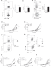
Fig. 1
Fig. 1
NLRP3 is involved in Th17 differentiation. A IFNγ production detected by cytometry in Th17 cells differentiated in vitro with TGF-β and IL-6 for 72 h from naïve CD4 T cells isolated from CD4Nlrp3-/- (Th17Nlrp3-/-) mice and their littermate controls (Th17wt). B Quantification of IFNγ detected by ELISA in the same samples as in (A). C IFNγ and IL-17 production was detected by flow cytometry under the same conditions as in (A). Tumor growth (n = 5) of B16F10 (D) and LLC1 (E) cells in C57BL/6 WT mice treated with an IL-4 blocking antibody (anti-IL-4) or a control Ig and in CD4Nlrp3-/- mice and their littermate controls. IFNγ production detected by cytometry in CD4+ T cells (F) and Th17 cells (CCR6 + RORγt+Foxp3-) (G) from B16F10 tumors. B16F10 tumor growth (n = 5) in CD4Nlrp3-/- mice and controls treated with or without digoxin (Digo) (H, I) and in C57Bl6 WT, Rorc-/-, and CD4Nlrp3-/- mice and their respective controls (J). The data shown are representative of 3 independent in vivo experiments or 5 independent in vitro experiments. Statistical significance was determined by 2-way ANOVA and Tukey’s multiple comparison tests (D, E, HJ) or the Mann‒Whitney test (AC, F, G). * <0.05, **<0.01, ***<0.005
Fig. 2
Fig. 2
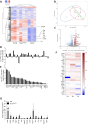
NLRP3 deficiency favors a Th17 inflammatory profile. WT inflammatory Th17 cells (Th17i), WT regulatory Th17 cells (Th17r), and Nlrp3-deficient regulatory Th17 cells (Th17Nlrp3-/-) were differentiated in vitro for 72 h (A–C, E) or 24 h (D). A Heatmap representing hierarchical clustering of RNA-seq data. B Principal component analysis (PCA) of the same data as in (A). Blue: triplicates of regulatory WT Th17 cells; red: triplicates of inflammatory WT Th17 cells; green: triplicates of regulatory Th17Nlrp3-/- cells. C Volcano plot showing the distribution of differentially expressed genes from the same data as in (A), with upregulated genes in red, downregulated genes in blue, and the inflammatory gene signature in green. Differential expression of genes associated with the inflammatory profile of Th17 cells according to the RNA-seq data after 24 h (D) and 72 h (E) of in vitro differentiation. F Heatmap representing the differential expression of the Th17 inflammatory profile evaluated by RT‒qPCR over a time course of 24 h, 72 h and 7 days of in vitro differentiation. G Differential expression of the inflammatory profile of Th17 cells isolated from B16F10 tumors from CD4Nlrp3-/- mice and their controls was evaluated via RT‒qPCR. Th17 cells were defined as CD4 + CCR6+Foxp3- cells. The data presented are pooled from 3 independent experiments. A Unsupervised hierarchical clustering was performed via Gene Cluster 3.0 software, and the results were visualized with Treeview viewer. Gene expression was normalized and mean-centered. Hierarchical clustering was conducted via the correlation measure and complete linkage analysis. D, E Cuffdiff analysis. G Statistical significance was determined via 2-way ANOVA. Asterisks indicate significant differences
Fig. 3
Fig. 3
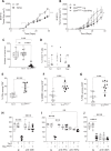
Th17 cells deficient in NLRP3 lose their immunosuppressive functions. AC B16F10 tumor cells were injected subcutaneously into CD4Nlrp3-/- mice (n = 10) and their littermate controls (Lit) with or without an anti-CD8a neutralizing antibody or a control Ig. A Tumor growth. B, C Analysis of CD8+ TILs by flow cytometry. B CD8+ frequency, C expression of IFNγ, granzyme B (GZB), and CD107a (CD107) and Ki67 staining. DG Naïve CD4+ T cells were isolated from CD4Nlrp3-/- mice and their littermate controls and differentiated into regulatory Th17 cells for 72 h (Th17wt, Th17Nlrp3-/-). CD8+ T lymphocytes were isolated from OTI mice and cultured alone or with Th17 cells at increasing CD8:Th17 ratios (1:1, 2:1, or 10:1). The production of IFNγ, D Granzyme B, E and TNFα, F and proliferation, G were evaluated via flow cytometry. H CD8+ T cells were isolated from B16F10 tumors from CD4Nlrp3-/- mice and their littermate controls and cocultured with B16F10 cells for 24 h. The B16F10 mortality rate was assessed by flow cytometry of CD45- cells. The data shown are representative of 3 independent in vivo experiments or 5 independent in vitro experiments. Statistical significance was determined by 2-way ANOVA, Tukey’s multiple comparison test (A) and the Mann‒Whitney test (BH). * <0.05, **<0.01, ***<0.005
Fig. 4
Fig. 4
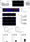
NLRP3 inflammasome-independent functions are involved in Th17 cells. A Ifnγ mRNA levels in Nlrp3-deficient Th17 cells (Th17Nlrp3-/-) and their controls (Th17wt) after 24 h of in vitro polarization, treated or not (NT) with pharmacological inhibitors of SMAD3 (SIS3), PI3K (LY294002), P38 (SB203580), ROCK (Y27632) or JNK (JNK Inhibitor II). B Immunofluorescence staining of pSMAD3 in Th17 cells differentiated in vitro for 24 h from naïve CD4Nlrp3-/- deficient mice (Th17Nlrp3-/-) and their littermate controls (Th17wt). C Cells were differentiated as described previously (Th0, Th17WT, and Th17Nlrp3-/-) for 24 h, lysed, and analyzed via Western blotting with the indicated antibodies. Immunofluorescence staining (D) and quantification (E) of pSMAD3 in Th17WT or Th17Nlrp3-/- cells during short-term kinetics. F Proximity of NLRP3 and pSMAD3 (upper panel) and NLRP3 and TGF-βR1 (lower panel) detected by proximity ligation assay in Th17 cells differentiated for 24 h in vitro from naïve CD4Nlrp3-/- deficient mice (Th17Nlrp3-/-) and their littermate controls (Th17wt). G Cells were differentiated as described in (C), harvested and lysed, followed by immunoprecipitation with an anti-TGF-β-RI antibody and Western blot analysis with the indicated antibodies. H Relative Foxp3 expression in Th17 cells treated with or without the SMAD3 inhibitor SIS3 either from the start of differentiation or after 1 day of differentiation. I Ifnγ relative expression under the same conditions as in (H). pSMAD3 enrichment at putative pSMAD3 binding sites on the Ifnγ (J), csf2 (K) and Tnfα (L) promoters by chromatin immunoprecipitation (ChIP) assay in Th17 cells differentiated for 1 h in vitro from naïve CD4Nlrp3-/- deficient mice (Th17Nlrp3-/-) and their littermate controls (Th17wt). The data shown are representative of 4–5 independent experiments. Statistical significance was determined by the Mann‒Whitney test. * <0.05, **<0.01, ***<0.005
Fig. 5
Fig. 5
NLRP3 deficiency inhibits the transdifferentiation of Th17 cells into Treg cells. Proportion of CD4+ T-cell subsets (Th1, Th2, Th17, and Treg) in subcutaneous B16F10 (A) and LLC1 (B) tumors from CD4Nlrp3-/- mice and their littermate controls (n = 5). Naïve CD4+ T cells isolated from CD4Nlrp3-/- mice and their littermate controls were differentiated in vitro into Th17 cells. The expression of RORγt and FOXP3 was detected by cytometry over a time course on days 0, 1, 2, 3 (C) and 6 (D). EH Th17 cells were differentiated from naïve CD4+ T cells isolated from CD45.2 OTII WT or Nlrp3-deficient mice. These cells were transferred into WT CD45.1 mice (EG) or into CD4Rorc-/- mice bearing B16‒OVA lung tumors (H). Forty-eight hours later, the frequencies of Treg cells present in the lungs were analyzed by cytometry among CD4+ T cells (E) (n = 8), among CD45.2+ and CD45.2− cells (n = 5) (F), and among RORγt + cells (G, H) (n = 5 in G and n = 8 in H). Growth of subcutaneously injected B16F10 melanoma tumors in CD4Nlrp3-/- mice (I) and their littermate controls (J) not treated with an anti-CD25 blocking antibody (n = 5). The data are representative of 3 independent experiments. Statistical significance was determined by the Mann‒Whitney test (A‒H), 2-way ANOVA and Tukey’s multiple comparison test (I‒J). * <0.05, **<0.01, ***<0.005
Fig. 6
Fig. 6
Thus, targeting NLRP3 in Th17 cells has therapeutic value. A Tumor growth of subcutaneously injected B16F10 melanoma in Nlrp3flox/flox × CD4CreERT2 mice treated or not treated with tamoxifen (brackets indicate the days of tamoxifen administration) (n = 5). B Tumor growth of CD4Nlrp3-/- mice and their littermate controls subcutaneously injected with B16F10 melanoma cells. CD4Nlrp3-/- mice were treated twice a week with an anti-IFNγ blocking antibody or a control Ig (n = 5). C B16-OVA lung tumor foci 13 days after intravenous injection of OTII Th17 (Th17) cells or OTII Nlrp3-deficient Th17 cells (Th17Nlrp3-/-) (n = 12). D Proportion of CD4+ T-cell subsets (Th1, Th17, and Treg) in the lungs of B16-OVA tumor-bearing mice 13 days after intravenous injection of OTII Th17 (white squares) or OTII Nlrp3-deficient Th17 cells (black squares) (n = 5). EG (n = 12), Analysis of CD8+ cells from the lungs of C mice via flow cytometry. D IFNγ production, E granzyme B production (F), and TNFα production. HJ B16-OVA lung tumor foci in mice treated with OTII Th17 (Th17) or OTII Nlrp3-deficient Th17 cells (Th17Nlrp3-/-) with or without (H, n = 10) anti-CD8 blocking antibody injected twice a week, (I, n = 6) anti-IFNγ or anti-TNFα blocking antibodies injected twice a week or (J, n = 10) anti-CD25 blocking antibody injected twice a week. The data are representative of 3 independent experiments. Statistical significance was determined by Tukey’s multiple comparison test (A, B) and 2-way ANOVA (CJ). * <0.05, **<0.01, ***<0.005, ****0.0001

References

    1. Yang XO, Pappu BP, Nurieva R, Akimzhanov A, Kang HS, Chung Y, et al. T helper 17 lineage differentiation is programmed by orphan nuclear receptors ROR alpha and ROR gamma. Immunity. 2008;28:29–39. - PMC - PubMed
    1. Miyahara Y, Odunsi K, Chen W, Peng G, Matsuzaki J, Wang RF. Generation and regulation of human CD4+ IL-17-producing T cells in ovarian cancer,. Proc Natl Acad Sci USA. 2008;105:15505–10. - PMC - PubMed
    1. Sfanos KS, Bruno TC, Maris CH, Xu L, Thoburn CJ, DeMarzo AM, et al. Phenotypic analysis of prostate-infiltrating lymphocytes reveals TH17 and Treg skewing. Clin Cancer Res. 2008;14:3254–61. - PMC - PubMed
    1. Su X, Ye J, Hsueh EC, Zhang Y, Hoft DF, Peng G. Tumor microenvironments direct the recruitment and expansion of human Th17 cells. J Immunol. 2010;184:1630–41. - PubMed
    1. Alizadeh D, Katsanis E, Larmonier N. The multifaceted role of Th17 lymphocytes and their associated cytokines in cancer. Clin Dev Immunol. 2013;2013:957878. - PMC - PubMed

MeSH terms

LinkOut - more resources