. 2025 Jun;97(6):1067-1073.
doi: 10.1002/ana.27231. Epub 2025 Apr 8.

Neuropathological Evidence of Reduced Amyloid Beta and Neurofibrillary Tangles in Multiple Sclerosis Cortex

Affiliations

Neuropathological Evidence of Reduced Amyloid Beta and Neurofibrillary Tangles in Multiple Sclerosis Cortex

J Pansieri et al. Ann Neurol. 2025 Jun.

Abstract

Multiple sclerosis (MS) and Alzheimer's disease are neurodegenerative diseases with age-related disability accumulation. In MS, inflammation spans decades, whereas AD is characterized by Aβ plaques and neurofibrillary tangles (NFT). Few studies explore accumulation of amyloids in MS. We examined Aβ deposition and NFT density in temporal and frontal cortices from postmortem MS (n = 75) and control (n = 66) cases. Compared with controls, MS cases showed reduced Aβ, especially in those aged <65 years, and reduced NFT, notably in cases aged >65 years. Aβ deposition predicted greater NFT density both in MS cases and controls. MS-related factors may affect Aβ/NFT deposition and/or clearance, offering new therapeutic insights for both diseases. ANN NEUROL 2025;97:1067-1073.


Conflict of interest statement

Nothing to report.

Figures
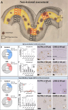
FIGURE 1
FIGURE 1
Amyloids are reduced in multiple sclerosis (MS) compared with control cortex. (A) 4G8 and AT8 expression was quantified in the nonlesional gray matter (NLGM) using predefined spaced trajectories (each 1 mm, when possible) perpendicular to the subpial surface of the cortex. For each trajectory, 2 fields of view (black and red squares) of the same size were applied to NLGM layers, with the exception of layer III, in which 4 fields of view were used due to the larger size of this layer. A sensitivity analysis was perfomed on layer II–III only (red squares). Using this method, >20,000 fields of view were quantified for each marker. (B–H) Aβ deposits in MS and controls. (B) A trend toward reduced number of MS cases compared with controls showing Aβ deposition was observed. (C–E) Quantitation of 4G8 expression in cortical layers comparing NLGM from frontal and temporal cortices in controls and MS cases shows a reduced Aβ deposition in younger than median age MS cases compared with controls. Particular attention on layer II–III (insert), the predominant site of Aβ deposition, shows the same trend. (F–H) No difference in Aβ deposition comparing NLGM from frontal and temporal cortices was found comparing older controls and MS cases. (I–O) Neurofibrillary tangles (NFTs) density in MS cases and controls. (I) A trend toward reduced number of MS cases compared with controls showing NFTs was observed. (J) NFTs were rarely found in young MS cases and control cases. (K–O) NFT density in cortical layers comparing NLGM from frontal and temporal cortices in controls and MS cases shows a reduced NFT density in cortical layers in older than median age MS cases compared with controls. Four cases (control, n = 2; MS, n = 2) were not assessed for tau expression due to tissue availability (generalized linear models; data presented as mean ± standard error of the mean; *p < 0.05; **p < 0.01; scale bar: 200 μm). NLGM, nonlesional grey matter. [Color figure can be viewed at www.annalsofneurology.org]
FIGURE 2
FIGURE 2
Aβ burden and demyelination in multiple sclerosis (MS) cortex. (A) MS lesions were defined by complete loss of myelin. In cases with demyelination, the entire lesional area was delineated for each lesion and a field of view (FOV) in the core of the lesion was used (black square 1). When available, 2 additional FOVs of the same size matched for cortical layer location on each side of the lesion were used for border, perilesional, and nonlesional areas as follows: border region—from lesion edge up to 340 μm of adjacent nonlesional grey matter (NGLM; black squares 2–3); perilesional region—340 680 μm from lesion edge (black squares 4–5); NLGM region >680 μm from lesion edge (black squares 6–7). Given the preferential deposition of Aβ in layers II and III, we focused our comparative analyses of 4G8 expression in and outside of lesions affecting theses cortical layers. (B) Proportion of MS cases (n = 75) showing demyelination. (C) Proportion of demyelinated lesions (n = 174) depending on their location in the grey matter layers and white matter (WM). (D) Distribution of lesional area size on the total demyelinated area in white matter and grey matter observed in our MS cohort. (E–G) Representative field of view in a demyelinated lesion identified by (E) PLP staining and (F) corresponding area with 4G8 staining. Pink FOV represent the total demyelinated plaque area, whereas squared FOVs represent the lesion core (light gray), border‐plaque area (light blue), periplaque area (light green), and nonlesional gray matter (light red) in corresponding cortical layers. (G) Quantitation of Aβ deposition (4G8 expression) comparing demyelinated lesions affecting the layer II–III and corresponding nonlesional gray matter layers (n = 156) shows a reduced Aβ deposition in lesional and border‐plaque areas (ANOVA and post‐hoc paired t‐test; data presented as mean ± standard error of the mean; *p < 0.05; **p < 0.01; ****p < 0.0001; scale bar: 500 μm). [Color figure can be viewed at www.annalsofneurology.org]

References

    1. Sloane PD, Zimmerman S, Suchindran C, et al. The public health impact of Alzheimer's disease, 2000–2050: potential implication of treatment advances. Annu Rev Public Health 2002;23:213–231. - PubMed
    1. Brier MR, Li Z, Ly M, et al. “Brain age” predicts disability accumulation in multiple sclerosis. Ann Clin Transl Neurol 2023;10:990–1001. - PMC - PubMed
    1. DeLuca GC, Ebers GC, Esiri MM. Axonal loss in multiple sclerosis: a pathological survey of the corticospinal and sensory tracts. Brain 2004;127:1009–1018. - PubMed
    1. Lassmann H. Mechanisms of neurodegeneration shared between multiple sclerosis and Alzheimer's disease. J Neural Transm 2011;118:747–752. - PubMed
    1. Zeydan B, Lowe VJ, Reichard RR, et al. Multiple sclerosis is associated with lower amyloid but normal tau burden on PET. Alzheimers Dement 2020;16:e039179.

Substances