[Preprint]. 2025 Mar 24:rs.3.rs-6109657.
doi: 10.21203/rs.3.rs-6109657/v1.

ADAPT-3D: Accelerated Deep Adaptable Processing of Tissue for 3-Dimensional Fluorescence Tissue Imaging for Research and Clinical Settings

Affiliations

ADAPT-3D: Accelerated Deep Adaptable Processing of Tissue for 3-Dimensional Fluorescence Tissue Imaging for Research and Clinical Settings

Daniel D Lee et al. Res Sq. .

Update in

Abstract

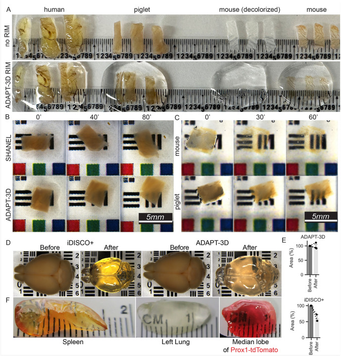
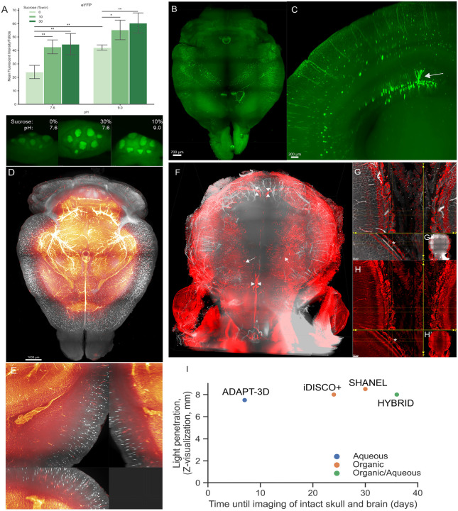
Light sheet microscopy and preparative clearing methods that improve light penetration in 3D tissues have revolutionized imaging in biomedical research. While most clearing methods focus on removing molecules that scatter light, the methods generally involve immersing tissues in solutions that minimize refraction of light to enhance detection of fluorescent signal deeper into tissues. Here, we developed a new tissue preparative method called ADAPT-3D with broad applicability across species and tissue types. This method enables efficient antibody staining and detection of endogenous fluorophores and offers advantages in terms of speed at which tissue staining and clearing is achieved. In about 4 days from tissue harvest to imaging, human intestinal tissue could be Axed, decolored and delipidated to remove light-interfering substances and stained with antibodies for imaging. In the intact mouse skull and brain, involving an 8-day protocol from tissue harvest to completion of imaging, the aqueous and non-shrinking ADAPT-3D method allowed the specialized channels between skull and underlying tissue to be detected without meningeal tearing. Overall, ADAPT-3D provides a highly versatile preparative method for 3D fixed tissue imaging with superior time savings, sensitivity and preservation of tissue morphology compared with previously described methods.


Conflict of interest statement

Declaration of Interests: Washington University and D.D.L., D.L.D., R.S.C., B.Z., and G.J.R. have filed a provisional patent on ADAPT-3D. ADAPT-3D solutions are being commercially developed by Leinco Technologies, Inc., Saint Louis, Missouri (https://www.leinco.com). J. K. is cofounder of Rho Bio that aims to develop therapeutics for the lymphatic vasculature. Other authors declare no competing interests.

Figures

Figure 1
Figure 1. Use of ADAPT-3D on rendering tissues transparent to the eye without shrinkage.
A) Illustration of the impact on tissue transparency to the eye after application of ADAPT-3D RIM solution to triplicate samples of fixed human, piglet, and mouse colons for 10 minutes without decolorization or delipidation. In group noted (decolorized), decolorization solution was applied overnight for mouse colon before immersion in ADAPT-3D RIM for 60 minutes. B) Effect of decolorization solution on full-thickness pieces of piglet colon using SHANEL or ADAPT-3D as a function of time over two 40-minute intervals. C) Effect of the ADAPT-3D RIM solution as a function of time illustrated on fixed mouse or piglet colon samples following decolorization and delipidation for 60 minutes, respectively. D) Before and after top-down view of fixed whole mouse brains by stereoscope following iDISCO+ method and RIM in ethyl cinnamate for 4 hours or application of ADAPT-3D decolorization for 48 hours days, delipidation for 36 hours, and RIM solutions for 4 hours. E) Area of the brain surface images measured in duplicate samples after application of the iDISCO+ method or ADAPT-3D decolorization, delipidation, and RIM solutions as mentioned in D. Symbols on the graph depict paired samples before or after. F) Demonstration of mouse spleen or lung clearing using ADAPT-3D, with decolorization for 48 hours and delipidation for 36 hours, with one liver lobe sample derived from Prox1ERCre × TdTomatofl/fl mouse that was decolorized for 72 hours and delipidated for 36 hours followed by RIM incubation overnight.
Figure 2
Figure 2. Effect of ADAPT-3D on retaining intensities of endogenous fluorescent reporter proteins and visualization across skull-brain interface.
A) Comparison of fluorescent intensities of CD11c-eYFP follicles in Peyer’s patches of mouse ileum following different fixative conditions (n = 4–5 follicles per condition, two-way ANOVA followed by Tukey test, *p-value ≤ 0.05, **p-value ≤ 0.01) and captured by stereomicroscopy. B) 3D whole-mount projection of a brain from 16 weeks old mouse expressing CD11c-eYFP which was decolorized, delipidated, applied in refractive index matching solution followed by imaging with light sheet microscopy. C) 500-micron coronal projection from whole brain of CD11c-eYFP from (B). White arrow points to a CD11c-positive neuron. D) 3D whole-mount projection of a brain from ChAT-CreER × TdTomatofl/fl mouse injected retro-orbitally with Lectin-Dylight649 to label vasculature. E) Extended display near ventricle from d displaying preservation of fine dendrites from neurons. F) Whole mount projection of Lyve1CreER × TdTomatofl/fl mouse injected with i.v. with Lectin-Dylight649 and CD31-Alexa Fluor647. G and H) Extended display displaying skull channels. Asterisk depicts consecutive skull channels emanating across skull to leptomeninges. I) A graphical depiction of the relationship between depth of light penetration in intact mouse brains and skull imaged using light-sheet microscopy compared with the reported times in the literature for other protocols listed on the graph or the 7-day time frame determined here for ADAPT-3D.
Figure 3
Figure 3. Effect of ADAPT-3D on finicky antigens and compatibility with deep immunolabeling.
A) Maximum intensity projections of tight junctions (arachnoid barrier: occludin in red and claudin-11 in green, endothelial-cell specific: claudin-5 in grey) found in leptomeninges from mouse imaged by confocal microscopy. B) Extended display showing en face and z-side projections of mouse ileum that was immunolabeled with alpha smooth muscle actin (yellow), lymphatic vasculature (LYVE-1, magenta), myeloid cells (S100A9, cyan), and nuclei (DAPI, grey) followed by imaging with confocal microscopy. C) Extended display showing en face and z-side projections of ileum from a 16-week-old mouse that expresses TnfΔARE, a model of ileitis. D) Extended display showing en face, z-side, and 3D projections of fixed human ileum applied with decolorization, delipidation, immunolabeling with CD163 (green), IBA1 (red), and nuclei (DAPI, grey) followed by refractive index matching.

References

    1. Daetwyler S, Fiolka RP: Light-sheets and smart microscopy, an exciting future is dawning. Commun Biol 2023, 6:502. - PMC - PubMed
    1. Chung K, Deisseroth K: CLARITY for mapping the nervous system. Nat Methods 2013, 10:508–13. - PubMed
    1. Chung K, Wallace J, Kim SY, Kalyanasundaram S, Andalman AS, Davidson TJ, Mirzabekov JJ, Zalocusky KA, Mattis J, Denisin AK, Pak S, Bernstein H, Ramakrishnan C, Grosenick L, Gradinaru V, Deisseroth K: Structural and molecular interrogation of intact biological systems. Nature 2013, 497:332–7. - PMC - PubMed
    1. Yang B, Treweek JB, Kulkarni RP, Deverman BE, Chen CK, Lubeck E, Shah S, Cai L, Gradinaru V: Single-cell phenotyping within transparent intact tissue through whole-body clearing. Cell 2014, 158:945–58. - PMC - PubMed
    1. Nudell V, Wang Y, Pang Z, Lal NK, Huang M, Shaabani N, Kanim W, Teijaro J, Maximov A, Ye L: HYBRiD: hydrogel-reinforced DISCO for clearing mammalian bodies. Nat Methods 2022, 19:479–85. - PMC - PubMed

Publication types

LinkOut - more resources