[Preprint]. 2025 Mar 26:rs.3.rs-6081213.
doi: 10.21203/rs.3.rs-6081213/v1.

Targeting senescence in Amyotrophic Lateral Sclerosis: senolytic treatment improves neuromuscular function and preserves cortical excitability in a TDP-43Q331K mouse model

Affiliations

Targeting senescence in Amyotrophic Lateral Sclerosis: senolytic treatment improves neuromuscular function and preserves cortical excitability in a TDP-43Q331K mouse model

Jose A Viteri et al. Res Sq. .

Abstract

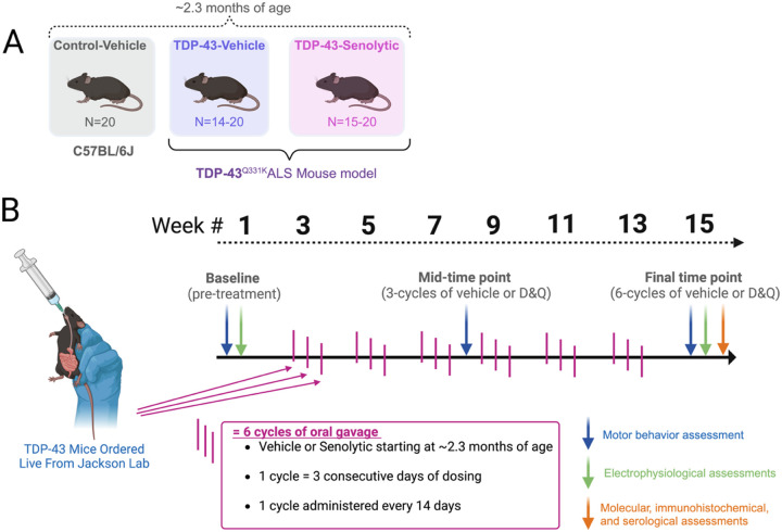
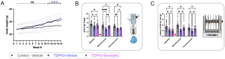
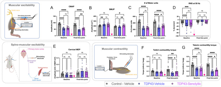
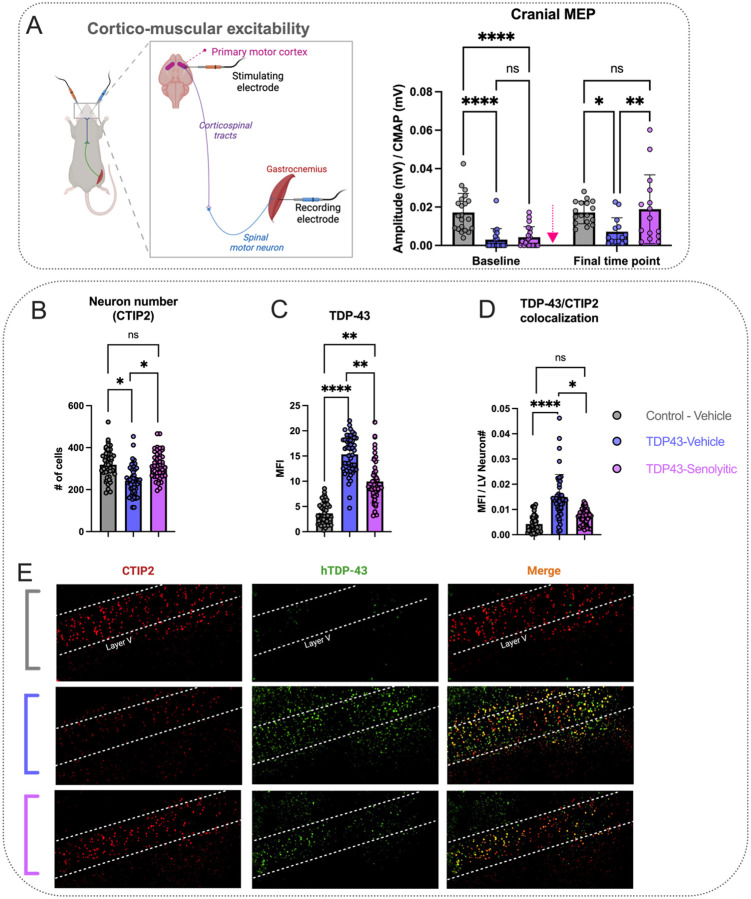
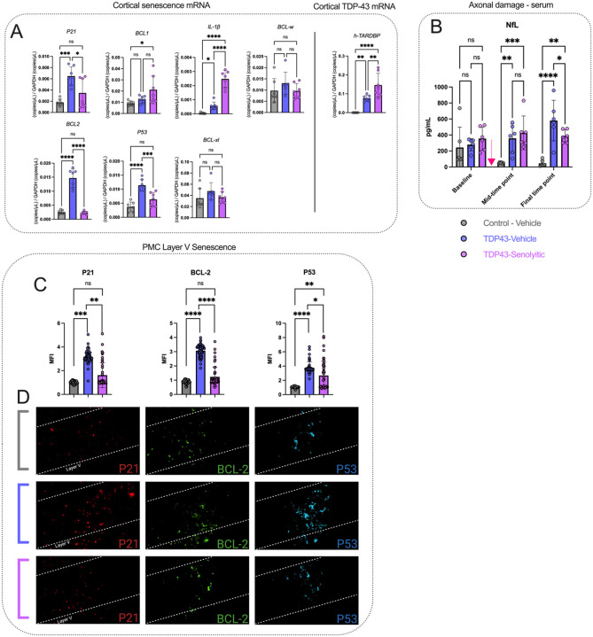
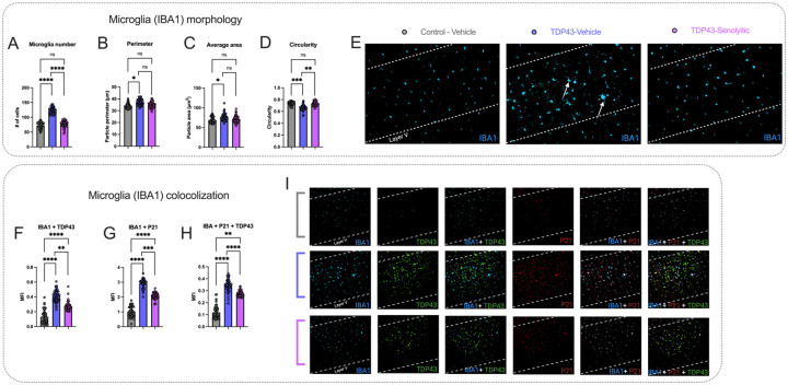
Amyotrophic lateral sclerosis (ALS) is a fatal neurodegenerative disorder marked by progressive motor neuron degeneration in the primary motor cortex (PMC) and spinal cord. Aging is a key factor in ALS onset and progression, with evidence suggesting that biological aging-a process involving cellular decline- far outpaces chronological aging in ALS. This promotes senescent cell accumulation-marked by irreversible cell-cycle arrest, impaired apoptosis, and chronic inflammation-disrupting tissue homeostasis and impairing neuronal support functions. Thus, targeting senescence presents a novel therapeutic strategy for ALS. Here, we investigated the senolytic combination Dasatinib and Quercetin (D&Q) in TDP-43Q331K ALS mice. D&Q improved neuromuscular function and reduced plasma neurofilament light chain, a biomarker of axonal damage. The most pronounced improvement was the improved cortical excitability, accompanied by reductions in senescence and TDP-43 in the PMC. These findings highlight the potential of senolytics to mitigate ALS-related dysfunction, supporting their viability as a therapeutic strategy.

Keywords: ALS; aging; motor cortex; neuromuscular; senescence; senolytics.


Conflict of interest statement

DISCLOSURES The authors have no conflicts of interest to report.

Figures

Figure 1
Figure 1. Experimental design and timeline for evaluating the effects of senolytic treatment in TDP-43 ALS mice.
(A) Experimental groups included control-vehicle mice (C57BL/6J, N = 20), TDP-43Q331K vehicle-treated mice (N = 14–20), and TDP-43Q331K senolytic-treated mice (N = 15–20). All mice were approximately ~ 2.3 months old at the start of the study. Senolytic-treated groups received a combination of Dasatinib and Quercetin (D&Q), while vehicle groups received the equivalent vehicle solution. (B) Experimental timeline showing three main time points of assessment. D&Q or vehicle treatment began at approximately ~ 2.3 months of age (after baseline measures were performed), administered via oral gavage in 6 cycles, with each cycle consisting of 3 consecutive days of dosing every 14 days. Assessments included motor behavior (blue arrows), in-vivo electrophysiology (green arrows), and molecular, immunohistochemical, and serological assessments (orange arrows), conducted at different combinations at baseline (pre-treatment; ~2.3 months of age), a mid-time point (3 cycles of vehicle or senolytics; ~4.5 months of age), and at a final time point (6 cycles of vehicle or senolytics; ~6 months of age). The experimental design highlights longitudinal dosing and assessments to evaluate senolytic efficacy.
Figure 2
Figure 2. Senolytic treatment improves motor function in TDP-43Q331K ALS mice.
(A) Longitudinal body weight measurements over 15 weeks of the study. No significant differences were observed between TDP-43-vehicle and TDP-43-senolytic mice at any time point, though both groups exhibited significantly higher body weights compared to control-vehicle mice after week 11. Data are presented as group means ± SD. (B) All-limb grip strength normalized to body weight (grams/grams of body weight) was assessed across three time points. At the mid-time point, after three cycles of senolytic treatment, TDP-43-senolytic mice showed significantly improved grip strength compared to TDP-43-vehicle mice, but this improvement was lost by the final time point. Control-vehicle mice consistently exhibited higher grip strength than both TDP-43 groups. Grip strength was measured using a force transducer, as illustrated in the inset diagram. (C) Latency to fall during the rotarod test, normalized to body weight (seconds/grams of body weight), was measured to evaluate motor coordination. TDP-43-senolytic mice exhibited improved rotarod performance compared to TDP-43-vehicle mice at the mid-time point and at the final time point, but their performance remained lower than control-vehicle mice. Rotarod setup is illustrated in the inset diagram. Significance: *P < 0.05, **P < 0.01, ****P < 0.0001, ns = not significant. Data are presented as means ± SD. The pink arrow indicates when mice received either vehicle or D&Q.
Figure 3
Figure 3. Senolytic treatment improves neuromuscular excitability and contractility in TDP-43Q331KALS mice.
(A) TDP-43-senolytic mice exhibited significant improvements in compound muscle action potential (CMAP, a measure of summated muscle excitability) amplitudes compared to TDP-43-vehicle mice at the final time point, though values remained lower than control-vehicle mice. (B) Single motor unit potential (SMUP, a measure of NMJ collateral sprouting and remodeling) amplitudes were significantly higher in TDP-43-senolytic mice compared to TDP-43-vehicle mice at the final time point. (C) TDP-43-senolytic mice showed increased motor unit numbers (MUNE, estimated from CMAP and MUNE values) compared to TDP-43-vehicle mice at the final time point but remained lower than control-vehicle mice. (D) While both TDP-43 groups exhibited RNS (Repetitive nerve stimulation decrement at 50 Hz, a measure that quantified NMJ transmission) decrements at baseline, TDP-43-senolytic mice showed no differences in RNS decrement compared to TDP-43-vehicle or control-vehicle groups at the final time point. (E) TDP-43-senolytic mice exhibited no significant improvements in cervical motor-evoked potentials (MEPs, a measure of corticospinal-muscular connectivity) compared to other groups at both time points. (F) TDP-43-senolytic mice exhibited significant improvements in twitch contractility (a measure of instantaneous force production) compared to TDP-43-vehicle mice at the final time point, though values remained lower than controls. (G) TDP-43-senolytic mice displayed enhanced tetanic contractility (a measure of sustained force production) compared to TDP-43-vehicle mice at the final time point, but performance did not reach control-vehicle levels. Illustrations depict the experimental setup for neuromuscular excitability (top-left), cervical MEPs (bottom-left), and neuromuscular contractility (bottom-middle) assessments, showing electrode placements and recording methods. Significance: *P < 0.05, **P < 0.01, ****P < 0.0001, ns = not significant. Data are presented as means ± SD. A pink arrow indicates when mice received either vehicle or D&Q.
Figure 4
Figure 4. Senolytic treatment enhances cortico-muscular excitability and neuron number preservation in TDP-43Q331K ALS mice.
(A) Cranial motor-evoked potentials (MEPs), reflecting cortico-muscular excitability and connectivity, were significantly improved in TDP-43-senolytic mice compared to TDP-43-vehicle mice at the final time point and were no longer significantly different from those in control-vehicle mice. The illustration depicts the experimental setup for cranial MEPs, showing stimulation of the PMC and recording of muscle responses from the gastrocnemius. (B) CTIP2-positive neurons (red), a marker for layer neurons, are reduced in TDP-43-vehicle mice compared to controls. Senolytic treatment significantly increases CTIP2-positive neuronal density compared to TDP-43-vehicle mice, and levels are not significantly different from controls. (C) Human-specific TDP-43 (hTDP-43) expression is significantly elevated in TDP-43-vehicle mice compared to controls. Senolytic treatment reduces hTDP-43 mean fluorescence intensity (MFI) relative to TDP-43-vehicle mice but does not restore levels to those of controls. (D) Colocalization analysis of CTIP2 and hTDP-43 shows significantly increased overlap in TDP-43-vehicle mice, indicating elevated TDP-43 pathology in layer V neurons. Senolytic treatment significantly reduces TDP-43/CTIP2 colocalization compared to TDP-43-vehicle mice, though it remains elevated relative to controls. (E) Representative images show CTIP2, hTDP-43, and merged staining in layer V. Significance: *P < 0.05, **P < 0.01, ****P < 0.0001, ns = not significant. Data are presented as means ± SD. A pink arrow indicates when mice received either vehicle or D&Q. If no pink arrow is present, measurements were done terminally at the final time point (Panels B-D).
Figure 5
Figure 5. Senolytic treatment reduces cortical senescence and TDP-43 expression, while stabilizing systemic axonal damage in TDP-43Q331K mice.
(A) Cortical senescence markers (P21, BCL1, IL-lβ, BCL-w, BCL2, P53, and Bcl-xL) were quantified using digital PCR and normalized to GAPDH. TDP-43-vehicle mice exhibited elevated levels of P21, BCL2, P53, and IL1β compared to controls. Treatment with senolytics significantly reduced P21, BCL2, and P53 levels compared to TDP-43-vehicle mice normalizing these markers to control levels, while IL-lβ remained elevated. BCL1 was elevated in TDP-43-senolytic mice compared to controls but showed no significant difference from TDP-43-vehicle mice. Cortical TDP-43 expression, assessed via human TDP-43 (h-TARDBP) transcript levels, was significantly elevated in TDP-43-vehicle and TDP-43-senolytic mice compared to controls, with TDP-43-senolytic mice showing further increases. (B) Axonal damage, represented by serum neurofilament light (NfL) levels, was significantly elevated in TDP-43-vehicle mice compared to controls at the mid-time point and at the final time point. Senolytic treatment significantly reduced NfL levels at the final time point compared to TDP-43-vehicle mice, though levels remained higher than controls. (C-D) P21 (red), a marker of cell-cycle arrest, is significantly increased in TDP-43-vehicle mice compared to controls. Senolytic treatment significantly reduces P21 expression, and normalizes levels compared to controls. BCL2 (green), an anti-apoptotic protein associated with senescence, is significantly increased in TDP-43-vehicle mice compared to controls. Senolytic treatment significantly reduces BCL2 expression, and normalizes levels compared to controls. P53 (cyan), a tumor suppressor and regulator of cellular senescence, is significantly elevated in TDP-43-vehicle mice compared to controls. Senolytic treatment significantly reduces P53 expression, but levels remain elevated compared to controls. Representative images (bottom) show P21, BCL2, and P53 staining in layer V. Significance: *P < 0.05, **P < 0.01, ****P < 0.0001, ns = not significant. Data are presented as means ± SD. A pink arrow indicates when mice received either vehicle or D&Q. If no pink arrow is present, measurements were done terminally at the final time point (Panels A, C, and D).
Figure 6
Figure 6. Senolytic treatment reduces microglial activation, senescence, and TDP-43 colocalization in TDP-43Q331K mice.
(A-E). TDP-43-vehicle mice showed increased microglial density (A), larger cell perimeter (B), and increased cell area (C) compared to controls. Senolytics reduced microglial density and restored morphology parameters to control levels. Circularity (D) was reduced in TDP-43-vehicle mice compared to controls and improved with senolytics, normalizing to control levels. (E) Representative immunofluorescence images of IBA1 staining in layer V show increased microglial numbers and altered morphology in TDP-43-vehicle mice (white arrows), compared to controls. These changes are reduced in TDP-43-senolytic mice. (F-I). IBA1+TDP43 (F) IBA1+P21 (G) and IBA1+P21+TDP43 (H) colocalization showed significant increases in TDP-43-vehicle mice compared to controls. Senolytic treatment reduced colocalization across all markers, though all values remained higher than controls. (I) Representative images of IBA1+TDP43, IBA1+P21, and IBA1+P21+TDP43 colocalization show increased signals in TDP-43-vehicle mice, which decrease with senolytic treatment. Significance: *P < 0.05, **P < 0.01, ****P < 0.0001, ns = not significant. Data are presented as means ± SD. All measurements were done terminally at the final time point.

Similar articles

References

    1. Masrori P, Van Damme P (2020) Amyotrophic lateral sclerosis: a clinical review. Eur J Neurol 27:1918–1929 - PMC - PubMed
    1. Dashtmian AR, Darvishi FR, Arnold WD (2024) Chronological and Biological Aging in Amyotrophic Lateral Sclerosis and the Potential of Senolytic Therapies. Cells ;13 - PMC - PubMed
    1. Das MM, Svendsen CN (2015) Astrocytes show reduced support of motor neurons with aging that is accelerated in a rodent model of ALS. Neurobiol Aging 36:1130–1139 - PubMed
    1. Cuollo L, Antonangeli F, Santoni A, Soriani A (2020) The Senescence-Associated Secretory Phenotype and Age-Related Diseases. Biology (Basel) 9:1–16 - PMC - PubMed
    1. Hamczyk MR, Nevado RM, Barettino A, Fuster V, Andrés V (2020) Biological Versus Chronological Aging: JACC Focus Seminar. J Am Coll Cardiol 75:919–930 - PubMed

Publication types

LinkOut - more resources