Case Reports
. 2025 Mar 7;17(3):e80219.
doi: 10.7759/cureus.80219. eCollection 2025 Mar.

Middle Cerebral Artery Aneurysm and Distal Anterior Cerebral Artery (DACA) Aneurysms Related to Azygos and an Unusual Single Pericallosal Artery Variant: A Case Report

Affiliations
Case Reports

Middle Cerebral Artery Aneurysm and Distal Anterior Cerebral Artery (DACA) Aneurysms Related to Azygos and an Unusual Single Pericallosal Artery Variant: A Case Report

Victor R Chavez-Herrera et al. Cureus. .

Abstract

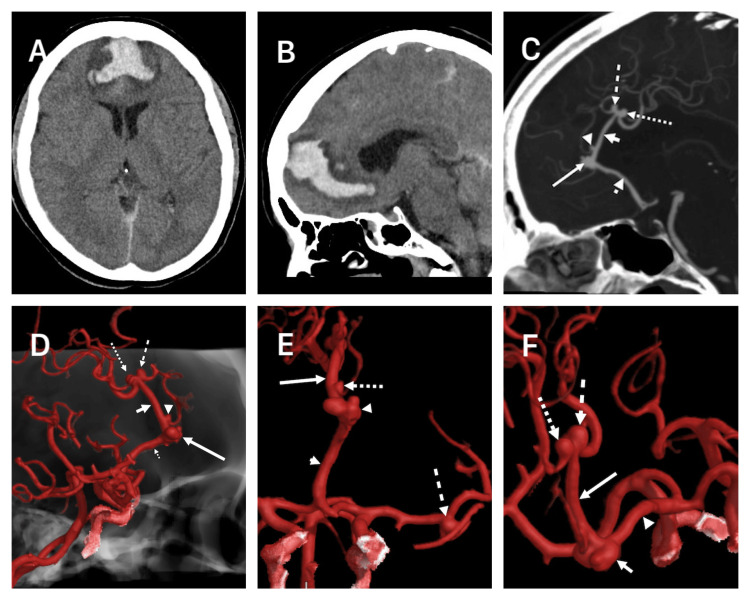
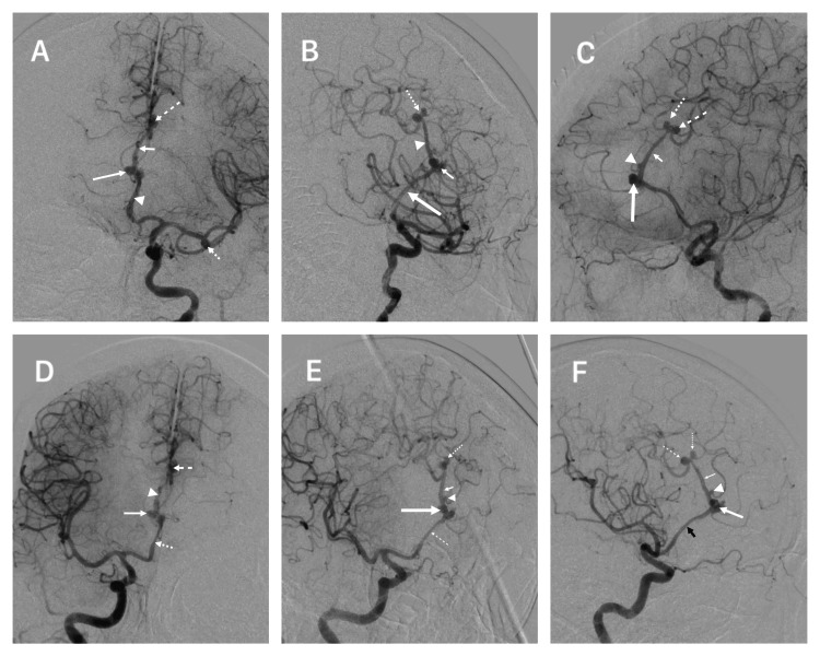
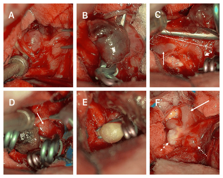
The authors describe the case of multiple rare distal anterior cerebral artery (DACA) aneurysms related to a middle cerebral artery (MCA) aneurysm, an azygos, and an undescribed anterior cerebral artery (ACA) anatomical variation. A 61-year-old woman in a severe clinical state was diagnosed with subarachnoid hemorrhage (SAH) secondary to a ruptured anterior A3 DACA aneurysm. The patient also had unruptured kissing superior A3 DACA aneurysms, an atherosclerotic DACA aneurysm between the anterior and superior DACA aneurysms, and an unruptured MCA left aneurysm. Two pericallosal anatomical variations were seen: an A2 azygos and a rare single post-bifurcation (callosomarginal artery) pericallosal artery. All aneurysms were clipped using a single-staged left craniotomy with interhemispheric, subfrontal, and transsylvian access. This study demonstrates a patient with rare anatomical ACA variants and multiple complex aneurysms treated in a single-stage craniotomy.

Keywords: aneurysm; anterior cerebral artery variant; daca; mirror aneurysm; pericallosal artery.


Conflict of interest statement

Human subjects: Consent for treatment and open access publication was obtained or waived by all participants in this study. Conflicts of interest: In compliance with the ICMJE uniform disclosure form, all authors declare the following: Payment/services info: All authors have declared that no financial support was received from any organization for the submitted work. Financial relationships: All authors have declared that they have no financial relationships at present or within the previous three years with any organizations that might have an interest in the submitted work. Other relationships: All authors have declared that there are no other relationships or activities that could appear to have influenced the submitted work.

Figures

Figure 1
Figure 1. CT images and 3D reconstructed angioCT.
(A) Axial CT showing an interhemispheric-frontal hematoma. (B) Sagittal CT showing the interhemispheric hematoma extending subcallosally. (C) Sagittal AngioCT evidencing four DACAa, an anterior A3 (large arrow), two “kissing” superior A3 (dashed and dotted arrows), and a small A3 between the anterior and superior A3 (arrowhead); azygos A2 ACA variation (dotted arrowhead) and the single A3 pericallosal artery ACA variation (small arrow). (D) Lateral angioCT reconstruction showing multiple DACAa, the large anterior A3 (long arrow), “kissing” superior A3 (long dashed and dotted arrows), small A3 (arrowhead), azygos (short dotted arrow), and the single pericallosal A3 artery (short arrow). (E) Anteroposterior angioCT showing the azygos (short arrow), single pericallosal artery A3 trunk (long arrow), and three aneurysms, the large anterior A3 DACA (arrowhead), the small A3 DACA (dotted arrow), and the MCA (dashed arrow). (F) Superior view angioCT of the “kissing” superior A3 DACAa (dashed and dotted arrows), single A3 pericallosal artery (long arrow), large lobulated anterior A3 DACAa (short arrow), and the azygos variant. CT: computed tomography, DACAa: distal anterior cerebral artery aneurysm, ACA: anterior cerebral artery, MCA: middle cerebral artery.
Figure 2
Figure 2. Digital subtracted angiography
(A) Left anteroposterior DSA showing an anterior A3 DACAa (long arrow), mirror DACAa (dashed arrow), MCA aneurysm (dotted arrow), azygos variant (arrowhead) and single pericallosal artery variant (short arrow). (B) Left oblique DSA showing an anterior A3 DACAa (short arrow), mirror DACAa (dotted arrow), azygos variant (long arrow), and single pericallosal artery variant (arrowhead). (C) Left lateral DSA showing an anterior A3 DACAa (long arrow), mirror DACAa (dotted and dashed arrows), small A3 DACAa (arrowhead), and single pericallosal artery variant (short arrow). (D) Right anteroposterior DSA showing anterior A3 DACAa (long arrow), mirror DACAa (dashed arrow), azygos variant (dotted arrow), and single pericallosal artery variant (arrowhead). (E) Right oblique DSA showing an anterior A3 DACAa (long arrow), mirror DACAa (dotted arrow), azygos variant (dashed arrow), single pericallosal artery variant (short arrow), and a small A3 DACAa (arrowhead). (F) Right lateral DSA showing an anterior A3 DACAa (large arrow), mirror DACAa (dotted and dashed arrows), small A3 DACAa (arrowhead), and single pericallosal artery variant (small arrow). DSA: digital subtracted angiography, DACAa: distal anterior cerebral artery aneurysm, MCA: middle cerebral artery
Figure 3
Figure 3. Operative images
(A) MCA aneurysm. (B) Large anterior A3 DACAa with a preliminary miniclip. (C) Neck of the large anterior A3 DACAa (dashed line), azygos variant (arrow), single pericallosal artery A3 variant (dotted arrow). (D) Anterior A3 DACAa with multiple clips and a remaining lobule portion (arrow). (E) Small A3 DACAa clipped with significant atheroma plaques. (F) “Kissing” DACAa (dashed and dotted arrows) and the single pericallosal artery variant (arrow). MCA: middle cerebral artery, DACAa: distal anterior cerebral artery aneurysm

Similar articles

References

    1. Lehecka M, Dashti R, Lehto H, Kivisaari R, Niemelä M, Hernesniemi J. In: Surgical Management of Cerebrovascular Disease. Acta Neurochirurgica Supplementum. Vol. 107. Vienna: Springer; 2010. Distal anterior cerebral artery aneurysms; pp. 15–26. - PubMed
    1. Anatomic features of distal anterior cerebral artery aneurysms: a detailed angiographic analysis of 101 patients. Lehecka M, Porras M, Dashti R, Niemelä M, Hernesniemi JA. Neurosurgery. 2008;63:219–229. - PubMed
    1. Detailed description of the anterior cerebral artery anomalies observed in a cadaver population. Cilliers K, Page BJ. Ann Anat. 2016;208:1–8. - PubMed
    1. The anatomical variation of the distal anterior cerebral artery: an angiographic study in a Greek population sample. Chrissicopoulos C, Mavrovounis G, Piagkou M, et al. Cureus. 2024;16:0. - PMC - PubMed
    1. The change of location of the anterior cerebral artery in angiographic imaging [Article in German] Fischer E. Zentralbl Neurochir. 1938;3:300–312.

Publication types

LinkOut - more resources