[Preprint]. 2025 Mar 25:arXiv:2503.17600v2.

Imaging Intravoxel Vessel Size Distribution in the Brain Using Susceptibility Contrast Enhanced MRI

Affiliations

Imaging Intravoxel Vessel Size Distribution in the Brain Using Susceptibility Contrast Enhanced MRI

Natenael B Semmineh et al. ArXiv. .

Abstract

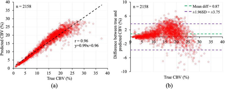
Vascular remodelling is inherent to the pathogenesis of many diseases including cancer, neurodegeneration, fibrosis, hypertension, and diabetes. In this paper, a new susceptibility-contrast based MRI approach is established to non-invasively image intravoxel vessel size distribution (VSD), enabling a more comprehensive and quantitative assessment of vascular remodelling. The approach utilizes high-resolution light-sheet fluorescence microscopy images of rodent brain vasculature, simulating gradient echo sampling of free induction decay and spin echo (GESFIDE) MRI signals for the three-dimensional vascular networks, and training a deep learning model to predict cerebral blood volume (CBV) and VSD from GESFIDE signals. The results from ex vivo experiments demonstrated strong correlation (r = 0.96) between the true and predicted CBV. High similarity between true and predicted VSDs was observed (mean Bhattacharya Coefficient = 0.92). With further in vivo validation, intravoxel VSD imaging could become a transformative preclinical and clinical tool for interrogating disease and treatment induced vascular remodelling.


Figures

Fig. 1.
Fig. 1.
Two representative VOIs (a,b) extracted from LSFM image of a mouse-brain vasculature with similar CBV and mean radius but different VSDs (c).
Fig. 2.
Fig. 2.
The Pearson correlation (a) and Bland-Altman plot (b) of true and predicted CBV (n=2,158).
Fig. 3.
Fig. 3.
Color-coded maps of true (a) and predicted (b) CBV values over an entire axial slice of mouse whole brain LSFM image. The residual map of the true and predicted CBV values is shown in (c).
Fig. 4.
Fig. 4.
The true (green) and predicted (red) VSDs for 12 VOIs with CBV varying from 2.5 to 24%. Significant overlap between the true and predicted VSDs are noticeable despite the true VSD being noisy and of varying shape.
Fig. 5.
Fig. 5.
The results of quantitative evaluation for the VSDE. (a) Distribution of the BC values against the true mean radius for the test VOIs (n=2,158). (b) The Bland-Altman plot of difference between true and predicted mean radius. (c) Same as (b) but for the difference between true mean radius and VSI.
Fig. 6.
Fig. 6.
Qualitative results of VSD prediction on an entire axial slice of mouse whole brain LSFM image. (a,b) Color-coded coefficient of variance (CV) maps of the true (a) and predicted (b) VSDs. (c) The residual map of the true and predicted CV maps. (d) The map of BC values measured between the true and predicted VSDs.
Fig. 7.
Fig. 7.
Qualitative results of mean vessel radius and VSI computation on an entire axial slice of mouse whole brain LSFM image. (a-c) Color-coded maps of the true (a) and predicted (b) mean radius and VSI (c) values. (d,e) The residual map of true and predicted mean radius and the true mean radius and VSI.
Fig. 8.
Fig. 8.
Steps involved in animal preparation and LSFM imaging of a rat brain. (a) A rat brain after skull removal. (b) Same brain after tissue clearing. (c) Three-dimensional (3D) rendition of an entire rat brain vasculature along with a zoomed in volume of interest (VOI) for better representation of the highlighted vessels. Note that, large vessels have hollow lumen (white arrow) as lectin only stains the vessel walls. This approach enables the visualization of blood vessels down to capillary size.
Fig. 9.
Fig. 9.
Steps involved in binary segmentation of the vasculature from a LSFM image and computation of the VSD. (a) Axial view of a LSFM VOI with an array size of 123 × 123 × 123. (b) Segmented vasculature from (a) after contrast limited adaptive histogram equalization (CLAHE) and binary thresholding. (c) Same as (b) but after applying morphological hole filling to close the hollow lumen of the large vessels. (d) 3D rendition of the maximally connected segmented vascular network. (e) Skeleton of (d) where individual vessels are uniquely labelled by distinct colours. (f) Color-coded rendition of voxel-wise radius map of (d). (g) True VSD of (d) computed as the normalized histogram of vessel radius values with a bin size of 1 μm. See text for details.
Fig. 10.
Fig. 10.
Example VOIs (1st column) extracted from LSFM images with varying CBV and the corresponding VSD (2nd column) and ratio of simulated pre- and post-contrast GESFIDE signals (3rd column). Variations in signal at a specific echo time result from disparities in vessel size, orientation, and CBV.
Fig. 11.
Fig. 11.
The DL framework used to predict the VSD from a given GESFIDE signal. (a) Two FCNs called the CBVE and VSDE are trained simultaneously to predict the CBV and the VSD from the GESFIDE signal, respectively. (b,c) The network architecture of the CBVE (b) and VSDE (c); see text for details.

References

    1. Carmeliet P and Jain RK, “Angiogenesis in cancer and other diseases,” Nature, 407, 249–57, 2000. - PubMed
    1. Gatenby RA and Gillies RJ, “A microenvironmental model of carcinogenesis,” Nat Rev Cancer, 8, 56–61, 2008. - PubMed
    1. Hanahan D and Weinberg RA, “Hallmarks of cancer: the next generation,” Cell, 144, 646–74, 2011. - PubMed
    1. Nagy JA, Dvorak AM, and Dvorak HF, “Vascular hyperpermeability, angiogenesis, and stroma generation,” Cold Spring Harb Perspect Med, 2, a006544, 2012, . - PMC - PubMed
    1. Ross R, “Atherosclerosis--an inflammatory disease,” N Engl J Med, 340, 115–26, 1999. - PubMed

Publication types

LinkOut - more resources