[Preprint]. 2025 Mar 25:2025.03.21.644649.
doi: 10.1101/2025.03.21.644649.

X-Linked Cancer-Associated Polypeptide (XCP) from lncRNA1456 Cooperates with PHF8 to Regulate Gene Expression and Cellular Pathways in Breast Cancer

Affiliations

X-Linked Cancer-Associated Polypeptide (XCP) from lncRNA1456 Cooperates with PHF8 to Regulate Gene Expression and Cellular Pathways in Breast Cancer

Shrikanth S Gadad et al. bioRxiv. .

Abstract

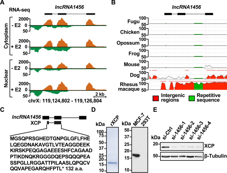
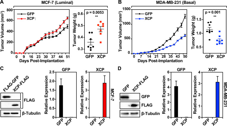
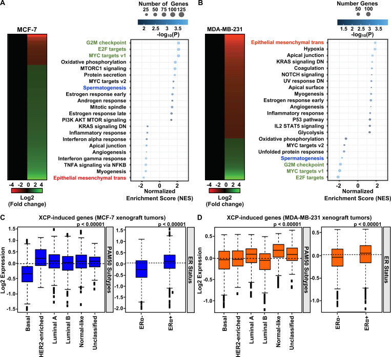
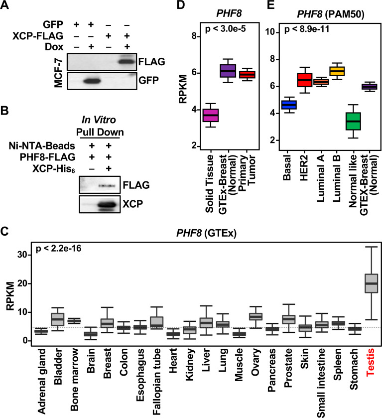
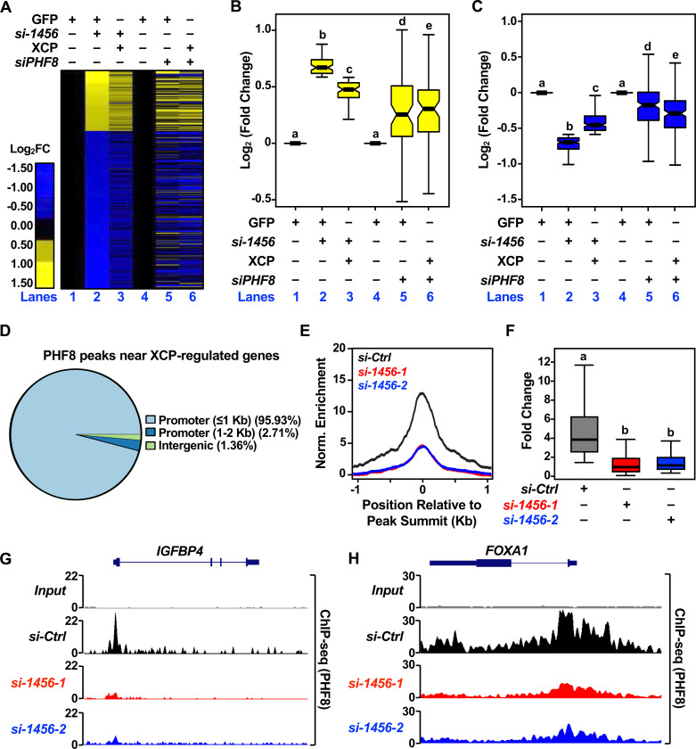
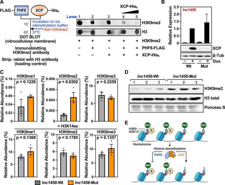
Recent studies have demonstrated that a subset of long "noncoding" RNAs (lncRNAs) produce functional polypeptides and proteins. In this study, we discovered a 132 amino acid protein in human breast cancer cells named XCP (X-linked Cancer-associated Polypeptide), which is encoded by lncRNA1456 (a.k.a. RHOXF1P3), a transcript previously thought to be noncoding. lncRNA1456 is a pancreas- and testis-specific RNA whose gene is located on chromosome X. We found that the expression of lncRNA1456 and XCP are highly upregulated in the luminal A, luminal B, and HER2 molecular subtypes of breast cancer. XCP modulates both estrogen-dependent and estrogen-independent growth of breast cancer cells by regulating cancer pathways, as shown in cell and xenograft models. XCP shares some homology with homeodomain-containing proteins and interacts with the histone demethylase plant homeodomain finger protein 8 (PHF8), which is also encoded by an X-linked gene. Mechanistically, XCP stimulates the histone demethylase activity of PHF8 to regulate gene expression in breast cancer cells. These findings identify XCP as a coregulator of transcription and emphasize the need to interrogate the potential functional roles of open reading frames originating from noncoding RNAs.

Keywords: PHF8; X-Linked Cancer-Associated Polypeptide (XCP); breast cancer; cancer cell growth; chromatin; histone demethylase; lncRNA.


Conflict of interest statement

Declaration of interests The authors have no competing interests to declare.

Figures

Figure 1.
Figure 1.. Characterization of lncRNA1456 and its encoded peptide, XCP.
A) Genome browser view for the lncRNA1456 locus from fractionated RNA-seq data. B) ECR browser tracks showing conservation of lncRNA1456 across different species. C) Diagram showing the putative lncRNA1456- encoded peptide from exons 1 and 2 and amino acid sequence. D) Coomassie gel showing recombinant XCP peptide (left) and its endogenous detection in MCF-7 cells by Western blot using a derived XCP antibody (right). E) Western blot showing reduced levels of endogenous XCP peptide upon siRNA-mediated knockdown of lncRNA1456 RNA in MCF-7 cells. [See also Figure S1]
Figure 2.
Figure 2.. Expression of lncRNA1456 RNA and XCP peptide in human normal and breast cancer tissues.
A) GTEx survey of lncRNA1456 RNA expression across normal human tissues. Observed differences are significant as determined by an ANOVA comparison of the means (p-value < 2.2 e-16). Insets show immunohistochemical staining of pancreas and testis using antibody against XCP. B) Graph showing lncRNA1456 RNA expression in normal solid tissue (NT; TCGA), normal breast tissue (N; GTEx) and malignant primary breast tumors (T; TCGA) (left) and lncRNA1456 RNA expression in malignant primary breast tumors (TCGA) stratified into subtypes (B: basal; H: HER2; LA/LB: luminal A/B; NL: normal like) using PAM50 gene set analysis compared to normal breast tissue (N; GTEx) (right). Observed differences are significant as determined by an ANOVA comparison of the means. C) Representative immunohistochemical staining for XCP peptide in normal versus malignant (IDC) breast tissue. D) Frequency of XCP peptide expression across non-malignant (NM) and malignant plus metastatic (MM) breast tissues based on IHC staining intensity scores. E) Expression scores based on IHC of XCP in non-malignant (left) and malignant plus metastatic (right) breast cancer samples, stratified by ER status. Each bar represents the mean ± SEM; For Non-Mal, ER(−) n=8 and ER(+) n=36; For Mal+Met, ER(−) n=53 and ER(+) n=89. Significance was calculated using unpaired t-test.
Figure 3.
Figure 3.. XCP modulates tumor growth in a context-dependent manner.
A and B) Xenograft assays showing subcutaneous tumor growth of MCF-7 (A) or MDA-MB-231 (B) breast cancer cells ectopically expressing dox-inducible FLAG-GFP or XCP-FLAG (left). Tumor weight at end of experiment is shown (right). Animals injected with MCF-7 cells had E2-pellet implantations at the back of the neck to promote growth of MCF-7 cells in vivo. All animals were fed doxycycline in their chow. Each point represents the mean ± SEM. For MCF-7, GFP n=7 and XCP n=7; For MDA-MB-231, GFP n=8 and XCP n=8. Significance was calculated using unpaired t-test. C and D) Expression of FLAG-GFP or XCP-FLAG in tumor tissue was validated from MCF-7 (C) or MDA-MB-231 (D) xenografts tumors by Western blot (left) and RT-qPCR (right). Each bar represents the mean + SEM. For MCF-7, GFP n=5 and XCP n=5; For MDA-MB-231, GFP n=8 and XCP n=8.
Figure 4.
Figure 4.. XCP modulates gene expression and regulates cellular pathways in a context-dependent manner.
A and B) Heatmaps (left) and Gene Ontology analysis (right) showing XCP-mediated gene expression changes in MCF-7 (A) and MDA-MB-231 (B) xenograft tumors. C and D) Box plots showing gene set analysis of XCP-induced genes and their expression levels stratified into breast cancer subtypes using PAM50 gene set analysis (left) and stratified by ER status (right) in MCF-7 (C) and MDA-MB-231 (D) xenograft tumors. Observed differences are significant as determined by an ANOVA comparison of the means (P value < 0.00001).
Figure 5.
Figure 5.. XCP interacts with the demethylase PHF8.
A) Western blot showing expression of GFP and FLAG-tagged XCP used for pull down from MCF-7 whole cell lysates. B) Western blot showing interaction of PHF8 and XCP confirmed in an in vitro pull down assay using recombinant PHF8 and XCP. C) GTEx survey of PHF8 expression across normal human tissues. Observed differences are significant as determined by an ANOVA comparison of the means (p-value < 2.2 e-16). D) Graph showing PHF8 RNA expression in normal solid tissue (TCGA), normal breast tissue (GTEx) and malignant primary breast tumors (TCGA). Observed differences are significant as determined by an ANOVA comparison of the means. E) PHF8 RNA expression in malignant primary breast tumors (TCGA) stratified into subtypes using PAM50 gene set analysis compared to normal breast tissue (GTEx). Observed differences are significant as determined by an ANOVA comparison of the means. [See also Figure S2]
Figure 6.
Figure 6.. XCP modulates gene expression through its interaction with the demethylase PHF8.
A) Heatmap from RNA-seq data showing the regulation of 239 genes upon siRNA-mediated knockdown of lncRNA1456 RNA or PHF8 mRNA and co-expression of XCP-FLAG. B and C) Box plots quantifying the upregulated (B) and downregulated (C) gene expression changes observed in heatmap of 239 XCP-regulated genes (A). Each bar represents the mean ± SEM; Bars marked with different letters are significantly different from each other, Wilcox rank sum test. D) Pie chart showing the distribution of PHF8 peaks at the 239 XCP-regulated genes. E and F) Metagene plot (E) and box plot (F) showing the reduced enrichment of PHF8 binding upon siRNA-mediated knockdown of lncRNA1456. Each bar represents the mean ± SEM; Bars marked with different letters are significantly different from each other, Wilcox rank sum test. G and H) Browser tracks showing reduced PHF8 binding near promoter of IGFBP4 and FOXA1 genes. [See also Figure S3]
Figure 7.
Figure 7.. XCP directly modulates the demethylase activity of PHF8.
A) Diagram showing the flow of in vitro demethylase reactions using recombinant PHF8-FLAG, His6-XCP and H3K9me2 peptide (left). Dot bot showing the demethylase activity of PHF8 on H3K9me2 peptide in the presence or absence of XCP (right). B) RT-qPCR (top) and Western blot (bottom) showing dox-inducible ectopic expression of full length lncRNA1456 in its wild-type form, which expresses the XCP peptide, or an ATG mutant version which abolishes XCP peptide production. Each bar represents the mean + SEM; n= 3. C) Bar graphs showing global changes in histone post-translational modifications assayed mass spectrometry of MCF-7 histones. Each bar represents the mean ± SEM; n=3. Significance calculated by 1-sided Welch’s t test. D) Western blot analysis of MCF-7 histone preparation used for mass spectrometry analysis (C). E) Diagram summarizing model: XCP, encoded by lncRNA1456, interacts with PHF8, a histone demethylase, and directly regulates its activity to modulate gene expression.

Similar articles

References

    1. Djebali S, Davis CA, Merkel A, Dobin A, Lassmann T, Mortazavi A, Tanzer A, Lagarde J, et al. (2012) Landscape of transcription in human cells. Nature 489:101–108. - PMC - PubMed
    1. Lin MF, Jungreis I, Kellis M (2011) PhyloCSF: a comparative genomics method to distinguish protein coding and non-coding regions. Bioinformatics 27:i275–282. - PMC - PubMed
    1. Scheidler CM, Kick LM, Schneider S (2019) Ribosomal Peptides and Small Proteins on the Rise. Chembiochem 20:1479–1486. - PubMed
    1. Huarte M (2015) The emerging role of lncRNAs in cancer. Nat Med 21:1253–1261. - PubMed
    1. Barczak W, Carr SM, Liu G, Munro S, Nicastri A, Lee LN, Hutchings C, Ternette N, et al. (2023) Long non-coding RNA-derived peptides are immunogenic and drive a potent anti-tumour response. Nat Commun 14:1078. - PMC - PubMed

Publication types