[Preprint]. 2025 Mar 24:2025.03.24.644916.
doi: 10.1101/2025.03.24.644916.

Studies on Gene Enhancer with KSHV mini-chromatin

Affiliations

Studies on Gene Enhancer with KSHV mini-chromatin

Tomoki Inagaki et al. bioRxiv. .

Abstract

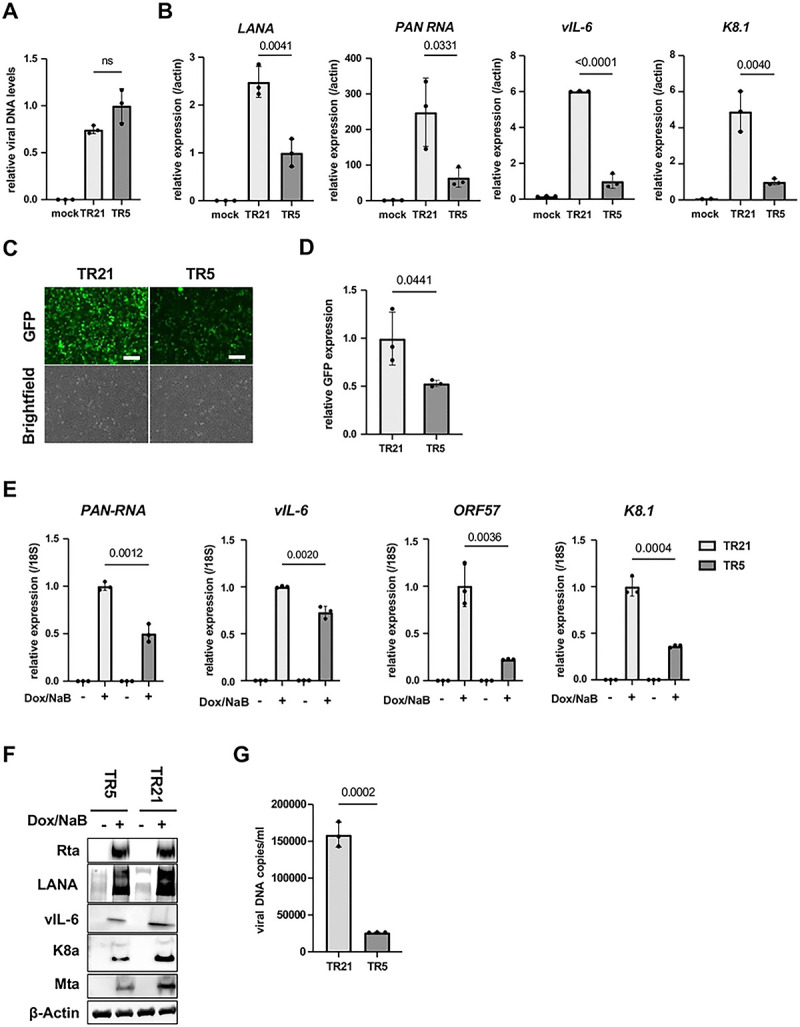
Kaposi's sarcoma-associated herpesvirus (KSHV) genome contains a terminal repeats (TR) sequence. Previous studies demonstrated that KSHV TR functions as a gene enhancer for inducible lytic gene promoters. Gene enhancers anchor bromodomain-containing protein 4 (BRD4) at specific genomic region, where BRD4 interacts flexibly with transcription-related proteins through its intrinsically disordered domain and exerts transcription regulatory function. Here, we generated recombinant KSHV with reduced TR copy numbers and studied BRD4 recruitment and its contributions to the inducible promoter activation. Reducing the TR copy numbers from 21 (TR21) to 5 (TR5) strongly attenuated viral gene expression during de novo infection and impaired reactivation. The EF1α promoter encoded in the KSHV BAC backbone also showed reduced promoter activity, suggesting a global attenuation of transcription activity within TR5 latent episomes. Isolation of reactivating cells confirmed that the reduced inducible gene transcription from TR-shortened DNA template and is mediated by decreased efficacies of BRD4 recruitment to viral gene promoters. Separating the reactivating iSLK cell population from non-responders showed that reactivatable iSLK cells harbored larger LANA nuclear bodies (NBs) compared to non-responders. The cells with larger LANA NBs, either due to prior transcription activation or TR copy number, supported KSHV reactivation more efficiently than those with smaller LANA NBs. With auxin-inducible LANA degradation, we confirmed that LANA is responsible for BRD4 occupancies on latent chromatin. Finally, with purified fluorescence-tagged proteins, we demonstrated that BRD4 is required for LANA to form liquid-liquid phase-separated dots. The inclusion of TR DNA fragments further facilitated the formation of larger BRD4-containing LLPS in the presence of LANA, similar to the "cellular enhancer dot" formed by transcription factor-DNA bindings. These results suggest that LANA binding to TR establishes an enhancer domain for infected KSHV episomes. The strength of this enhancer, regulated by TR length or transcription memory, determines the outcome of KSHV replication.


Conflict of interest statement

Competing interests: YI declares a competing interest relating to founding role for VGN Bio Inc. All other authors declare they have no competing interests.

Figures

Figure 1.
Figure 1.. Generation of recombinant KSHV BAC with different numbers of TR copies. (A). Schematic diagram of KSHV BAC16 with different copy numbers of TR.
The kanamycin cassette with I-SceI recognition sequence, along with the TR homologous sequence, was generated by PCR with pEP-Kan plasmid as a template. This DNA fragment was cloned into KSHV BAC16 by homologous recombination. The kanamycin cassette was deleted by recombination with induction of I-SceI in bacteria by incubation with L-arabinose. Correct insertion of the different copy numbers of TR was confirmed by PstI digestions. (B). Confirmation of TR copies and BAC integrity. Purified KSHV BAC16 was digested by PstI and subjected to electrophoresis. Agarose gel was stained with Ethidium bromide (EtBr) (left panel). The TR-containing fragments were probed with Southern blotting with a fluorescently labelled TR probe (right panel). The numbers in the Southern blotting represent the length (base pair) of the TR fragment after PstI digestion. The 1 kb DNA ladder is shown. (C). Fluorescence images of iSLK cells with different copy numbers of TR. KSHV BAC16 with different copy numbers of TR were transfected into iSLK cells. The mock indicates iSLK cells without KSHV BAC16. Scales: 300μm. (D). Proportion of GFP positive cells in iSLK cells with different copy numbers of TR. iSLK cells harboring KSHV BAC16 with different copy numbers of TR were cultured under hygromycin, and the proportion of GFP positive cells was calculated using flow cytometry. E. Mean fluorescence intensity (MFI) of iSLK cells with different copy numbers of TR. MFI in the FITC channel was determined using FlowJo v10 software.
Figure 2.
Figure 2.. KSHV inducible gene expression is positively regulated in a TR-dependent manner. (A). Relative viral DNA levels in KSHV-infected iSLK cells.
iSLK cells were infected with TR5-KSHV or TR21-KSHV for 24 hours and viral DNA levels were determined by quantitative PCR (qPCR). GAPDH expression was used for internal control. Relative viral DNA levels in TR5-KSHV iSLK cells was set as 1. Data was analyzed using a two-sided unpaired Student’s t-test and shown as mean ± SD. (B). KSHV gene expression in iSLK cells during de novo infection. Relative RNA levels of the indicated viral genes were determined by Reverse Transcription qPCR (RT-qPCR).18S rRNA expression was used for internal control. Relative gene expression in TR5-KSHV iSLK cells was set as 1. Data was analyzed using two-sided unpaired Student’s t test and shown as mean ± SD. (C). Fluorescence and brightfield images of iSLK cells infected with TR5-KSHV or TR21-KSHV. The iSLK cells were infected with the equal amount of TR5-KSHV and TR21-KSHV followed by selection with hygromycin. Scales: 300μm. (D). Relative GFP expression in iSLK cells infected with TR5-KSHV or TR21-KSHV. Total RNAs were harvested from iSLK cells infected with the TR5-KSHV or TR21-KSHV. 18S rRNA expression was used for internal control, and relative GFP expression was normalized by the viral genome. Relative GFP expression in TR21-KSHV-infected iSLK cells was set as 1. Data was analyzed using two-sided unpaired Student’s t test and shown as mean ± SD. (E). Relative KSHV gene expression in iSLK cells infected with TR5-KSHV or TR21-KSHV. Each iSLK cell was reactivated by sodium butyrate and doxycycline. Total RNAs were purified two days after reactivation. 18S rRNA was used for internal control, and relative gene expression in TR21 iSLK cells after reactivation was set as 1. Data was analyzed using two-sided unpaired Student’s t-test and shown as mean ± SD. (F). Immunoblotting of KSHV latent protein (LANA) and lytic proteins (Rta, vIL-6, K8α, Mta) in TR5-KSHV or TR21-KSHV-infected iSLK cells. iSLK cells were reactivated by sodium butyrate and doxycycline and total cell lysates were prepared two days after reactivation and subjected to immunoblotting with specific antibodies. β-actin was used as a loading control. (G). KSHV virion production after reactivation of TR5-KSHV or TR21-KSHV-infected iSLK cells. Virions in the supernatant four days after reactivation by sodium butyrate and doxycycline were treated with DNaseI, followed by DNA purification. Results are shown as viral copies per milliliter. ORF6 was used to quantify the viral DNA. The data was analyzed using a two-sided unpaired Student’s t-test and shown as mean ± SD.
Figure 3.
Figure 3.. BRD4 regulates KSHV inducible gene expression in the TR dependent manner. (A). Immunofluorescence image of the LANA protein in TR5-KSHV or TR21-KSHV-infected iSLK cells.
Anti-LANA rat monoclonal antibody and alexa-647 (secondary) goat anti-rat antibody were used for staining LANA in TR5- or TR21-KSHV-infected iSLK cells and Cy5 channel was used for its detection. LANA dots (upper panels) and DAPI (lower panels) are shown. Scales: 50 μm. (B). Perimeter of each LANA dots. The size of each LANA dot in TR5- or TR21-KSHV-infected iSLK cells was calculated by imageJ (ver.1.53). Data was analyzed using a two-sided unpaired Student’s t-test and shown as mean ± SD. (C). Representative images of proximity ligation assay (PLA) for BRD4 and LANA interaction in TR5- or TR21-KSHV-infected iSLK cells. TR5- or TR21-KSHV-infected iSLK cells were subjected to PLA using antibodies against BRD4 (1:100 dilution) and LANA (1:100 dilution). Red dots indicate BRD4-LANA interactions. Scales: 20 μm. (D). Quantification of PLA dots. Each PLA dot in TR5- or TR21-KSHV-infected iSLK cells was calculated by imageJ (ver.1.53). (E). Enrichment of BRD4 and H3K27Ac in TR5- or TR21-KSHV-infected iSLK cells. TR5 or TR21-KSHV-infected iSLK cells were subjected to CUT&RUN analysis with anti-BRD4 (1:100 dilution) or anti-H3K27Ac (1:100 dilution) antibodies to examine their occupancy on the specific promoter regions in the KSHV genome. The control region was the coding region of the ORF23, which has been shown not to accumulate BRD4. Normal Rabbit IgG antibody was used as a negative control. Data was analyzed using a two-sided unpaired Student’s t-test and shown as mean ± SD. (F). Immunoblotting assay of BRD4 and KSHV proteins. r.219 iSLK cells were reactivated by doxycycline and sodium butyrate one day after MZ1 (100 nM) treatment. Two days after reactivation, total cell lysates were collected for immunoblotting to detect BRD4 and KSHV proteins (vIL-6 and K-Rta). β-actin was used as a loading control. (G). Virion production in r.219 iSLK cells with or without MZ1 treatment. Virions in the supernatant four days after r.219 iSLk cells reactivated by sodium butyrate and doxycycline were treated with DNaseI, followed by DNA purification. MZ1 or DMSO were added one day before reactivation. Results are shown as viral copies per microliter. Data was analyzed using a two-sided unpaired Student’s t-test. (H). Proportion of GFP positive cells after r.219 KSHV infection. r.219 iSLK cells were reactivated by sodium butyrate and doxycycline one day after MZ1 or DMSO treatment. Four days after reactivation, an equal amount of supernatant was added to iSLK cells, and the proportion of GFP-positive cells was determined by flow cytometry one day after infection. (I). Experimental flow of LANA degradation. mAID-LANA KSHV BAC16 iSLK cells were treated with indole-3-acetic acid (IAA) (5 μM) for four hours, followed by reactivation with sodium butyrate and doxycycline for one day to examine the effect of LANA on BRD4 accumulation on the KSHV genome. (J). Immunoblotting assay of KSHV gene expression in LANA AID iSLK cells with or without IAA treatment. One day after reactivation, total cell lysates were collected for immunoblotting to detect KSHV proteins (LANA and K-Rta). β-actin was used as a loading control. (K). BRD4 occupancy in mAID-LANA KSHV BAC16 iSLK cells. One day after reactivation. BRD4 enrichment on the Ori-RNA promoter and TR region was determined using CUT&RUN. Normal Rabbit IgG antibody was used as a negative control. Data was analyzed using a two-sided unpaired Student’s t-test and shown as mean ± SD.
Figure 4.
Figure 4.. Establishment of cBu-1 BAC16 for the isolation of reactivated cell population. (A). Schematic model of the isolation of reactivated cells using chicken Bu-1 (cBu-1) BAC16 system.
cBu-1 external domain sequence along with GSG-P2A peptide sequence was inserted as an N-terminal fusion with vIL-6 protein. After reactivation by doxycycline and sodium butyrate, cBu-1 is expressed on the cell surface only in the vIL-6 expressed cells, and anti-cBu-1 antibody can bind to cBu-1 protein for isolation. (B). Representative FACS profiles of iSLK cells with cBu-1 BAC (cBu-1 iSLK cells). Two days after reactivation by sodium butyrate and doxycycline, cells were fixed by 2% paraformaldehyde and permeabilized by 0.1% Triton X-100. Cells were then stained by vIL-6 antibody and cBu-1 antibody for two hours at room temperature. After washing with PBS, cells were incubated with secondary antibodies (APC for vIL-6 and PE for cBu-1). (C). Brightfield image of cBu-1-expressed cells and non-expressed cells. Two days after reactivation, cells were incubated with a biotin-conjugated anti-cBu-1 antibody (10 μg/ml) for one hour at room temperature. Cells were washed with PBS twice, followed by incubation with streptavidin magnetic beads for one hour at room temperature. Cells were then observed on the cover glass. Scales: 20 μm. (D). Immunoblotting of KSHV lytic proteins (ORF59 and vIL-6) in cBu-1 iSLK cells with or without cBu-1 enrichment after reactivation. cBu-1 iSLK cells were reactivated by sodium butyrate and doxycycline for two days, and cBu-1 expressed cells (IP) were then enriched with cBu1 monoclonal antibody (10 μg/ml). The cell population that did not express cBu-1 was also harvested as flow thorough at the same time (FL). β-actin was used as a loading control. (E). vIL-6 expression of cBu-1 expressed cells (IP) and non-expressed cells (FL) after isolation by cBu-1 antibody. 18S rRNA expression was used for internal control. Relative gene expression in FL population was set as 1. Data was analyzed using a two-sided unpaired Student’s t-test and shown as mean ± SD. (F). Experimental design of the isolation of reactivated cells by cBu-1 system for the following analysis. cBu-1 virus with different copy numbers of TR (cBu-1 TR21 or cBu-1 TR6) were infected with iSLK cells and cultured with hygromycin. One day after reactivation by sodium butyrate and doxycycline, cBu-1 expressed cells (IP), and cBu-1 non-expressed cells (FL) were sorted by magnetic beads for the following analysis. (G). Relative KSHV gene expression in iSLK cells after cBu-1 enrichment. cBu-1 TR21- or cBu-1 TR6-KSHV-infected iSLK cells were reactivated by sodium butyrate and doxycycline. One day after reactivation, reactivated cells were isolated according to cBu-1 expression, and total RNAs were purified to determine the viral gene expression. 18S rRNA was used as an internal standard for normalization, and relative gene expression was normalized by the viral genome. Relative gene expression in cBu-1 TR6-KSHV-infetced iSLK cells was set as 1. Data was analyzed using two-sided unpaired Student’s t test and shown as mean ± SD. (H). BRD4 and H3K27Ac occupancy in cBu-1 TR21- or cBu-1 TR6-KSHV-infected iSLK cells after cBu-1 enrichment. cBu-1 TR21- or cBu-1 TR6-KSHV-infected iSLK cells were reactivated by sodium butyrate and doxycycline. One day after reactivation, reactivated cells were isolated according to cBu-1 expression, and BRD4 or H3K27Ac enrichment of the Ori-RNA promoter TR, and vIL-6 promoter region was determined. Normal Rabbit IgG antibody was used as a negative control. Data was analyzed using a two-sided unpaired Student’s t-test and shown as mean ± SD.
Figure 5.
Figure 5.. LANA NBs functions as transcription memory. (A). Experimental workflow of the isolation of reactivated cells by cBu-1 system for the following analysis.
Two days after reactivation by sodium butyrate and doxycycline, cBu-1 expressed cells (IP) and cBu-1 non-expressed cells (FL) were sorted by magnetic beads. Cells were then cultured for ten days for the following analysis. (B). Immunofluorescence analysis showing LANA dots in IP and FL population. Anti-LANA rat monoclonal antibody and alexa-647 (secondary) goat anti-rat antibody were used for staining LANA in IP and FL population from cBu-1 TR21-KSHV-infected iSLK cells. Cy5 channel was used for its detection. LANA dots (upper panels) and DAPI (lower panels) are shown. Scales: 20 μm. (C). Quantification of LANA dots. The size of each LANA dot in IP and FL population was determined and calculated by imageJ (ver.1.53). (D). Relative KSHV gene expression in IP and FL population. IP and FL population were cultured for ten days, followed by reactivation by sodium butyrate and doxycycline. Two days after reactivation, total RNAs were purified to determine the viral gene expression. 18S rRNA was used as an internal standard for normalization, and relative gene expression was normalized by the viral genome. Data was analyzed using two-sided unpaired Student’s t test and shown as mean ± SD. (E). Experimental workflow of the isolation and the comparison of reactivated cBu-1 TR21- and TR6-KSHV infected iSLK cells. cBu-1 TR21 or cBu-1 TR6 virus were infected with iSLK cells and preapred stably infected cells with hyglomycin selection. Two days after reactivation, cBu-1 expressed cells were sorted by magnetic beads. Cells were then cultured for ten days for the following analysis. (F). Relative KSHV gene expression in cBu-1 TR21- and cBu-1 TR6-KSHV infected iSLK cells. cBu-1 expressed population in cBu-1 TR21- and TR6-KSHV infected iSLK cells were cultured for ten days, followed by reactivation. Re-seeded cells were reactivated again, and total RNAs were purified to determine the viral gene expression. 18S rRNA was used as an internal standard for normalization, and relative gene expression was normalized by the viral genome. Data was analyzed using two-sided unpaired Student’s t test and shown as mean ± SD. (G). BRD4 and H3K27Ac occupancy in cBu-1 TR21- and cBu-1 TR6-KSHV-infected iSLK cells. BRD4 or H3K27Ac enrichment of the Ori-RNA promoter and TR region was determined using CUT&RUN. Normal Rabbit IgG antibody was used as a negative control. Data was analyzed using a two-sided unpaired Student’s t-test and shown as mean ± SD.
Figure 6.
Figure 6.. BRD4 and LANA protein form LLPS. (A). Representative image of fluorescent proteins.
Indicated proteins were purified with Flag-beads and imaged under UV light. From left to right (up): mCherry-BRD4 and EGFP-LANA (bottom) mCherry and EGFP proteins. (B). SDS-PAGE analysis of purified proteins. Molecular weight markers (kDa) are indicated on the left. (C). Representative images of LANA LLPS with or without BRD4. EGFP-LANA (1 μM) was mixed with mCherry-BRD4 protein (1 μM) or mCherry protein (1 μM). Images were taken five minutes after the mixture of the proteins. Scales: 20 μm. (D). Representative images of LANA and BRD4 LLPS in the presence or absence of NaCl or 1,6 HD. EGFP-LANA (1 μM) and mCherry-BRD4 (1 μM) were mixed with sodium chloride (NaCl) (1 M) or 1,6-hexanediol (1,6HD) (10%) for ten minutes, and images were taken under fluorescence microscopy. Scales: 10 μm. (E). Representative images of LANA and BRD4 LLPS in the presence or absence of the purified TR fragment. EGFP-LANA (1 μM) were incubated with purified TR21 DNA fragment (50 ng) or KSHV genome lacking TR (≃ 16kbp, 50 ng) for ten minutes at room temperature. EGFP-LANA were then mixed with mCherry-BRD4 protein (1 μM) for five minutes at room temperature. Scales: 20 μm. (F) Quantitative measurements of EGFP-LANA and mCherry-BRD4 LLPS droplet. Droplets were determined and calculated with roundness > 0.5 by using ImageJ (ver.1.53). Data was analyzed using two-sided unpaired Student’s t test and shown as mean ± SD.
Figure 7.
Figure 7.. Schematic model of TR functions as an inducible enhancer for KSHV lytic gene transcription.
KSHV LANA binds TR sequence specific manner and LANA recruits BRD4 with protein-protein interactions. The LANA forms LLPS in presence of BRD4, TR DNA fragment serves as a platform to recruit multiple copies of LANA to facilitate larger BRD4 containing LLPS formation. The larger LLPS (enhancer) increases a chance to interact with inducible promoters tightly packed in unique region.

References

    1. Lagunoff M, Ganem D. The structure and coding organization of the genomic termini of Kaposi’s sarcoma-associated herpesvirus. Virology. 1997;236(1):147–54. doi: 10.1006/viro.1997.8713. - DOI - PubMed
    1. Russo JJ, Bohenzky RA, Chien MC, Chen J, Yan M, Maddalena D, et al. Nucleotide sequence of the Kaposi sarcoma-associated herpesvirus (HHV8). Proc Natl Acad Sci U S A. 1996;93(25):14862–7. Epub 1996/12/10. doi: 10.1073/pnas.93.25.14862. - DOI - PMC - PubMed
    1. Ohsaki E, Ueda K. Kaposi’s Sarcoma-Associated Herpesvirus Genome Replication, Partitioning, and Maintenance in Latency. Front Microbiol. 2012;3:7. Epub 20120124. doi: 10.3389/fmicb.2012.00007. - DOI - PMC - PubMed
    1. Uppal T, Banerjee S, Sun Z, Verma SC, Robertson ES. KSHV LANA--the master regulator of KSHV latency. Viruses. 2014;6(12):4961–98. Epub 20141211. doi: 10.3390/v6124961. - DOI - PMC - PubMed
    1. Izumiya Y, Algalil A, Espera JM, Miura H, Izumiya C, Inagaki T, et al. Kaposi’s sarcoma-associated herpesvirus terminal repeat regulates inducible lytic gene promoters. J Virol. 2024;98(2):e0138623. Epub 2024/01/19. doi: 10.1128/jvi.01386-23. - DOI - PMC - PubMed

Publication types