[Preprint]. 2025 Mar 26:2025.03.24.645063.
doi: 10.1101/2025.03.24.645063.

A method for the detection and enrichment of endogenous cereblon substrates

Affiliations

A method for the detection and enrichment of endogenous cereblon substrates

Hannah C Lloyd et al. bioRxiv. .

Update in

  • A method for the detection and enrichment of endogenous cereblon substrates.
    Lloyd HC, Li Y, Payne NC, Zhao Z, Xu W, Kroupova A, Zollman D, Long T, Kabir F, Chen M, Freeman R, Feng EY, Xi SY, Hsu YC, Ciulli A, Mazitschek R, Woo CM. Lloyd HC, et al. Cell Chem Biol. 2025 Aug 21;32(8):1028-1041.e13. doi: 10.1016/j.chembiol.2025.07.002. Epub 2025 Aug 8. Cell Chem Biol. 2025. PMID: 40782806

Abstract

C-Terminal cyclic imides are posttranslational modifications on proteins that are recognized and removed by the E3 ligase substrate adapter cereblon (CRBN). Despite the observation of these modifications across the proteome by mass spectrometry-based proteomics, an orthogonal and generalizable method to visualize the C-terminal cyclic imide would enhance detection, sensitivity, and throughput of endogenous CRBN substrate characterization. Here we develop an antibody-like reagent, termed "cerebody," for visualizing and enriching C-terminal cyclic imide-modified proteins. We describe the engineering of CRBN derivatives to produce cerebody and use it to identify CRBN substrates by Western blot and enrichment from whole cell and tissue lysates. CRBN substrates identified by cerebody enrichment are mapped, validated, and further characterized for dependence on the C-terminal cyclic imide modification. These methods will accelerate the characterization of endogenous CRBN substrates and their regulation.


Conflict of interest statement

Declaration of Interest R.M. and N.C.P. are inventors on patent applications related to the CoraFluor TR-FRET technology used in this work. The Woo Lab receives or has received sponsored research support from Amgen, Ono Pharmaceuticals, and Merck. The Ciulli laboratory receives or has received sponsored research support from Almirall, Amgen, Amphista Therapeutics, Boehringer Ingelheim, GlaxoSmithKline, Eisai, Merck KGaA, Nurix Therapeutics, Ono Pharmaceuticals, and Tocris-Biotechne. A.C. is a co-founder and shareholder of Amphista Therapeutics, a company that is developing targeted protein degradation therapeutic platforms.

Figures

Extended Data Figure 1.
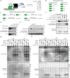
Extended Data Figure 1.. Phage display of TBD and application of mutations to CRBNmidi.
(a) Illustration of association TR-FRET assay. (b) Overview of PCMT1-catalyzed C-terminal cyclic imide formation on GLUL. (c) Overview of eSrtA reaction to engineer semisynthetic GFPs used in this study. (d) Western blot of GFP-PFQYKcN using CRBN/DDB1 in place of a primary antibody and using α-GFP antibody. (e) Workflow of TBD evolution.
Extended Data Figure 2.
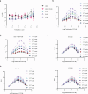
Extended Data Figure 2.. Optimization of cerebody linker from CRBNmidi.
(a) KDs of CRBNmidi mutations based on linker screen over 9 freeze-thaw cycles. (b) Signal to background ratios of CRBN midi with the native GSG linker across 9 freeze thaw cycles and different thal-FITC concentrations. (c) Signal to background ratios of CRBN midi with the native GSG linker and the mutation corresponding to P424A across 9 freeze thaw cycles and different thal-FITC concentrations. (d) Signal to background ratios of CRBN midi with an RFG linker across 9 freeze thaw cycles and different thal-FITC concentrations. (e) Signal to background ratios of CRBN midi with a RYD linker across 9 freeze thaw cycles and different thal-FITC concentrations. (f) Signal to background ratios of CRBN midi with a YYD linker across 9 freeze thaw cycles and different thal-FITC concentrations.
Extended Data Figure 3.
Extended Data Figure 3.. Optimization of cerebody from CRBNmidi-RYD
(a) Signal to background ratios of different CRBNmidi constructs. (b) Calculated KD values of CRBN midi mutations based on phage display over 9 freeze thaw cycles. (c) Western blotting of different concentrations of GFP-AcN with CRBNmidi mutations in 5% milk or 5% BSA blocking buffer.
Extended Data Figure 4.
Extended Data Figure 4.. Western blot with cerebody.
(a) Overview of C-terminal cyclic imide modified proteins. (b) Western blots of serial dilutions of GSS-cN, GFP-PFQYKcN, and GLUL incubated with PCMT1 for 14 h spiked in HEK293T lysate using different concentrations of cerebody in 5% milk in TBST as the primary antibody. (c) Cerebody blot of HEK293T WT and PCMT1 KO lysates with and without PCMT1 incubation.
Extended Data Figure 5.
Extended Data Figure 5.. Cerebody enrichment.
(a) Blot of GLUL with or without PCMT1 incubation that was spiked into HEK293T lysate prior to cerebody enrichment. (b) Blot of cerebody enrichment of HEK293T WT and PCMT1 KO cells with or without PCMT1 incubation for RPS20.
Figure 1.
Figure 1.. Development of a methodology to detect, image, and enrich C-terminal cyclic imides.
(a) Thalidomide, lenalidomide, and derivatives induce substrates to CRBN for ubiquitination and subsequent degradation. (b) Linear schematic of domains in CRBN. (c) Structures of thalidomide and lenalidomide. (d) Endogenous substrates bearing C-terminal cyclic imides are recognized for ubiquitinylation by CRBN, leading to their subsequent degradation. (e) Structure of the C-terminal cyclic imide degron from cyclization of asparagine (cN) or glutamine (cQ). (f–g) C-Terminal cyclic imides can be formed by protein damage (f) or enzymatic (g) mechanisms. (h) Applications of an ideal “cerebody” for CRBN substrate recognition by Western blot or affinity enrichment.
Figure 2.
Figure 2.. Development of cerebody, an antibody-like reagent for detecting C-terminal cyclic imides.
(a) Full length human CRBN, the thalidomide binding domain, CRBNmidi, and cerebody. Changes from CRBNmidi to cerebody highlighted in pink. (b) Dissociation constant (KD) of indicated constructs to dose response of thal-FITC by TR-FRET. (c) Relative dissociation constants of TBD, CRBN/DDB1, and CRBNmidi to thal-FITC and GFP-GLUL treated with PCMT1. (d) Linker region of CRBNmidi with visualization of the RYD linker (yellow) overlaid on the crystal structure of CRBNmidi bound to lenalidomide (maroon, PDB 8RQA). (e) Sampling of lowest predicted linker energies by computational modeling. Experimentally examined linkers are highlighted in red. (f) Calculated signal to background ratios of CRBNmidi with noted linker residues against thal-FITC (78 nM) across 9 freeze-thaw cycles. (g) Calculated signal to background ratios of CRBNmidi with RYD linker containing the indicated mutation against thal-FITC (78 nM) across 9 freeze-thaw cycles.
Figure 3.
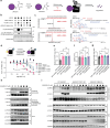
Figure 3.. Western blotting for C-terminal cyclic imides using cerebody reagent.
(a) Illustration of Western blot protocol with cerebody. (b) Engineered GFP displaying C-terminal cyclic imide or matched control generated by SrtA. (c) Schematic of CRBN substrate GLUL-cN generated by PCMT1 with associated controls. (d) Limit of detection of cerebody measured by serial dilution of GFP-PFQYKcN. (e) Cerebody detection of the indicated engineered GFP construct at 8 × 10⁻¹² mol. (f) Cerebody blot of recombinant GLUL or GLUL N373A following treatment with PCMT1 or inactive PCMT1 S60A control. (g) Cerebody blot of mouse brain and liver lysates with or without PCMT1 incubation (14 h). Bands appearing following PCMT1 incubation highlighted with yellow asterisks. (h) Cerebody blot of mouse spleen, kidney, and cardiac tissue lysates with or without PCMT1 incubation. Bands appearing following PCMT1 incubation highlighted with yellow asterisks.
Figure 4.
Figure 4.. Affinity enrichment of candidate CRBN substrates with cerebody from HEK293T cell lysates.
(a) Illustration of affinity enrichment procedure with cerebody. (b) Mass spectrometry analysis of HEK293T WT lysates with or without PCMT1 treatment. Green dots represent proteins with C-terminal asparagine. (c) Mass spectrometry analysis of HEK293T PCMT1 KO cells with or without PCMT1 treatment. Green dots represent proteins with C-terminal asparagine. (d) Western blot of proteins following affinity enrichment with cerebody. HEK293T WT or PCMT1 KO were treated with or without PCMT1 for 14 h.
Figure 5.
Figure 5.. Analysis of mouse tissue lysates following PCMT1 treatment and cerebody enrichment.
(a–c) Mass spectrometry analysis following cerebody enrichment of (a) brain, (b) liver, and (c) spleen tissue with or without PCMT1 incubation (14 h). Green dots represent proteins with C-terminal asparagine. (d–f) Western blot for GLUL following cerebody enrichment of (d) brain, (e) liver, and (f) spleen tissue. (g–h) Mass spectrometry analysis following cerebody enrichment of (g) kidney and (h) cardiac tissue with or without PCMT1 incubation (14 h). Green dots represent proteins with C-terminal asparagine. (i–j) Western blot for GLUL following cerebody enrichment of (i) kidney and (i) cardiac tissue.
Figure 6.
Figure 6.. UROD is an endogenous substrate of CRBN.
(a) Illustration of C-terminal cyclic imide formation on UROD leading to CRBN recognition and subsequent degradation. (b) Cerebody blot of UROD and UROD N367A incubated with or without PCMT1 or PCMT1 S60A for 19 h. (c) Selected ion monitoring and MS2 fragmentation spectra of the C-Terminal peptide of UROD treated with PCMT1 (17.5 h) and digested by Glu-C. (d) TR-FRET of UROD and UROD N367A following incubation with PCMT1 or PCMT1 S60A (17.5 h) by competitive displacement of thal-FITC against CRBN/DDB1. (e–f) Flow cytometry analysis of GFP-UROD incubated with PCMT1 (17.5 h) and electroporated into HEK293T WT (e) and CRBN KO (f) cells 6 h after electroporation. (g) Flow cytometry analysis of GFP-UROD following electroporation to HEK293T WT cells after 42 h. (h) Cerebody enrichment of HEK293T WT cells treated with MLN4924 or DMSO for 24 h prior to harvesting with DMSO or lenalidomide present during enrichment. (i) Blot of cerebody enrichment in HEK293T WT, PCMT1 KO, and CRBN KO cells with and without MLN4924 treatment (24 h) prior to harvesting.

Similar articles

References

    1. Higgins J. J., Pucilowska J., Lombardi R. Q., Rooney J. P. A mutation in a novel ATP-dependent Lon protease gene in a kindred with mild metal retardation. Neurology 63, 1927–1931 (2004). - PMC - PubMed
    1. Ito T. et al. Identification of a primary target of thalidomide teratogenicity. Science 327, 1345–1350 (2010). 10.1126/science.1177319 - DOI - PubMed
    1. Lupas A. N., Zhu H. & Korycinski M. The thalidomide-binding domain of cereblon defines the CULT domain family and is a new member of the beta-tent fold. PLoS Comput Biol 11, e1004023 (2015). 10.1371/journal.pcbi.1004023 - DOI - PMC - PubMed
    1. Kronke J. et al. Lenalidomide causes selective degradation of IKZF1 and IKZF3 in multiple myeloma cells. Science 343, 301–305 (2014). 10.1126/science.1244851 - DOI - PMC - PubMed
    1. Lu G. et al. The myeloma drug lenalidomide promotes the cereblon-dependent destruction of Ikaros proteins. Science 343, 305–309 (2014). 10.1126/science.1244917 - DOI - PMC - PubMed

Publication types

LinkOut - more resources