

The novel house dust mite allergen Der p 39 exacerbates atopic dermatitis-like inflammation in mice by inducing skin barrier dysfunction
- PMID: 40196722
- PMCID: PMC11973690
- DOI: 10.1016/j.waojou.2025.101036
The novel house dust mite allergen Der p 39 exacerbates atopic dermatitis-like inflammation in mice by inducing skin barrier dysfunction
Abstract
Background: House dust mite (HDM) allergens can induce or exacerbate allergic inflammation, including atopic dermatitis (AD). Substances that damage the epithelial barrier can trigger or worsen AD. The mechanism by which the novel HDM allergen Der p 39 induces allergic inflammation remains unclear. Our aim was to investigate the effects of Der p 39 on AD-like inflammation and associated mechanisms.
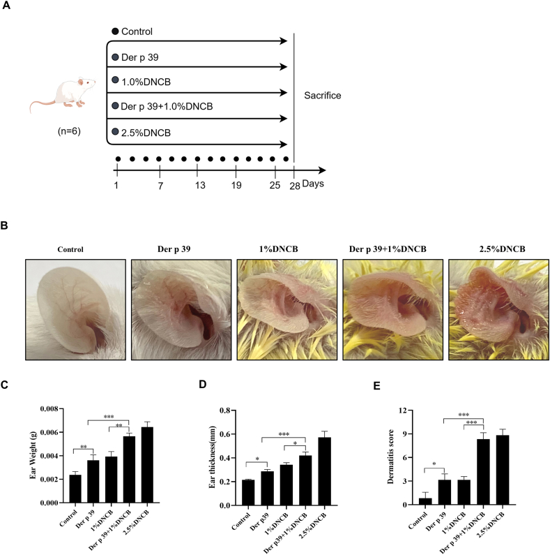
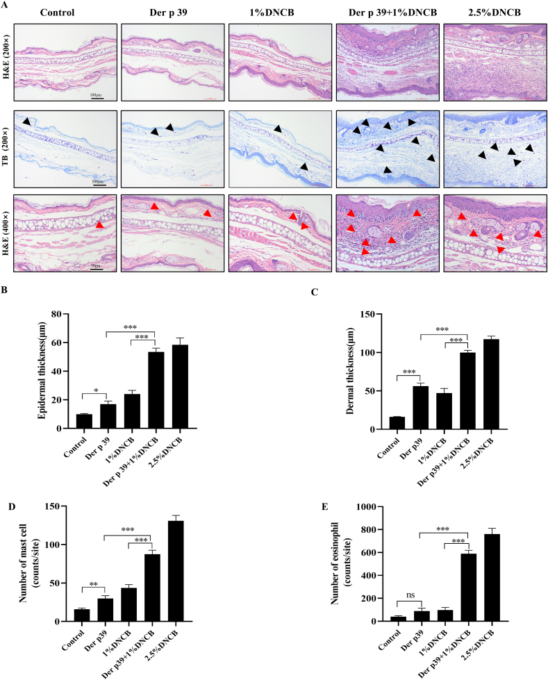
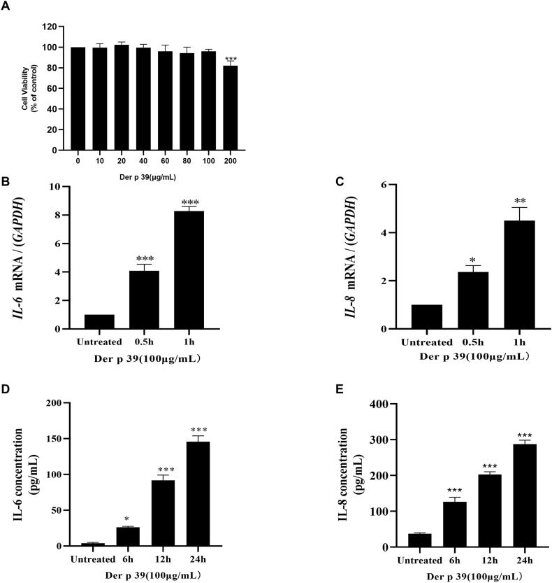
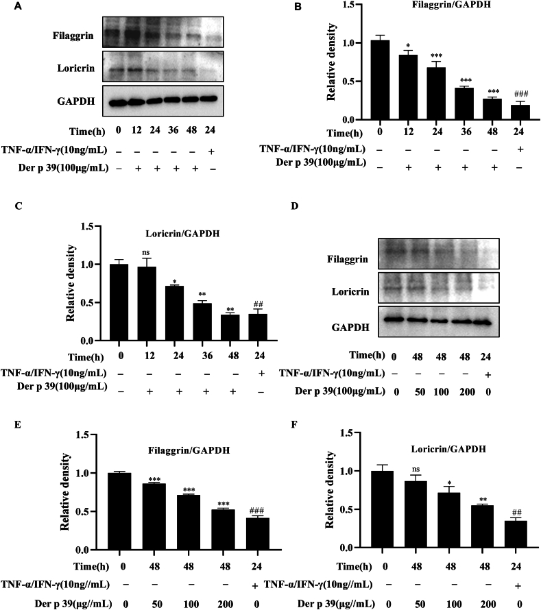
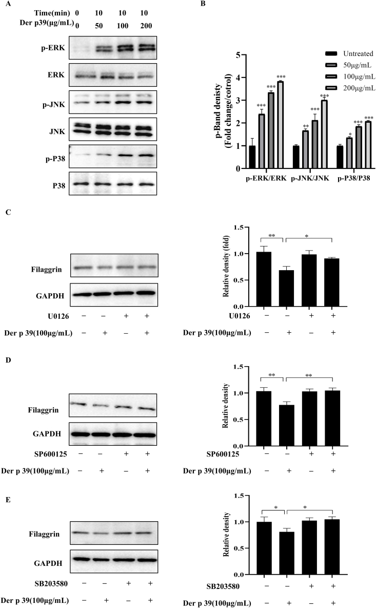
Methods: Dinitrochlorobenzene (DNCB) and Der p 39 were utilized to establish AD model mice. Inflammation severity was evaluated with physiological and morphological assays. The effects of Der p 39 on inflammatory cytokine release and skin barrier protein expression were examined in HaCaT cells (human epidermal keratinocytes). Mitogen-activated protein kinase (MAPK) activation was examined by western blots. MAPK inhibitors were employed to assess MAPK involvement in filaggrin expression.
Results: Der p 39 worsened allergic inflammation (tissue thickness) in murine ears pretreated with 1% DNCB. Compared to controls, Der p 39-sensitized tissues showed epidermal and dermal thickening with elevated numbers of mast cells and eosinophils in inflammatory lesions. Der p 39 increased transcription and production of pro-inflammatory interleukins (ILs), down-regulated expression of the skin barrier proteins filaggrin and loricrin, and upregulated phosphorylation of ERK, JNK and p38 in HaCaT cells. Inhibition of MAPK signaling rescued filaggrin expression in Der p 39-treated cells.
Conclusions: The HDM allergen Der p 39 enhances allergic inflammation and promotes MAPK pathway-mediated epidermal barrier dysfunction, suggesting that Der p 39 may possess pathogenic and clinically relevant immunomodulatory potential.
Keywords: Atopic dermatitis; Der p 39; House dust mite; Skin barrier; Skin inflammation.
© 2025 The Author(s).
Conflict of interest statement
The authors declare no competing interests.
Figures
Similar articles
-
Pathogenic Mechanism of Der p 38 as a Novel Allergen Homologous to RipA and RipB Proteins in Atopic Dermatitis.Front Immunol. 2021 Oct 8;12:646316. doi: 10.3389/fimmu.2021.646316. eCollection 2021. Front Immunol. 2021. PMID: 34691014 Free PMC article.
-
Non-IgE-reactive allergen peptides deteriorate the skin barrier in house dust mite-sensitized atopic dermatitis patients.Front Cell Dev Biol. 2023 Aug 22;11:1240289. doi: 10.3389/fcell.2023.1240289. eCollection 2023. Front Cell Dev Biol. 2023. PMID: 37675143 Free PMC article.
-
Anti-inflammatory effects of Eclipta prostrata Linné on house dust mite-induced atopic dermatitis in vivo and in vitro.J Ethnopharmacol. 2022 Jun 28;292:115233. doi: 10.1016/j.jep.2022.115233. Epub 2022 Mar 26. J Ethnopharmacol. 2022. PMID: 35346812
-
The effect of immunotherapy on cross-reactivity between house dust mite and other allergens in house dust mite -sensitized patients with allergic rhinitis.Expert Rev Clin Immunol. 2021 Sep;17(9):969-975. doi: 10.1080/1744666X.2021.1968834. Epub 2021 Aug 18. Expert Rev Clin Immunol. 2021. PMID: 34388949 Review.
-
A human-SCID mouse model for allergic immune response bacterial superantigen enhances skin inflammation and suppresses IgE production.J Invest Dermatol. 1998 Mar;110(3):224-31. doi: 10.1046/j.1523-1747.1998.00119.x. J Invest Dermatol. 1998. PMID: 9506440 Review.
References
-
- Miller J.D. The role of dust mites in allergy. Clin Rev Allergy Immunol. 2019 Dec 1;57(3):312–329. - PubMed
-
- Vrtala S. Allergens from house dust and storage mites. Allergo J Int. 2022 Dec 1;31(8):267–271.
-
- Huang H.J., Sarzsinszky E., Vrtala S. House dust mite allergy: the importance of house dust mite allergens for diagnosis and immunotherapy. Mol Immunol. 2023 Jun 1;158:54–67. - PubMed
LinkOut - more resources
Full Text Sources
Research Materials
Miscellaneous
