

Effects of high-flow oxygen therapy on oxygenation in dogs undergoing diagnostic bronchoscopy
- PMID: 40196814
- PMCID: PMC11974253
- DOI: 10.3389/fvets.2025.1545427
Effects of high-flow oxygen therapy on oxygenation in dogs undergoing diagnostic bronchoscopy
Abstract
Introduction: Hypoxemia is a common complication during bronchoscopy and bronchoalveolar lavage (BAL). High-Flow Oxygen Therapy (HFOT) has been used to improve oxygenation and prevent periods of hypoxemia in people undergoing bronchoscopy.
Objective: The main objective of this study was to evaluate the effect of HFOT on oxygenation in dogs undergoing diagnostic bronchoscopy compared to a traditional oxygen supplementation method (TOT). A secondary objective was to assess potential HFOT-related complications.
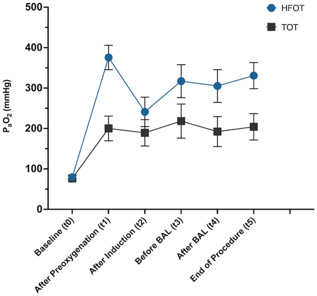
Methods: Prospective randomized clinical trial. Dogs presented for diagnostic bronchoscopy were randomly assigned to receive either HFOT or TOT using nasal cannulas during the bronchoscopic procedure. Oxygenation was monitored through PaO2 measurements taken at seven time points: baseline (t0), after preoxygenation (t1), post-induction (t2), pre- and post-BAL sampling (t3 and t4), at the end of the procedure (t5), and 1 h after bronchoscopy (t6). Pre- and post-procedure thoracic radiographs were assessed for air leak syndrome or aerophagia.
Results: 20 privately owned dogs presented for diagnostic bronchoscopy were included in the study (HFOT group: n = 10, TOT group: n = 10). Baseline characteristics and physiological parameters did not differ significantly between groups. Five dogs in each group showed hypoxemia (PaO2 < 80 mmHg) at baseline with 1/5 in each group having PaO2 < 60 mmHg. HFOT improved oxygenation throughout the procedure, with a significant increase in PaO2 observed after preoxygenation (P = 0.001) and at the end of the procedure (P = 0.013). Additionally, only 1/10 dogs in the HFOT group experienced hypoxemia during bronchoscopy compared to 5/10 dogs in the TOT group, and patients in the HFOT achieved numerically higher PaO2 values across all time points during the procedure (t1-t5). No serious adverse events related to HFOT were observed, although aerophagia occurred in both groups without necessitating intervention.
Conclusion: HFOT can improve oxygenation and prevent episodes of hypoxemia in dogs undergoing bronchoscopy compared to traditional oxygen supplementation methods.
Keywords: bronchoscopy; dogs; high flow oxygen therapy; hypoxemia; oxygen supplementation; oxygenation.
Copyright © 2025 Ortlieb, Bender, Schneider, Tacke and Hassdenteufel.
Conflict of interest statement
The authors declare that the research was conducted in the absence of any commercial or financial relationships that could be construed as a potential conflict of interest.
Figures


Similar articles
-
High flow oxygen therapy versus conventional oxygen therapy in dogs and cats undergoing bronchoscopy and bronchoalveolar lavage: a pilot study.Front Vet Sci. 2024 May 24;11:1360017. doi: 10.3389/fvets.2024.1360017. eCollection 2024. Front Vet Sci. 2024. PMID: 38855409 Free PMC article.
-
Retrospective evaluation of the effect of high flow oxygen therapy delivered by nasal cannula on PaO2 in dogs with moderate-to-severe hypoxemia.J Vet Emerg Crit Care (San Antonio). 2016 Jul;26(4):598-602. doi: 10.1111/vec.12495. Epub 2016 Jun 22. J Vet Emerg Crit Care (San Antonio). 2016. PMID: 27333466
-
Pre-oxygenation with high-flow oxygen through the nasopharyngeal airway compared to facemask on carbon dioxide clearance in emergency adults: a prospective randomized non-blinded clinical trial.Eur J Trauma Emerg Surg. 2024 Jun;50(3):1051-1061. doi: 10.1007/s00068-023-02418-2. Epub 2023 Dec 26. Eur J Trauma Emerg Surg. 2024. PMID: 38148421 Free PMC article. Clinical Trial.
-
High-flow nasal cannula for reducing hypoxemic events in patients undergoing bronchoscopy: A systematic review and meta-analysis of randomized trials.PLoS One. 2021 Dec 1;16(12):e0260716. doi: 10.1371/journal.pone.0260716. eCollection 2021. PLoS One. 2021. PMID: 34851996 Free PMC article.
-
The effectiveness of transnasal high flow nasal cannula in bronchoscopy under sedation: a systematic review and meta-analysis.Front Med (Lausanne). 2024 Jul 10;11:1428431. doi: 10.3389/fmed.2024.1428431. eCollection 2024. Front Med (Lausanne). 2024. PMID: 39050533 Free PMC article.
References
LinkOut - more resources
Full Text Sources